The high-affinity IFN-gamma receptor complex is made up of two type I membrane proteins, IFN-gamma R1 (IFN-gamma R alpha ) and IFN-gamma R2 (IFN-gamma R beta ). Both proteins are members of the type II cytokine receptor family and share approximately 52% amino acid sequence identity. IFN-gamma R1 is the ligand-binding subunit that is necessary and sufficient for IFN-gamma binding and receptor internalization. IFN-gamma R2 is required for IFN-gamma signaling but does not bind IFN-gamma by itself. Human IFN-gamma R1 cDNA encodes a 499 amino acid (aa) residue protein with a 17 aa signal peptide, a 228 aa extracellular domain, a 23 aa transmembrane domain, and a 221 aa intracellular domain. Human and mouse IFN-gamma R1 share 52% amino acid sequence identity and bind IFN-gamma in a species-specific manner. IFN-gamma R1 is constitutively expressed in most cell types. Soluble IFN-gamma R1 that binds IFN-gamma has been detected in biological fluids.
Human IFN-gamma R1/CD119 Antibody (92101)
R&D Systems | Catalog # MAB6731

Key Product Details
Species Reactivity
Validated:
Human
Cited:
Human
Applications
Validated:
Western Blot, Neutralization, Flow Cytometry, CyTOF-ready
Cited:
Western Blot, Neutralization, Flow Cytometry, Neutralizing
Label
Unconjugated
Antibody Source
Monoclonal Mouse IgG1 Clone # 92101
Loading...
Product Specifications
Immunogen
Mouse myeloma cell line NS0-derived recombinant human IFN-gamma R1
Glu18-Gly245
Accession # P15260.1
Glu18-Gly245
Accession # P15260.1
Specificity
Detects human IFN-gamma R1 in direct ELISAs and Western blots. In direct ELISAs, this antibody does not cross-react with recombinant mouse IFN‑ gamma R1, recombinant human (rh) IFN-gamma R2, or rhIL-10 R beta.
Clonality
Monoclonal
Host
Mouse
Isotype
IgG1
Endotoxin Level
<0.10 EU per 1 μg of the antibody by the LAL method.
Scientific Data Images for Human IFN-gamma R1/CD119 Antibody (92101)
IFN‑ gamma Inhibition of EMCV-induced Cytopathy and Neutralization by Human IFN‑ gamma R1/CD119 Antibody.
Recombinant Human IFN-gamma (Catalog # 285-IF) reduces the Encephalomyocarditis Virus (EMCV)-induced cytopathy in the HeLa human cervical epithelial carcinoma cell line in a dose-dependent manner (orange line). Inhibition of EMCV activity elicited by Recombinant Mouse Anti-Human IFN-gamma (2 ng/mL) is neutralized (green line) by increasing concentrations of Human IFN-gamma R1/CD119 Monoclonal Antibody (Catalog # MAB6731). The ND50 is typically 0.01-0.03 µg/mL.Applications for Human IFN-gamma R1/CD119 Antibody (92101)
Application
Recommended Usage
CyTOF-ready
Ready to be labeled using established conjugation methods. No BSA or other carrier proteins that could interfere with conjugation.
Flow Cytometry
2.5 µg/106 cells
Sample: Human whole blood monocytes
Sample: Human whole blood monocytes
Western Blot
1 µg/mL
Sample: Recombinant Human IFN-gamma R1/CD119 (Catalog # 673-IR)
Sample: Recombinant Human IFN-gamma R1/CD119 (Catalog # 673-IR)
Neutralization
Measured by its ability to neutralize IFN‑ gamma R1/CD119-mediated inhibition of EMCV-induced cytopathy in the HeLa human cervical epithelial carcinoma cell line. Meager, A. (1987) in Lymphokines and Interferons, a Practical Approach. Clemens, M.J. et al. (eds): IRL Press. 129. The Neutralization Dose (ND50) is typically 0.01-0.03 µg/mL in the presence of 2 ng/mL Recombinant Human IFN‑ gamma.
Reviewed Applications
Read 1 review rated 4 using MAB6731 in the following applications:
Flow Cytometry Panel Builder
Bio-Techne Knows Flow Cytometry
Save time and reduce costly mistakes by quickly finding compatible reagents using the Panel Builder Tool.
Advanced Features
- Spectra Viewer - Custom analysis of spectra from multiple fluorochromes
- Spillover Popups - Visualize the spectra of individual fluorochromes
- Antigen Density Selector - Match fluorochrome brightness with antigen density

Formulation, Preparation, and Storage
Purification
Protein A or G purified from ascites
Reconstitution
Reconstitute at 0.5 mg/mL in sterile PBS. For liquid material, refer to CoA for concentration.
Loading...
Formulation
Lyophilized from a 0.2 μm filtered solution in PBS with Trehalose. *Small pack size (SP) is supplied either lyophilized or as a 0.2 µm filtered solution in PBS.
Shipping
Lyophilized product is shipped at ambient temperature. Liquid small pack size (-SP) is shipped with polar packs. Upon receipt, store immediately at the temperature recommended below.
Stability & Storage
Use a manual defrost freezer and avoid repeated freeze-thaw cycles.
- 12 months from date of receipt, -20 to -70 °C as supplied.
- 1 month, 2 to 8 °C under sterile conditions after reconstitution.
- 6 months, -20 to -70 °C under sterile conditions after reconstitution.
Calculators
Background: IFN-gamma R1/CD119
References
- Bach, E.A. et al. (1997) Annu. Rev. Immunol. 15:563.
Long Name
Interferon gamma Receptor 1
Alternate Names
CD119, IFN-gR1, IFNgammaR1, IFNGR1
Gene Symbol
IFNGR1
UniProt
Additional IFN-gamma R1/CD119 Products
Product Documents for Human IFN-gamma R1/CD119 Antibody (92101)
Certificate of Analysis
To download a Certificate of Analysis, please enter a lot or batch number in the search box below.
Note: Certificate of Analysis not available for kit components.
Product Specific Notices for Human IFN-gamma R1/CD119 Antibody (92101)
For research use only
Citations for Human IFN-gamma R1/CD119 Antibody (92101)
 Powered by Bioz
Customer Reviews for Human IFN-gamma R1/CD119 Antibody (92101) (1)
4 out of 5
1 Customer Rating
Have you used Human IFN-gamma R1/CD119 Antibody (92101)?
Submit a review and receive an Amazon gift card!
$25/€18/£15/$25CAN/¥2500 Yen for a review with an image
$10/€7/£6/$10CAN/¥1110 Yen for a review without an image
Submit a review
Customer Images
Showing
1
-
1 of
1 review
Showing All
Filter By:
-
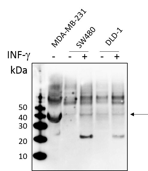
Application: Western BlotSample Tested: Cancer CellsSpecies: HumanVerified Customer | Posted 07/16/2018Cancer cells as indicated were treated with or without INF-g (10ng/ml) for 24h. Total cell lysates were subjected to western blot. PVDF membrane were probed with 1mm/ml Human Human IFN-gamma R1/CD119 Antibody (MAB6731). A specific band was detected for IFN-gamma R1/CD119 at approximately 43kDa. This experiment was conducted under reducing conditions

There are no reviews that match your criteria.
Protocols
Find general support by application which include: protocols, troubleshooting, illustrated assays, videos and webinars.
- 7-Amino Actinomycin D (7-AAD) Cell Viability Flow Cytometry Protocol
- Cellular Response to Hypoxia Protocols
- Extracellular Membrane Flow Cytometry Protocol
- Flow Cytometry Protocol for Cell Surface Markers
- Flow Cytometry Protocol for Staining Membrane Associated Proteins
- Flow Cytometry Staining Protocols
- Flow Cytometry Troubleshooting Guide
- Intracellular Flow Cytometry Protocol Using Alcohol (Methanol)
- Intracellular Flow Cytometry Protocol Using Detergents
- Intracellular Nuclear Staining Flow Cytometry Protocol Using Detergents
- Intracellular Staining Flow Cytometry Protocol Using Alcohol Permeabilization
- Intracellular Staining Flow Cytometry Protocol Using Detergents to Permeabilize Cells
- Propidium Iodide Cell Viability Flow Cytometry Protocol
- Protocol for Liperfluo
- Protocol for the Characterization of Human Th22 Cells
- Protocol for the Characterization of Human Th9 Cells
- Protocol: Annexin V and PI Staining by Flow Cytometry
- Protocol: Annexin V and PI Staining for Apoptosis by Flow Cytometry
- R&D Systems Quality Control Western Blot Protocol
- Troubleshooting Guide: Fluorokine Flow Cytometry Kits
- Troubleshooting Guide: Western Blot Figures
- Western Blot Conditions
- Western Blot Protocol
- Western Blot Protocol for Cell Lysates
- Western Blot Troubleshooting
- Western Blot Troubleshooting Guide
- View all Protocols, Troubleshooting, Illustrated assays and Webinars
Loading...
Associated Pathways

