
Key Product Details
Species Reactivity
Validated:
Human
Cited:
Human
Applications
Validated:
Western Blot, Flow Cytometry, CyTOF-ready
Cited:
Western Blot, ELISA Development (Capture)
Label
Unconjugated
Antibody Source
Polyclonal Goat IgG
Loading...
Product Specifications
Immunogen
S. frugiperda insect ovarian cell line Sf 21-derived recombinant human IL‑1 RAcP/IL‑1 R3
Ser21-Glu359
Accession # Q9NPH3
Ser21-Glu359
Accession # Q9NPH3
Specificity
Detects human IL‑1 RAcP/IL‑1 R3 in direct ELISAs and Western blots. In direct ELISAs, approximately 25% cross-reactivity with recombinant mouse IL-1 RAcP is observed.
Clonality
Polyclonal
Host
Goat
Isotype
IgG
Scientific Data Images for Human IL‑1 RAcP/IL‑1 R3 Antibody
Detection of IL‑1 RAcP/IL‑1 R3 in PBMC Monocytes by Flow Cytometry
Human peripheral blood monocytes were stained with Goat Anti-Human IL‑1 RAcP/IL‑1 R3 Antigen Affinity-purified Polyclonal Antibody (Catalog # AF676, filled histogram) or isotype control antibody (Catalog # AB-108-C, open histogram) followed by Phycoerythrin-conjugated Anti-Goat IgG Secondary Antibody (Catalog # F0107). View our protocol for Staining Membrane-associated Proteins.Applications for Human IL‑1 RAcP/IL‑1 R3 Antibody
Application
Recommended Usage
CyTOF-ready
Ready to be labeled using established conjugation methods. No BSA or other carrier proteins that could interfere with conjugation.
Flow Cytometry
2.5 µg/106 cells
Sample: Human peripheral blood monocytes
Sample: Human peripheral blood monocytes
Western Blot
0.1 µg/mL
Sample: Recombinant Human IL‑1 RAcP/IL‑1 R3 Fc Chimera (Catalog # 676-CP)
Sample: Recombinant Human IL‑1 RAcP/IL‑1 R3 Fc Chimera (Catalog # 676-CP)
Reviewed Applications
Read 2 reviews rated 5 using AF676 in the following applications:
Flow Cytometry Panel Builder
Bio-Techne Knows Flow Cytometry
Save time and reduce costly mistakes by quickly finding compatible reagents using the Panel Builder Tool.
Advanced Features
- Spectra Viewer - Custom analysis of spectra from multiple fluorochromes
- Spillover Popups - Visualize the spectra of individual fluorochromes
- Antigen Density Selector - Match fluorochrome brightness with antigen density

Formulation, Preparation, and Storage
Purification
Antigen Affinity-purified
Reconstitution
Reconstitute at 0.2 mg/mL in sterile PBS. For liquid material, refer to CoA for concentration.
Loading...
Formulation
Lyophilized from a 0.2 μm filtered solution in PBS with Trehalose. See Certificate of Analysis for details.
*Small pack size (-SP) is supplied either lyophilized or as a 0.2 µm filtered solution in PBS.
*Small pack size (-SP) is supplied either lyophilized or as a 0.2 µm filtered solution in PBS.
Shipping
Lyophilized product is shipped at ambient temperature. Liquid small pack size (-SP) is shipped with polar packs. Upon receipt, store immediately at the temperature recommended below.
Stability & Storage
Use a manual defrost freezer and avoid repeated freeze-thaw cycles.
- 12 months from date of receipt, -20 to -70 °C as supplied.
- 1 month, 2 to 8 °C under sterile conditions after reconstitution.
- 6 months, -20 to -70 °C under sterile conditions after reconstitution.
Calculators
Background: IL-1 RAcP/IL-1 R3
non‑ligand‑binding accessory component of the receptors for IL-1 alpha, IL-1 beta, and IL-33 (6, 7). Together with IRAK4 and MyD88, it generates a functional signaling complex with IL‑1 RI; by itself, it generates a non-signaling, but high-affinity binding complex with IL-1 RII (8). In addition, it interacts with ST2 on mast cells and Th2 T cells to create a functional IL-33 receptor complex (7). Mature human IL-1 RAcP is a type I transmembrane glycoprotein that is 550 amino acids in length. It contains a 347 amino acid (aa) extracellular region (aa 21-367), a 21 aa transmembrane segment, and a 182 aa cytoplasmic domain (9). The extracellular region shows three C2‑type Ig-like domains, the most membrane proximal of which is suggested to be responsible for dimerization with IL-1 RI (10). There are three alternative splice forms reported for IL-1 RAcP. One is transmembrane, and shows a 239 aa substitution for the C-terminal 122 amino acids (11). The other two are soluble; one shows a six aa substitution for aa 351-570, while a second shows a 45 aa substitution for aa 302-579 (12, 13). The soluble receptor isoforms appear to be inhibitory to IL-1 signaling. When present with soluble IL-1 RII, soluble IL-1 RAcP increases the IL-1 binding affinity of IL-1 RII more than 100-fold, thus neutralizing the effects of IL-1 (14). The human and mouse IL-1 RAcP precursors are 89% aa identical; within the extracellular region, they share 86% aa identity.
References
- Subramaniam, S. et al. (2004) Dev. Comp. Immunol. 28:415.
- Boraschi, D. and A. Tagliabue (2006) Vitam. Horm. 74:229.
- Dunne, A. and L.A.J. O'Neill (2003) Sci STKE. Feb 25;2003(171):re3.
- Huang, J. et al. (1997) Proc. Natl. Acad. Sci. USA 94:12829.
- Greenfeder, S. A. et al. (1995) J. Biol. Chem. 270:13757.
- Brikos, C. et al. (2007) Mol. Cell. Proteomics 6:1551.
- Chackerian, A.A. et al. (2007) J. Immunol. 179:2551.
- Lang, D. et al. (1998) J. Immunol. 161:6871.
- SwissProt. Accession # Q9NPH3.
- Yoon, D-Y. and C.A. Dinarello (1998) J. Immunol. 160:3170.
- Lu, H-L. et al. (2008) Mol. Immunol. 45:1374.
- Jensen, L.E. et al. (2000) J. Immunol. 164:5277.
- Jensen, L.E. and A.S. Whitehead (2003) Cell. Signal. 15:793.
- Smith, D.E. et al. (2003) Immunity 18:87.
Long Name
Interleukin 1 Receptor Accessory Protein
Alternate Names
IL-1 R3, IL-1RAcP, IL1RAcP, IL1RAP
Gene Symbol
IL1RAP
UniProt
Additional IL-1 RAcP/IL-1 R3 Products
Product Documents for Human IL‑1 RAcP/IL‑1 R3 Antibody
Certificate of Analysis
To download a Certificate of Analysis, please enter a lot or batch number in the search box below.
Note: Certificate of Analysis not available for kit components.
Product Specific Notices for Human IL‑1 RAcP/IL‑1 R3 Antibody
For research use only
Citations for Human IL‑1 RAcP/IL‑1 R3 Antibody
 Powered by Bioz
Customer Reviews for Human IL‑1 RAcP/IL‑1 R3 Antibody (2)
5 out of 5
2 Customer Ratings
Have you used Human IL‑1 RAcP/IL‑1 R3 Antibody?
Submit a review and receive an Amazon gift card!
$25/€18/£15/$25CAN/¥2500 Yen for a review with an image
$10/€7/£6/$10CAN/¥1110 Yen for a review without an image
Submit a review
Customer Images
Showing
1
-
2 of
2 reviews
Showing All
Filter By:
-
Application: ELISASample Tested: Serum and PlasmaSpecies: HumanVerified Customer | Posted 05/17/2019

-
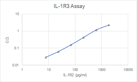
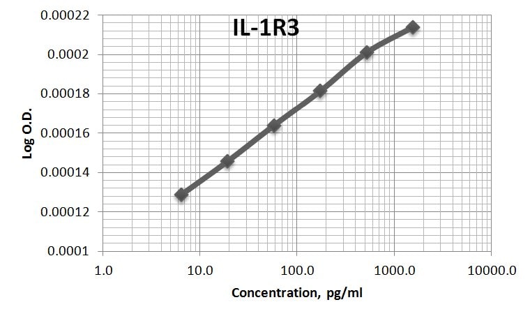
Application: ELISASample Tested: Serum and Plasma and Cell culture supernatantSpecies: HumanVerified Customer | Posted 11/09/2017AF676was used as both the capture and the detection antibody for the sandwich ELISA for sIL-1R3. The immunoassay standard was 676-CP. Assay had sensitivity of ~6pg/ml.
There are no reviews that match your criteria.
Protocols
Find general support by application which include: protocols, troubleshooting, illustrated assays, videos and webinars.
- 7-Amino Actinomycin D (7-AAD) Cell Viability Flow Cytometry Protocol
- Cellular Response to Hypoxia Protocols
- Extracellular Membrane Flow Cytometry Protocol
- Flow Cytometry Protocol for Cell Surface Markers
- Flow Cytometry Protocol for Staining Membrane Associated Proteins
- Flow Cytometry Staining Protocols
- Flow Cytometry Troubleshooting Guide
- Intracellular Flow Cytometry Protocol Using Alcohol (Methanol)
- Intracellular Flow Cytometry Protocol Using Detergents
- Intracellular Nuclear Staining Flow Cytometry Protocol Using Detergents
- Intracellular Staining Flow Cytometry Protocol Using Alcohol Permeabilization
- Intracellular Staining Flow Cytometry Protocol Using Detergents to Permeabilize Cells
- Propidium Iodide Cell Viability Flow Cytometry Protocol
- Protocol for Liperfluo
- Protocol for the Characterization of Human Th22 Cells
- Protocol for the Characterization of Human Th9 Cells
- Protocol: Annexin V and PI Staining by Flow Cytometry
- Protocol: Annexin V and PI Staining for Apoptosis by Flow Cytometry
- R&D Systems Quality Control Western Blot Protocol
- Troubleshooting Guide: Fluorokine Flow Cytometry Kits
- Troubleshooting Guide: Western Blot Figures
- Western Blot Conditions
- Western Blot Protocol
- Western Blot Protocol for Cell Lysates
- Western Blot Troubleshooting
- Western Blot Troubleshooting Guide
- View all Protocols, Troubleshooting, Illustrated assays and Webinars
Loading...
Associated Pathways


