
The interleukin-31 receptor A subunit (IL-31 RA), also known as gp130-Like Monocyte Receptor (GLM-R or GPL), is a ~100 kDa type I transmembrane glycoprotein that is classified as being a type I cytokine receptor (1, 2). A heterodimeric complex of IL-31 RA and the oncostatin M receptor (OSM-R) functions as the signaling receptor for IL-31 (3). Both subunits are inducibly expressed throughout the myelomonocytic lineage and are upregulated by interferon-gamma and bacterial lipopolysaccharides (1‑3). IL-31 RA is also expressed on keratinocytes, dorsal root ganglia neurons, and variably on lung epithelial cells (3‑6). The 732 amino acid (aa) IL-31 RA contains a 19 aa signal sequence, a 500 aa extracellular domain (ECD), a 21 aa transmembrane domain and a 192 aa cytoplasmic domain. The ECD shares 60%, 58%, 73% and 70% aa identity with mouse, rat, canine and bovine IL-31 RA ECD, respectively. Human IL-31 receptors do not respond to mouse IL-31 (7). The ECD contains five fibronectin type III domains; the first two contain four conserved cysteine residues and a WSXWS motif common to type I cytokine receptors (2). Twelve alternately spliced human IL-31 RA isoforms are known and range in size from 356‑745 amino acids. A long (745 aa) and a short (560 aa) transmembrane form are the predominant forms, and many cell lines express both forms (8). The long form, like the 732 aa form, signals by recruiting STAT3, 5 or 1, while the short form does not recruit STATs and inhibits IL-31 signaling. The ratio of these forms and their co-expression with OSM-R determines a cell’s response to IL‑31 (8). In both humans and transgenic mice, IL-31 from skin-homing Th2 cells may contribute to the pruritis (itching) associated with nonatopic dermatitis, especially in infected skin (3, 9, 10).
Human IL-31RA Antibody
R&D Systems | Catalog # AF2769


Scientific images may be resized, cropped, or adjusted for brightness or contrast for presentation purposes. These updates are limited to presentation and do not affect the underlying scientific interpretation of the data. Where available, additional source data may be provided upon request.
Key Product Details
Species Reactivity
Validated:
Cited:
Applications
Validated:
Cited:
Label
Antibody Source
Product Specifications
Immunogen
Ala20-Ser516
Accession # Q8NI17
Specificity
Clonality
Host
Isotype
Scientific Data Images for Human IL-31RA Antibody
Detection of IL‑31RA in U937 Human Cell Line by Flow Cytometry.
U937 human histiocytic lymphoma cell line was stained with Goat Anti-Human IL-31 RA Antigen Affinity-purified Polyclonal Antibody (Catalog # AF2769, filled histogram) or isotype control antibody (Catalog # AB-108-C, open histo-gram), followed by Phycoerythrin-conjugated Anti-Goat IgG Secondary Antibody (Catalog # F0107).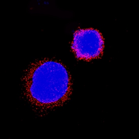
IL‑31 RA in K562 Human Cell Line.
IL-31 RA was detected in immersion fixed K562 human chronic myelogenous leukemia cell line using Goat Anti-Human IL-31 RA Antigen Affinity-purified Polyclonal Antibody (Catalog # AF2769) at 5 µg/mL for 3 hours at room temperature. Cells were stained using the NorthernLights™ 557-conjugated Anti-Goat IgG Secondary Antibody (red; Catalog # NL001) and counterstained with DAPI (blue). Specific staining was localized to cell surfaces and cytoplasm. View our protocol for Fluorescent ICC Staining of Non-adherent Cells.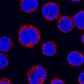
IL‑31 RA in THP‑1 Human Cell Line.
IL-31 RA was detected in immersion fixed THP-1 human acute monocytic leukemia cell line using Goat Anti-Human IL-31 RA Antigen Affinity-purified Polyclonal Antibody (Catalog # AF2769) at 15 µg/mL for 3 hours at room temperature. Cells were stained using the NorthernLights™ 557-conjugated Anti-Goat IgG Secondary Antibody (red; Catalog # NL001) and counterstained with DAPI (blue). Specific staining was localized to cytoplasm. View our protocol for Fluorescent ICC Staining of Non-adherent Cells.Applications for Human IL-31RA Antibody
CyTOF-ready
Flow Cytometry
Sample: U937 human histiocytic lymphoma cell line
Immunocytochemistry
Sample: Immersion fixed U937 human histiocytic lymphoma cell line, immersion fixed K562 human chronic myelogenous leukemia cell line, and immersion fixed THP-1 human acute monocytic leukemia cell line
Western Blot
Sample: Recombinant Human IL‑31 RA (Catalog # 2769-IL)
Flow Cytometry Panel Builder
Bio-Techne Knows Flow Cytometry
Save time and reduce costly mistakes by quickly finding compatible reagents using the Panel Builder Tool.
Advanced Features
- Spectra Viewer - Custom analysis of spectra from multiple fluorochromes
- Spillover Popups - Visualize the spectra of individual fluorochromes
- Antigen Density Selector - Match fluorochrome brightness with antigen density

Formulation, Preparation, and Storage
Purification
Reconstitution
Reconstitute at 0.2 mg/mL in sterile PBS. For liquid material, refer to CoA for concentration.
Formulation
Shipping
Stability & Storage
- 12 months from date of receipt, -20 to -70 °C as supplied.
- 1 month, 2 to 8 °C under sterile conditions after reconstitution.
- 6 months, -20 to -70 °C under sterile conditions after reconstitution.
Calculators
Background: IL-31RA
References
- Ghilardi, N. et al. (2002) J. Biol. Chem. 277:16831.
- Diveu, C. et al. (2003) J. Biol. Chem. 278:49850.
- Dillon, S. R. et al. (2004) Nat. Immunol. 5:752.
- Chattopadhyay, S. et al. (2007) J. Biol. Chem. 282:3014.
- Perrigoue, J. G. et al. (2007) J. Exp. Med. 204:481.
- Bando, T. et al. (2006) Neuroscience 142:1263.
- Broxmeyer, H. E. et al. (2007) Exp. Hematol. 35:78.
- Diveu, C. et al. (2004) Eur. Cytokine. Netw. 15:291.
- Bilsborough, J. et al. (2006) J. Allergy Clin. Immunol. 117:418.
- Sonkoly, E. et al. (2006) J. Allergy Clin. Immunol. 117:411.
Long Name
Alternate Names
Gene Symbol
UniProt
Additional IL-31RA Products
Product Documents for Human IL-31RA Antibody
Certificate of Analysis
To download a Certificate of Analysis, please enter a lot or batch number in the search box below.
Note: Certificate of Analysis not available for kit components.
Product Specific Notices for Human IL-31RA Antibody
For research use only
Related Research Areas
Citations for Human IL-31RA Antibody
 Powered by Bioz
Customer Reviews for Human IL-31RA Antibody
There are currently no reviews for this product. Be the first to review Human IL-31RA Antibody and earn rewards!
Have you used Human IL-31RA Antibody?
Submit a review and receive an Amazon gift card!
$25/€18/£15/$25CAN/¥2500 for a review with an image
$10/€7/£6/$10CAN/¥1110 for a review without an image
Submit a review
Protocols
Find general support by application which include: protocols, troubleshooting, illustrated assays, videos and webinars.
- 7-Amino Actinomycin D (7-AAD) Cell Viability Flow Cytometry Protocol
- Appropriate Fixation of IHC/ICC Samples
- Cellular Response to Hypoxia Protocols
- ClariTSA™ Fluorophore Kits
- Detection & Visualization of Antibody Binding
- Extracellular Membrane Flow Cytometry Protocol
- Flow Cytometry Protocol for Cell Surface Markers
- Flow Cytometry Protocol for Staining Membrane Associated Proteins
- Flow Cytometry Staining Protocols
- Flow Cytometry Troubleshooting Guide
- ICC Cell Smear Protocol for Suspension Cells
- ICC Immunocytochemistry Protocol Videos
- ICC for Adherent Cells
- Immunocytochemistry (ICC) Protocol
- Immunocytochemistry Troubleshooting
- Immunofluorescence of Organoids Embedded in Cultrex Basement Membrane Extract
- Immunohistochemistry (IHC) and Immunocytochemistry (ICC) Protocols
- Intracellular Flow Cytometry Protocol Using Alcohol (Methanol)
- Intracellular Flow Cytometry Protocol Using Detergents
- Intracellular Nuclear Staining Flow Cytometry Protocol Using Detergents
- Intracellular Staining Flow Cytometry Protocol Using Alcohol Permeabilization
- Intracellular Staining Flow Cytometry Protocol Using Detergents to Permeabilize Cells
- Preparing Samples for IHC/ICC Experiments
- Preventing Non-Specific Staining (Non-Specific Binding)
- Primary Antibody Selection & Optimization
- Propidium Iodide Cell Viability Flow Cytometry Protocol
- Protocol for Liperfluo
- Protocol for VisUCyte™ HRP Polymer Detection Reagent
- Protocol for the Characterization of Human Th22 Cells
- Protocol for the Characterization of Human Th9 Cells
- Protocol for the Fluorescent ICC Staining of Cell Smears - Graphic
- Protocol for the Fluorescent ICC Staining of Cultured Cells on Coverslips - Graphic
- Protocol for the Preparation and Fluorescent ICC Staining of Cells on Coverslips
- Protocol for the Preparation and Fluorescent ICC Staining of Non-adherent Cells
- Protocol for the Preparation and Fluorescent ICC Staining of Stem Cells on Coverslips
- Protocol for the Preparation of a Cell Smear for Non-adherent Cell ICC - Graphic
- Protocol: Annexin V and PI Staining by Flow Cytometry
- Protocol: Annexin V and PI Staining for Apoptosis by Flow Cytometry
- R&D Systems Quality Control Western Blot Protocol
- TUNEL and Active Caspase-3 Detection by IHC/ICC Protocol
- The Importance of IHC/ICC Controls
- Troubleshooting Guide: Fluorokine Flow Cytometry Kits
- Troubleshooting Guide: Western Blot Figures
- Western Blot Conditions
- Western Blot Protocol
- Western Blot Protocol for Cell Lysates
- Western Blot Troubleshooting
- Western Blot Troubleshooting Guide
- View all Protocols, Troubleshooting, Illustrated assays and Webinars