Human Integrin alpha 2b/CD41 Antibody (745201)
R&D Systems | Catalog # MAB7616

Key Product Details
Species Reactivity
Validated:
Human
Cited:
Human, Mouse, Chinese Hamster
Applications
Validated:
Western Blot, Flow Cytometry, CyTOF-ready
Cited:
Western Blot, ELISA Detection, Interferent Testing
Label
Unconjugated
Antibody Source
Monoclonal Mouse IgG1 Clone # 745201
Loading...
Product Specifications
Immunogen
heterodimer of human Integrin alpha 2B (Leu32-Arg993; R887L) Accession P08514 + human Integrin beta 3 (Gly27-Asp718) Accession P05106
Specificity
Detects human Integrin alpha 2b/CD41 in direct ELISAs.
In direct ELISAs, no cross-reactivity
with recombinant human Integrin alpha 5, alpha 8, alpha V, beta 3, beta 5, beta 6, recombinant mouse Integrin beta 2b or beta 3 is observed.
Clonality
Monoclonal
Host
Mouse
Isotype
IgG1
Scientific Data Images for Human Integrin alpha 2b/CD41 Antibody (745201)
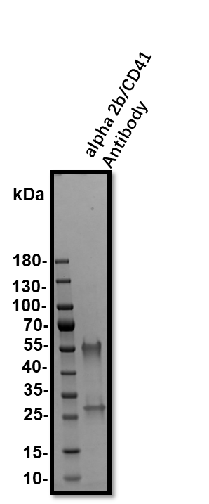
Detection of Human Integrin alpha 2b/CD41 by Western Blot.
Western blot shows lysates of human platelets. PVDF membrane was probed with 2 µg/mL of Mouse Anti-Human Integrin a2b/CD41 Monoclonal Antibody (Catalog # MAB7616) followed by HRP-conjugated Anti-Mouse IgG Secondary Antibody (HAF007). A specific band was detected for Integrin a2b/CD41 at approximately 130 kDa (as indicated). This experiment was conducted under reducing conditions and using Immunoblot Buffer Group 5.
Detection of Integrin alpha 2b/CD41 in Human Blood Platelets by Flow Cytometry.
Human peripheral blood platelets were stained with Fluorescein-conjugated Anti-Mouse Integrin a2b/CD41 (Clone P2) and either (A) Mouse IgG1Isotype Control (MAB002) or (B) Mouse Anti-Human Integrin a2b/CD41 Mono-clonal Antibody (Catalog # MAB7616) followed by Allophycocyanin-conjugated Anti-Mouse IgG Secondary Antibody (Catalog # F0101B). MAB7616 stains the same population as Clone P2.
Detection of Integrin alpha 2b/CD41 in Human Blood Platelets by Flow Cytometry.
Human peripheral blood platelets were stained with Mouse Anti-Human Integrin a2b/CD41 Mono-clonal Antibody (Catalog # MAB7616, filled histogram) or Mouse IgG1 Isotype Control (MAB002, open histogram) followed by Allophycocyanin-conjugated Anti-Mouse IgG Secondary Antibody (F0101B). View our protocol for Staining Membrane-associated Proteins.Applications for Human Integrin alpha 2b/CD41 Antibody (745201)
Application
Recommended Usage
CyTOF-ready
Ready to be labeled using established conjugation methods. No BSA or other carrier proteins that could interfere with conjugation.
Flow Cytometry
0.25 µg/106 cells
Sample: Human peripheral blood platelets
Sample: Human peripheral blood platelets
Western Blot
2 µg/mL
Sample: Human platelets
Sample: Human platelets
Reviewed Applications
Read 1 review rated 5 using MAB7616 in the following applications:
Flow Cytometry Panel Builder
Bio-Techne Knows Flow Cytometry
Save time and reduce costly mistakes by quickly finding compatible reagents using the Panel Builder Tool.
Advanced Features
- Spectra Viewer - Custom analysis of spectra from multiple fluorochromes
- Spillover Popups - Visualize the spectra of individual fluorochromes
- Antigen Density Selector - Match fluorochrome brightness with antigen density

Formulation, Preparation, and Storage
Purification
Protein A or G purified from hybridoma culture supernatant
Reconstitution
Sterile PBS to a final concentration of 0.5 mg/mL. For liquid material, refer to CoA for concentration.
Loading...
Formulation
Lyophilized from a 0.2 μm filtered solution in PBS with Trehalose. *Small pack size (SP) is supplied either lyophilized or as a 0.2 µm filtered solution in PBS.
Shipping
Lyophilized product is shipped at ambient temperature. Liquid small pack size (-SP) is shipped with polar packs. Upon receipt, store immediately at the temperature recommended below.
Stability & Storage
Use a manual defrost freezer and avoid repeated freeze-thaw cycles.
- 12 months from date of receipt, -20 to -70 °C as supplied.
- 1 month, 2 to 8 °C under sterile conditions after reconstitution.
- 6 months, -20 to -70 °C under sterile conditions after reconstitution.
Calculators
Background: Integrin alpha 2b/CD41
Alternate Names
CD41, GP2B, GPIIb, GTA, HPA3, ITGA2b
Gene Symbol
ITGA2B
Additional Integrin alpha 2b/CD41 Products
Product Documents for Human Integrin alpha 2b/CD41 Antibody (745201)
Certificate of Analysis
To download a Certificate of Analysis, please enter a lot or batch number in the search box below.
Note: Certificate of Analysis not available for kit components.
Product Specific Notices for Human Integrin alpha 2b/CD41 Antibody (745201)
For research use only
Related Research Areas
Citations for Human Integrin alpha 2b/CD41 Antibody (745201)
 Powered by Bioz
Customer Reviews for Human Integrin alpha 2b/CD41 Antibody (745201) (1)
5 out of 5
1 Customer Rating
Have you used Human Integrin alpha 2b/CD41 Antibody (745201)?
Submit a review and receive an Amazon gift card!
$25/€18/£15/$25CAN/¥2500 Yen for a review with an image
$10/€7/£6/$10CAN/¥1110 Yen for a review without an image
Submit a review
Customer Images
Showing
1
-
1 of
1 review
Showing All
Filter By:
-
Application: Western BlotSample Tested: Serum and PlasmaSpecies: HumanVerified Customer | Posted 09/22/2019

There are no reviews that match your criteria.
Protocols
Find general support by application which include: protocols, troubleshooting, illustrated assays, videos and webinars.
- 7-Amino Actinomycin D (7-AAD) Cell Viability Flow Cytometry Protocol
- Cellular Response to Hypoxia Protocols
- Extracellular Membrane Flow Cytometry Protocol
- Flow Cytometry Protocol for Cell Surface Markers
- Flow Cytometry Protocol for Staining Membrane Associated Proteins
- Flow Cytometry Staining Protocols
- Flow Cytometry Troubleshooting Guide
- Intracellular Flow Cytometry Protocol Using Alcohol (Methanol)
- Intracellular Flow Cytometry Protocol Using Detergents
- Intracellular Nuclear Staining Flow Cytometry Protocol Using Detergents
- Intracellular Staining Flow Cytometry Protocol Using Alcohol Permeabilization
- Intracellular Staining Flow Cytometry Protocol Using Detergents to Permeabilize Cells
- Propidium Iodide Cell Viability Flow Cytometry Protocol
- Protocol for Liperfluo
- Protocol for the Characterization of Human Th22 Cells
- Protocol for the Characterization of Human Th9 Cells
- Protocol: Annexin V and PI Staining by Flow Cytometry
- Protocol: Annexin V and PI Staining for Apoptosis by Flow Cytometry
- R&D Systems Quality Control Western Blot Protocol
- Troubleshooting Guide: Fluorokine Flow Cytometry Kits
- Troubleshooting Guide: Western Blot Figures
- Western Blot Conditions
- Western Blot Protocol
- Western Blot Protocol for Cell Lysates
- Western Blot Troubleshooting
- Western Blot Troubleshooting Guide
- View all Protocols, Troubleshooting, Illustrated assays and Webinars
Loading...
Associated Pathways

