Human Integrin alpha V beta 5 Antibody (P5H9)
R&D Systems | Catalog # MAB2528

Key Product Details
Species Reactivity
Validated:
Human
Cited:
Human, Transgenic Mouse
Applications
Validated:
Adhesion Blockade, Flow Cytometry, Immunocytochemistry, Immunoprecipitation, CyTOF-ready
Cited:
Immunohistochemistry, Neutralization, Flow Cytometry, Immunocytochemistry
Label
Unconjugated
Antibody Source
Monoclonal Mouse IgG1 Clone # P5H9
Loading...
Product Specifications
Immunogen
HT1080 human fibrosarcoma cell line
Specificity
Detects human Integrin alpha V beta 5. Recognizes the human Integrin alpha V beta 5 heterodimer and does not recognize the alpha V subunit in association with any other beta subunits.
Clonality
Monoclonal
Host
Mouse
Isotype
IgG1
Endotoxin Level
<0.10 EU per 1 μg of the antibody by the LAL method.
Scientific Data Images for Human Integrin alpha V beta 5 Antibody (P5H9)
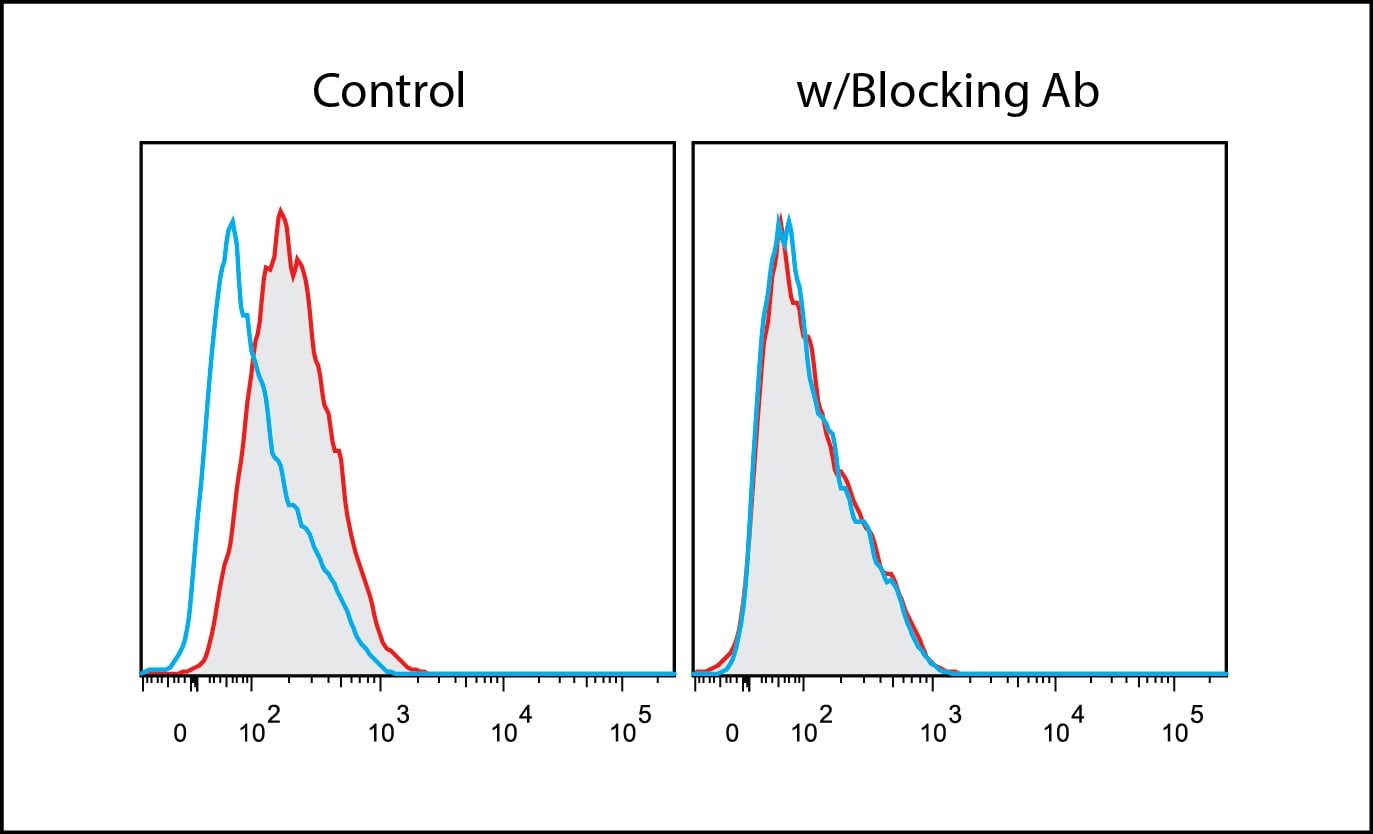
Integrin alpha V beta 5 in HT1080 Human Cell Line.
Integrin aV beta 5 was detected in immersion fixed HT1080 human fibrosarcoma cell line using Human Integrin aV beta 5 Monoclonal Antibody (Catalog # MAB2528) at 10 µg/mL for 3 hours at room temperature. Cells were stained using the NorthernLights™ 557-conjugated Anti-Mouse IgG Secondary Antibody (red; NL007) and counterstained with DAPI (blue). View our protocol for Fluorescent ICC Staining of Cells on Coverslips.
Detection of Human Integrin alpha V beta 5 by Immunocytochemistry/Immunofluorescence
alpha V beta 3 and EGFR mediate MBsome signaling. a–d HeLa cells were incubated with purified GFP-MBs for 3 h, followed by wash and another incubation for 24 h. Cells were then fixed and stained with anti-alpha V beta 3 (a), anti-alpha V beta 5 (b), anti-phospho-FAK (d) antibodies. Panels in c show control staining where primary antibodies were not added. Boxed regions mark the part of the image shown as a higher magnification image in the insets on the right. Scale bar is equivalent to 1 μm. e HeLa cells were co-incubated with purified GFP-MBs and EGF-Alexa647, followed by wash and another incubation for 24 h. Cells were then fixed and colocalization between MBs and EGF analyzed. Arrows point to MBsomes. f HeLa cells were co-incubated with purified GFP-MBs and non-labeled EGF, followed by wash and another incubation for 24 h. Cells were then fixed and stained with anti-phospho-EGFR antibodies. Arrows point to MBsomes. Scale bars in insets are equivalent to 2 μm. g HeLa cells were incubated with purified GFP-MBs. Cells were then washed and flow sorted to separate fractions with or without internalized GF-MBs. Equal number of cells from each fraction were then plated and incubated for 48 h in the presence or absence of 10 μm of EGFR inhibitor (erlotinib). Cells were then washed again and incubated for another 48 h followed by cell counting to determine the number of cells. Data shown are the means and standard deviations derived from three independent experiments (one-way ANOVA) Image collected and cropped by CiteAb from the following publication (https://pubmed.ncbi.nlm.nih.gov/31320617), licensed under a CC-BY license. Not internally tested by R&D Systems.
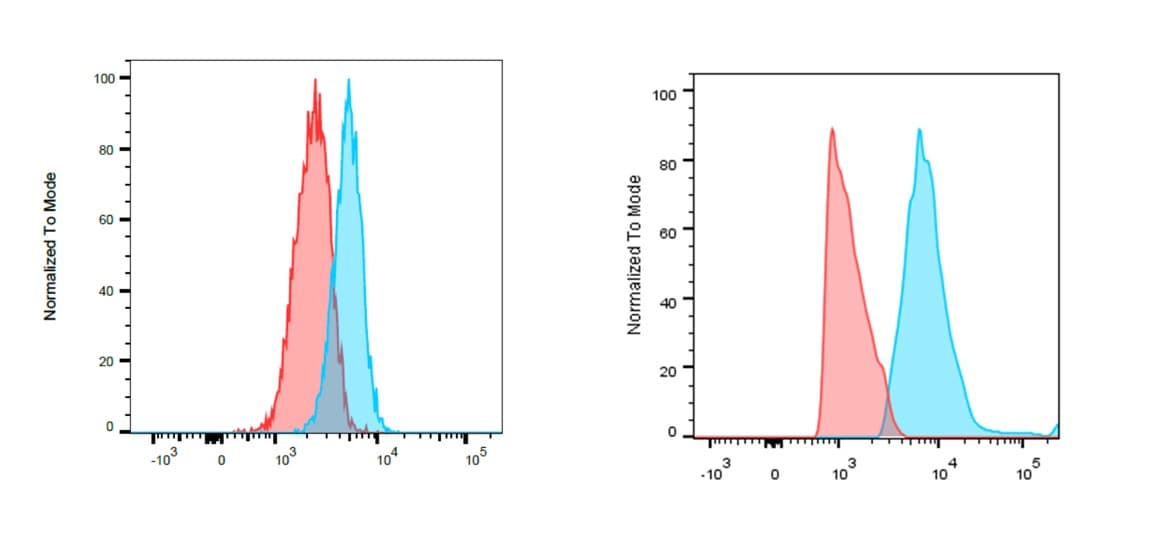
Detection of Integrin alpha V beta 5 in HT1080 cells by Flow Cytometry
HT1080 cells were stained with Mouse Anti-Human Integrin alpha V beta 5 Monoclonal Antibody (Catalog # MAB2528) isotype control antibody (Catalog # MAB002) followed by Allophycocyanin-conjugated Anti-Mouse IgG Secondary Antibody (Catalog # F0101B). View our protocol for Staining Membrane-associated Proteins.
Detection of Integrin alpha V beta 5 in MCF‑7 Human Cell Line by Flow Cytometry.
MCF-7 human breast cancer cell line was stained with Mouse Anti-Human Integrin aV beta 5 Monoclonal Antibody (Catalog # MAB2528, filled histogram) or isotype control antibody (IC002, open histogram) followed by Phycoerythrin-conjugated Anti-Mouse IgG Secondary Antibody (F0102B). View our protocol for Staining Membrane-associated Proteins.Applications for Human Integrin alpha V beta 5 Antibody (P5H9)
Application
Recommended Usage
Adhesion Blockade
Wayner, E.A. et al. (1991) J. Cell Biol. 113:919.
CyTOF-ready
Ready to be labeled using established conjugation methods. No BSA or other carrier proteins that could interfere with conjugation.
Flow Cytometry
0.25 µg/106 cells
Sample: HT1080 human fibrosarcoma cell line and MCF‑7 human breast cancer cell line
Sample: HT1080 human fibrosarcoma cell line and MCF‑7 human breast cancer cell line
Immunocytochemistry
8-25 µg/mL
Sample: Immersion fixed HT1080 human fibrosarcoma cell line, M21 human melanoma cell line, and H2981 human lung carcinoma cell line
Sample: Immersion fixed HT1080 human fibrosarcoma cell line, M21 human melanoma cell line, and H2981 human lung carcinoma cell line
Immunoprecipitation
Wayner, E.A. et al. (1991) J. Cell Biol. 113:919.
Reviewed Applications
Read 2 reviews rated 4.5 using MAB2528 in the following applications:
Flow Cytometry Panel Builder
Bio-Techne Knows Flow Cytometry
Save time and reduce costly mistakes by quickly finding compatible reagents using the Panel Builder Tool.
Advanced Features
- Spectra Viewer - Custom analysis of spectra from multiple fluorochromes
- Spillover Popups - Visualize the spectra of individual fluorochromes
- Antigen Density Selector - Match fluorochrome brightness with antigen density

Formulation, Preparation, and Storage
Purification
Protein A or G purified from hybridoma culture supernatant
Reconstitution
Reconstitute at 0.5 mg/mL in sterile PBS. For liquid material, refer to CoA for concentration.
Loading...
Formulation
Lyophilized from a 0.2 μm filtered solution in PBS with Trehalose. See Certificate of Analysis for details.
*Small pack size (-SP) is supplied either lyophilized or as a 0.2 µm filtered solution in PBS.
*Small pack size (-SP) is supplied either lyophilized or as a 0.2 µm filtered solution in PBS.
Shipping
Lyophilized product is shipped at ambient temperature. Liquid small pack size (-SP) is shipped with polar packs. Upon receipt, store immediately at the temperature recommended below.
Stability & Storage
Use a manual defrost freezer and avoid repeated freeze-thaw cycles.
- 12 months from date of receipt, -20 to -70 °C as supplied.
- 1 month, 2 to 8 °C under sterile conditions after reconstitution.
- 6 months, -20 to -70 °C under sterile conditions after reconstitution.
Calculators
Background: Integrin alpha V beta 5
Alternate Names
CD51, integrin subunit alpha V, ITGAV, MSK8, VNRA, VTNR
Entrez Gene IDs
3685 (Human)
Gene Symbol
ITGAV
Additional Integrin alpha V beta 5 Products
Product Documents for Human Integrin alpha V beta 5 Antibody (P5H9)
Certificate of Analysis
To download a Certificate of Analysis, please enter a lot or batch number in the search box below.
Note: Certificate of Analysis not available for kit components.
Product Specific Notices for Human Integrin alpha V beta 5 Antibody (P5H9)
For research use only
Related Research Areas
Citations for Human Integrin alpha V beta 5 Antibody (P5H9)
 Powered by Bioz
Customer Reviews for Human Integrin alpha V beta 5 Antibody (P5H9) (2)
4.5 out of 5
2 Customer Ratings
Have you used Human Integrin alpha V beta 5 Antibody (P5H9)?
Submit a review and receive an Amazon gift card!
$25/€18/£15/$25CAN/¥2500 Yen for a review with an image
$10/€7/£6/$10CAN/¥1110 Yen for a review without an image
Submit a review
Customer Images
Showing
1
-
2 of
2 reviews
Showing All
Filter By:
-
Application: Flow CytometrySample Tested: DU145 human prostate carcinoma cell line and PC-3 human prostate cancer cell lineSpecies: HumanVerified Customer | Posted 11/12/2025
 Bio-Techne ResponseThis review reflects a new species or application tested on a primary antibody.
 -
Application: Flow CytometrySample Tested: Mesenchymal stem cellsSpecies: HumanVerified Customer | Posted 01/10/2017
There are no reviews that match your criteria.
Protocols
Find general support by application which include: protocols, troubleshooting, illustrated assays, videos and webinars.
- 7-Amino Actinomycin D (7-AAD) Cell Viability Flow Cytometry Protocol
- Appropriate Fixation of IHC/ICC Samples
- Cellular Response to Hypoxia Protocols
- ClariTSA™ Fluorophore Kits
- Detection & Visualization of Antibody Binding
- Extracellular Membrane Flow Cytometry Protocol
- Flow Cytometry Protocol for Cell Surface Markers
- Flow Cytometry Protocol for Staining Membrane Associated Proteins
- Flow Cytometry Staining Protocols
- Flow Cytometry Troubleshooting Guide
- ICC Cell Smear Protocol for Suspension Cells
- ICC Immunocytochemistry Protocol Videos
- ICC for Adherent Cells
- Immunocytochemistry (ICC) Protocol
- Immunocytochemistry Troubleshooting
- Immunofluorescence of Organoids Embedded in Cultrex Basement Membrane Extract
- Immunohistochemistry (IHC) and Immunocytochemistry (ICC) Protocols
- Immunoprecipitation Protocol
- Intracellular Flow Cytometry Protocol Using Alcohol (Methanol)
- Intracellular Flow Cytometry Protocol Using Detergents
- Intracellular Nuclear Staining Flow Cytometry Protocol Using Detergents
- Intracellular Staining Flow Cytometry Protocol Using Alcohol Permeabilization
- Intracellular Staining Flow Cytometry Protocol Using Detergents to Permeabilize Cells
- Preparing Samples for IHC/ICC Experiments
- Preventing Non-Specific Staining (Non-Specific Binding)
- Primary Antibody Selection & Optimization
- Propidium Iodide Cell Viability Flow Cytometry Protocol
- Protocol for Liperfluo
- Protocol for VisUCyte™ HRP Polymer Detection Reagent
- Protocol for the Characterization of Human Th22 Cells
- Protocol for the Characterization of Human Th9 Cells
- Protocol for the Fluorescent ICC Staining of Cell Smears - Graphic
- Protocol for the Fluorescent ICC Staining of Cultured Cells on Coverslips - Graphic
- Protocol for the Preparation and Fluorescent ICC Staining of Cells on Coverslips
- Protocol for the Preparation and Fluorescent ICC Staining of Non-adherent Cells
- Protocol for the Preparation and Fluorescent ICC Staining of Stem Cells on Coverslips
- Protocol for the Preparation of a Cell Smear for Non-adherent Cell ICC - Graphic
- Protocol: Annexin V and PI Staining by Flow Cytometry
- Protocol: Annexin V and PI Staining for Apoptosis by Flow Cytometry
- TUNEL and Active Caspase-3 Detection by IHC/ICC Protocol
- The Importance of IHC/ICC Controls
- Troubleshooting Guide: Fluorokine Flow Cytometry Kits
- View all Protocols, Troubleshooting, Illustrated assays and Webinars
Loading...
Associated Pathways

