Human Integrin beta 3/CD61 Antibody
R&D Systems | Catalog # AF2266

Key Product Details
Species Reactivity
Validated:
Human
Cited:
Human, Mouse
Applications
Validated:
Western Blot, Flow Cytometry, Simple Western, CyTOF-ready
Cited:
Western Blot, Interferent Testing, Westen Blot
Label
Unconjugated
Antibody Source
Polyclonal Goat IgG
Loading...
Product Specifications
Immunogen
Chinese hamster ovary cell line CHO-derived recombinant human Integrin beta 3/CD61
Gly27-Asp718
Accession # P05106
Gly27-Asp718
Accession # P05106
Specificity
Detects human Integrin beta 3/CD61 in direct ELISAs and Western blots. In direct ELISAs, approximately 5% cross-reactivity with recombinant human (rh) Integrin beta 2 and recombinant mouse (rm) Integrin beta 6 is observed and less than 1% cross-reactivity with rhIntegrin beta 1 and rmIntegrin beta 4 is observed.
Clonality
Polyclonal
Host
Goat
Isotype
IgG
Scientific Data Images for Human Integrin beta 3/CD61 Antibody
Detection of Human Integrin beta 3/CD61 by Western Blot.
Western blot shows lysates of Jurkat human acute T cell leukemia cell line and DU145 human prostate carcinoma cell line. PVDF membrane was probed with 1 µg/mL of Goat Anti-Human Integrin beta 3/ CD61 Antigen Affinity-purified Polyclonal Antibody (Catalog # AF2266) followed by HRP-conjugated Anti-Goat IgG Secondary Antibody (HAF017). A specific band was detected for Integrin beta 3/CD61 at approximately 100 kDa (as indicated). This experiment was conducted under reducing conditions and using Immunoblot Buffer Group 1.
Detection of Human Integrin beta 3/CD61 by Simple WesternTM.
Simple Western shows lysates of Exosome Standards (HT‑29) (NBP3-11685) and U‑87 MG human glioblastoma/astrocytoma cell line, loaded at 0.5 mg/ml. A specific band was detected for Integrin beta 3/CD61 at approximately 116 kDa (as indicated) using 20 µg/mL of Goat Anti-Human Integrin beta 3/CD61 Antigen Affinity-purified Polyclonal Antibody (Catalog # AF2266). This experiment was conducted under reducing conditions and using the 12-230kDa separation system.Applications for Human Integrin beta 3/CD61 Antibody
Application
Recommended Usage
CyTOF-ready
Ready to be labeled using established conjugation methods. No BSA or other carrier proteins that could interfere with conjugation.
Flow Cytometry
0.25 µg/106 cells
Sample: Human peripheral blood mononuclear cells
Sample: Human peripheral blood mononuclear cells
Simple Western
20 µg/mL
Sample: Exosome Standards (HT-29) (Catalog # NBP3-11685) and U‑87 MG human glioblastoma/astrocytoma cell line
Sample: Exosome Standards (HT-29) (Catalog # NBP3-11685) and U‑87 MG human glioblastoma/astrocytoma cell line
Western Blot
1 µg/mL
Sample: Jurkat human acute T cell leukemia cell line and DU145 human prostate carcinoma cell line
Sample: Jurkat human acute T cell leukemia cell line and DU145 human prostate carcinoma cell line
Reviewed Applications
Read 3 reviews rated 4.7 using AF2266 in the following applications:
Flow Cytometry Panel Builder
Bio-Techne Knows Flow Cytometry
Save time and reduce costly mistakes by quickly finding compatible reagents using the Panel Builder Tool.
Advanced Features
- Spectra Viewer - Custom analysis of spectra from multiple fluorochromes
- Spillover Popups - Visualize the spectra of individual fluorochromes
- Antigen Density Selector - Match fluorochrome brightness with antigen density

Formulation, Preparation, and Storage
Purification
Antigen Affinity-purified
Reconstitution
Reconstitute at 0.2 mg/mL in sterile PBS. For liquid material, refer to CoA for concentration.
Loading...
Formulation
Lyophilized from a 0.2 μm filtered solution in PBS with Trehalose. See Certificate of Analysis for details.
*Small pack size (-SP) is supplied either lyophilized or as a 0.2 µm filtered solution in PBS.
*Small pack size (-SP) is supplied either lyophilized or as a 0.2 µm filtered solution in PBS.
Shipping
Lyophilized product is shipped at ambient temperature. Liquid small pack size (-SP) is shipped with polar packs. Upon receipt, store immediately at the temperature recommended below.
Stability & Storage
Use a manual defrost freezer and avoid repeated freeze-thaw cycles.
- 12 months from date of receipt, -20 to -70 °C as supplied.
- 1 month, 2 to 8 °C under sterile conditions after reconstitution.
- 6 months, -20 to -70 °C under sterile conditions after reconstitution.
Calculators
Background: Integrin beta 3/CD61
Alternate Names
CD61, GP3A, GPIIIA, INGRB3, ITGB3
Gene Symbol
ITGB3
UniProt
Additional Integrin beta 3/CD61 Products
Product Documents for Human Integrin beta 3/CD61 Antibody
Certificate of Analysis
To download a Certificate of Analysis, please enter a lot or batch number in the search box below.
Note: Certificate of Analysis not available for kit components.
Product Specific Notices for Human Integrin beta 3/CD61 Antibody
For research use only
Citations for Human Integrin beta 3/CD61 Antibody
 Powered by Bioz
Customer Reviews for Human Integrin beta 3/CD61 Antibody (3)
4.7 out of 5
3 Customer Ratings
Have you used Human Integrin beta 3/CD61 Antibody?
Submit a review and receive an Amazon gift card!
$25/€18/£15/$25CAN/¥2500 Yen for a review with an image
$10/€7/£6/$10CAN/¥1110 Yen for a review without an image
Submit a review
Customer Images
Showing
1
-
3 of
3 reviews
Showing All
Filter By:
-
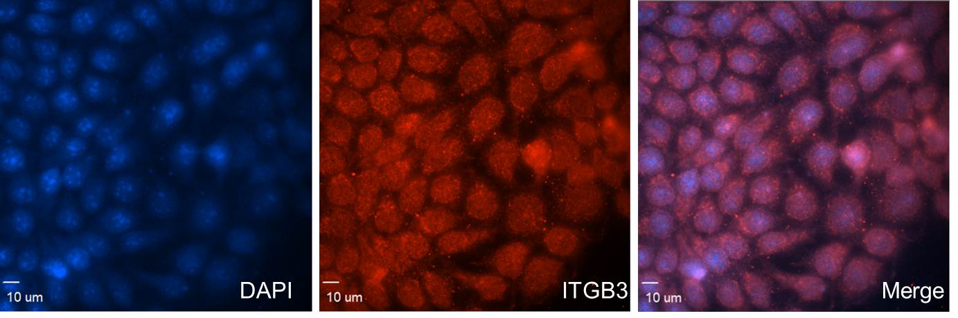
Application: ImmunocytochemistrySample Tested: SUM159Species: HumanVerified Customer | Posted 12/01/2022Immunofluorescence (IF): ITGB3 Antibody [AF2266 ] was used (1:100) for labeling and microscopy analysis of the distribution of ITGB3 proteins in SUM159 cells.

-
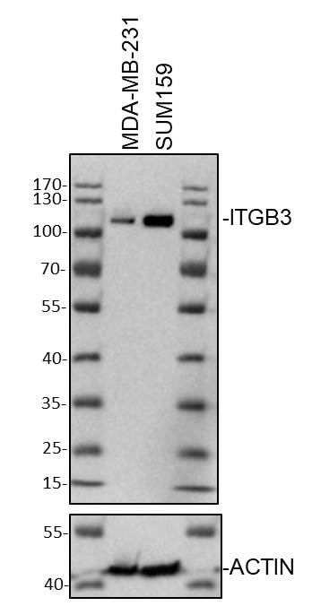
Application: Western BlotSample Tested: human MDAMB-231 whole cell lysate and SUM-159PTSpecies: HumanVerified Customer | Posted 10/20/2022Western Blot: whole cell lysates from MDA-MB-231 or SUM159PT cells was loaded with 50 ug/lane. 10% SDS-PAGE. ITGB3 Antibody (AF2266) was used for primary antibody: 1:1000, 4℃, overnight.
-
Application: Western BlotSample Tested: Cell LysatesSpecies: HumanVerified Customer | Posted 09/14/2019
There are no reviews that match your criteria.
Protocols
Find general support by application which include: protocols, troubleshooting, illustrated assays, videos and webinars.
- 7-Amino Actinomycin D (7-AAD) Cell Viability Flow Cytometry Protocol
- Cellular Response to Hypoxia Protocols
- Extracellular Membrane Flow Cytometry Protocol
- Flow Cytometry Protocol for Cell Surface Markers
- Flow Cytometry Protocol for Staining Membrane Associated Proteins
- Flow Cytometry Staining Protocols
- Flow Cytometry Troubleshooting Guide
- Intracellular Flow Cytometry Protocol Using Alcohol (Methanol)
- Intracellular Flow Cytometry Protocol Using Detergents
- Intracellular Nuclear Staining Flow Cytometry Protocol Using Detergents
- Intracellular Staining Flow Cytometry Protocol Using Alcohol Permeabilization
- Intracellular Staining Flow Cytometry Protocol Using Detergents to Permeabilize Cells
- Propidium Iodide Cell Viability Flow Cytometry Protocol
- Protocol for Liperfluo
- Protocol for the Characterization of Human Th22 Cells
- Protocol for the Characterization of Human Th9 Cells
- Protocol: Annexin V and PI Staining by Flow Cytometry
- Protocol: Annexin V and PI Staining for Apoptosis by Flow Cytometry
- R&D Systems Quality Control Western Blot Protocol
- Troubleshooting Guide: Fluorokine Flow Cytometry Kits
- Troubleshooting Guide: Western Blot Figures
- Western Blot Conditions
- Western Blot Protocol
- Western Blot Protocol for Cell Lysates
- Western Blot Troubleshooting
- Western Blot Troubleshooting Guide
- View all Protocols, Troubleshooting, Illustrated assays and Webinars


