The family of junctional adhesion molecules (JAM), comprising at least three members, are type I transmembrane receptors belonging to the immunoglobulin (Ig) superfamily (1, 2). These proteins are localized in the tight junctions between endothelial or epithelial cells. Some family members are also found on blood leukocytes and platelets. Human JAM-A, also known as platelet adhesion molecule 1 (PAM-1) and platelet F11 receptor (3), is predominantly expressed at intercellular junctions of both epithelial cells and endothelial cells (1‑4). It is also expressed on circulating blood cells including neutrophils, monocytes, platelets, erythrocytes and lymphocytes (5). Human JAM-A cDNA predicts a 299 amino acid (aa) residue precursor protein with a putative 27 aa signal peptide, a 210 aa extracellular region containing two Ig‑like V-subset domains, a 24 aa transmembrane domain and a 38 aa cytoplasmic domain. The human and mouse proteins share approximately 67% aa sequence homology. Human JAM-A also shares approximately 35% and 32% aa sequence homology with human JAM-B and JAM-C, respectively. JAM-A exhibits homophilic interactions to regulate tight junction assembly and modulate paracellular permeability. This homophilic interation also mediates platelet aggregation and adhesion to endothelial cells and may play a role in thrombosis (3). JAM-A binds heterotypically with the beta 2 integrin lymphocyte function-associated antigen-1 (LFA-1). This JAM‑A‑LFA‑1 interaction is involved in leukocyte adhesion and transmigration (6). JAM-A has also been shown to bind reovirus attachment protein sigma-1 to permit reovirus infection and signal virus-induced apoptosis (7).

Key Product Details
Species Reactivity
Validated:
Human
Cited:
Human
Applications
Validated:
Immunohistochemistry, Western Blot, Immunocytochemistry
Cited:
Immunohistochemistry, Western Blot, Flow Cytometry, Immunocytochemistry, Immunoprecipitation, ELISA Development, ELISA Development (Capture)
Label
Unconjugated
Antibody Source
Polyclonal Goat IgG
Loading...
Product Specifications
Immunogen
Mouse myeloma cell line NS0-derived recombinant human JAM-A
Ser28-Ala242
Accession # Q9Y624
Ser28-Ala242
Accession # Q9Y624
Specificity
Detects human JAM-A in direct ELISAs and Western blots. In direct ELISAs and Western blots, approximately 10% cross-reactivity with recombinant mouse JAM-A is observed.
Clonality
Polyclonal
Host
Goat
Isotype
IgG
Scientific Data Images for Human JAM-A Antibody
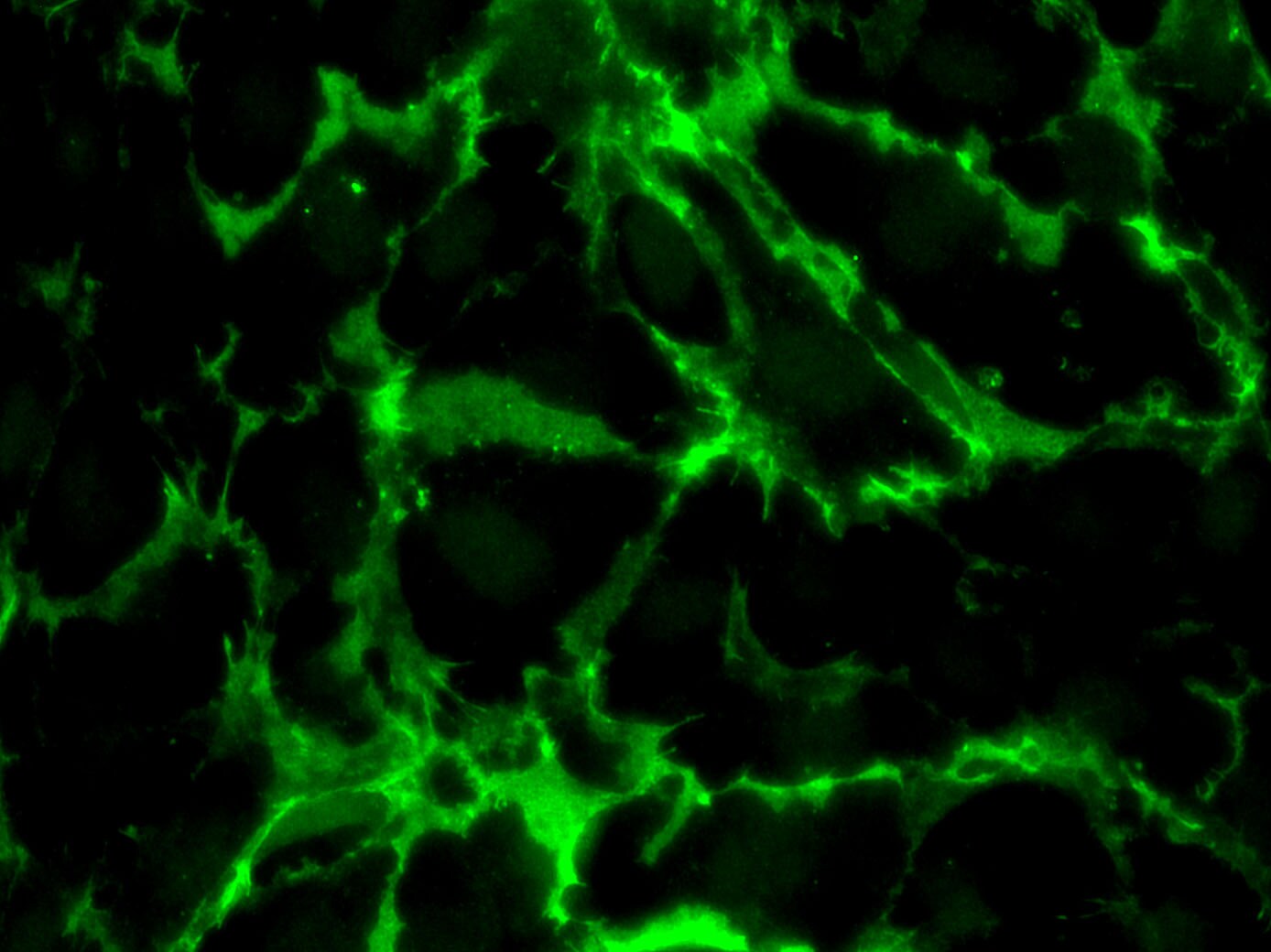
JAM‑A in MCF‑7 Human Cell Line.
JAM-A was detected in immersion fixed MCF-7 human breast cancer cell line using Goat Anti-Human JAM-A Antigen Affinity-purified Polyclonal Antibody (Catalog # AF1103) at 10 µg/mL for 3 hours at room temperature. Cells were stained using the NorthernLights™ 493-conjugated Anti-Goat IgG Secondary Antibody (green; Catalog # NL003) and counterstained with DAPI (blue). Specific staining was localized to intercellular junctions. View our protocol for Fluorescent ICC Staining of Cells on Coverslips.Applications for Human JAM-A Antibody
Application
Recommended Usage
Immunocytochemistry
5-15 µg/mL
Sample: Immersion fixed MCF‑7 human breast cancer cell line
Sample: Immersion fixed MCF‑7 human breast cancer cell line
Immunohistochemistry
5-15 µg/mL
Sample: Immersion fixed paraffin-embedded sections of human lung
Sample: Immersion fixed paraffin-embedded sections of human lung
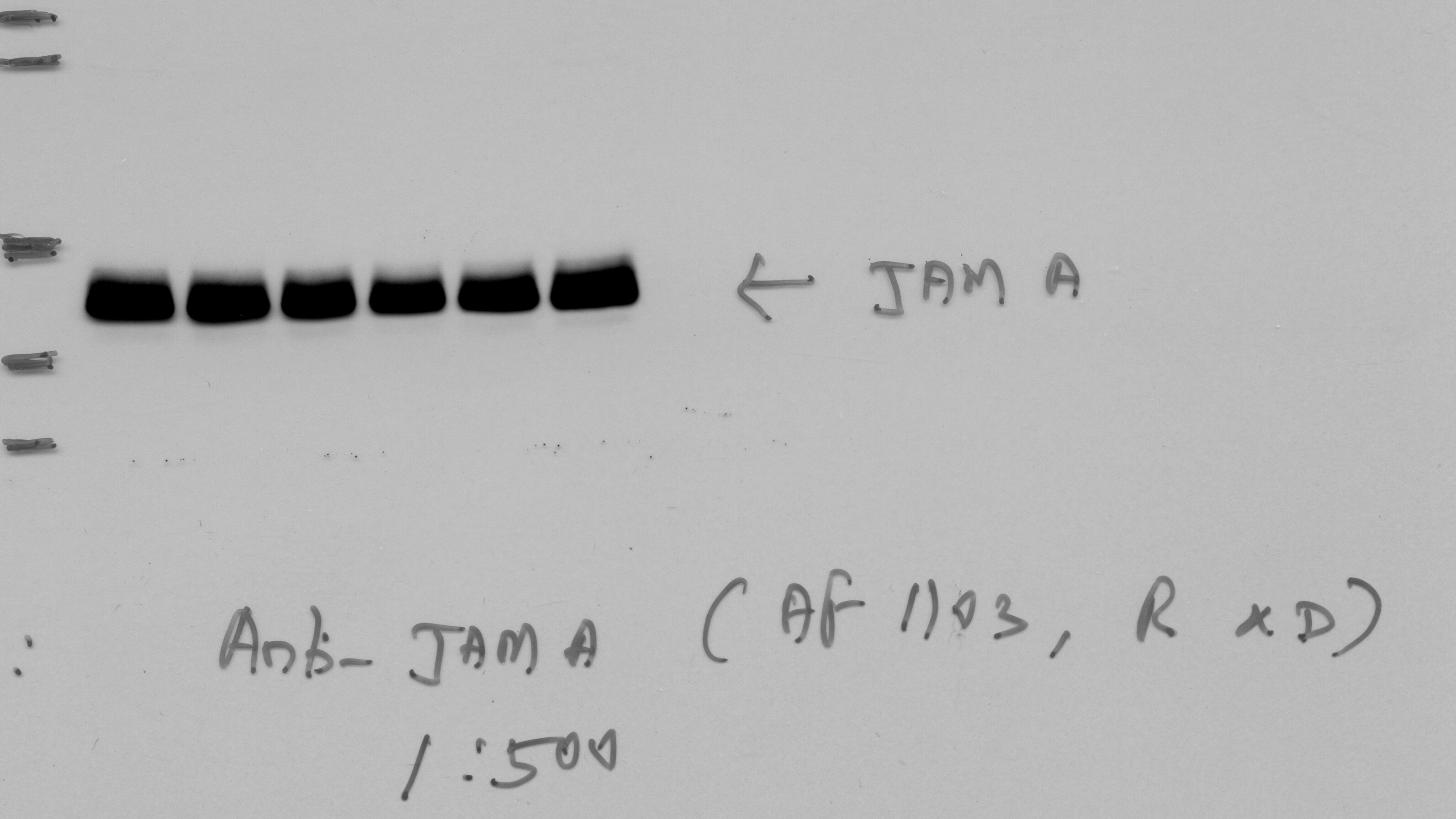
Western Blot
0.1 µg/mL
Sample: Recombinant Human JAM-A Fc Chimera (Catalog # 1103-JM)
Sample: Recombinant Human JAM-A Fc Chimera (Catalog # 1103-JM)
Reviewed Applications
Read 2 reviews rated 5 using AF1103 in the following applications:
Formulation, Preparation, and Storage
Purification
Antigen Affinity-purified
Reconstitution
Reconstitute at 0.2 mg/mL in sterile PBS. For liquid material, refer to CoA for concentration.
Loading...
Formulation
Lyophilized from a 0.2 μm filtered solution in PBS with Trehalose. *Small pack size (SP) is supplied either lyophilized or as a 0.2 µm filtered solution in PBS.
Shipping
Lyophilized product is shipped at ambient temperature. Liquid small pack size (-SP) is shipped with polar packs. Upon receipt, store immediately at the temperature recommended below.
Stability & Storage
Use a manual defrost freezer and avoid repeated freeze-thaw cycles.
- 12 months from date of receipt, -20 to -70 °C as supplied.
- 1 month, 2 to 8 °C under sterile conditions after reconstitution.
- 6 months, -20 to -70 °C under sterile conditions after reconstitution.
Calculators
Background: JAM-A
References
- Chavakis, T. et al. (2003) Thromb. Haemost. 89:13.
- Aurand-Lions, M. et al. (2001) Blood 98:3699.
- Sobocka, M.B. et al. (2000) Blood 95:2600.
- Martin-Padura, I. et al. (1998) J. Cell Biol. 142:117.
- Williams, L.A. et. al. (1999) Mol. Immunol. 36:1175.
- Ostermann, G. et al. (2002) Nature Immunol. 3:151.
- Barton, E.S. et al. (2001) Cell 104:441.
Long Name
Junctional Adhesion Molecule A
Alternate Names
CD321, F11R, JAMA, PAM-1
Gene Symbol
F11R
UniProt
Additional JAM-A Products
Product Documents for Human JAM-A Antibody
Certificate of Analysis
To download a Certificate of Analysis, please enter a lot or batch number in the search box below.
Note: Certificate of Analysis not available for kit components.
Product Specific Notices for Human JAM-A Antibody
For research use only
Citations for Human JAM-A Antibody
 Powered by Bioz
Customer Reviews for Human JAM-A Antibody (2)
5 out of 5
2 Customer Ratings
Have you used Human JAM-A Antibody?
Submit a review and receive an Amazon gift card!
$25/€18/£15/$25CAN/¥2500 Yen for a review with an image
$10/€7/£6/$10CAN/¥1110 Yen for a review without an image
Submit a review
Customer Images
Showing
1
-
2 of
2 reviews
Showing All
Filter By:
-
Application: Immunocytochemistry/ImmunofluorescenceSample Tested: Umbilical vein endothelial cellsSpecies: HumanVerified Customer | Posted 06/09/2021

-
Application: Western BlotSample Tested: Human Aortic Endothelial CellsSpecies: HumanVerified Customer | Posted 06/19/2017
There are no reviews that match your criteria.
Protocols
Find general support by application which include: protocols, troubleshooting, illustrated assays, videos and webinars.
- Antigen Retrieval Protocol (PIER)
- Antigen Retrieval for Frozen Sections Protocol
- Appropriate Fixation of IHC/ICC Samples
- Cellular Response to Hypoxia Protocols
- Chromogenic IHC Staining of Formalin-Fixed Paraffin-Embedded (FFPE) Tissue Protocol
- Chromogenic Immunohistochemistry Staining of Frozen Tissue
- ClariTSA™ Fluorophore Kits
- Detection & Visualization of Antibody Binding
- Fluorescent IHC Staining of Frozen Tissue Protocol
- Graphic Protocol for Heat-induced Epitope Retrieval
- Graphic Protocol for the Preparation and Fluorescent IHC Staining of Frozen Tissue Sections
- Graphic Protocol for the Preparation and Fluorescent IHC Staining of Paraffin-embedded Tissue Sections
- Graphic Protocol for the Preparation of Gelatin-coated Slides for Histological Tissue Sections
- ICC Cell Smear Protocol for Suspension Cells
- ICC Immunocytochemistry Protocol Videos
- ICC for Adherent Cells
- IHC Sample Preparation (Frozen sections vs Paraffin)
- Immunocytochemistry (ICC) Protocol
- Immunocytochemistry Troubleshooting
- Immunofluorescence of Organoids Embedded in Cultrex Basement Membrane Extract
- Immunofluorescent IHC Staining of Formalin-Fixed Paraffin-Embedded (FFPE) Tissue Protocol
- Immunohistochemistry (IHC) and Immunocytochemistry (ICC) Protocols
- Immunohistochemistry Frozen Troubleshooting
- Immunohistochemistry Paraffin Troubleshooting
- Preparing Samples for IHC/ICC Experiments
- Preventing Non-Specific Staining (Non-Specific Binding)
- Primary Antibody Selection & Optimization
- Protocol for Heat-Induced Epitope Retrieval (HIER)
- Protocol for Making a 4% Formaldehyde Solution in PBS
- Protocol for VisUCyte™ HRP Polymer Detection Reagent
- Protocol for the Fluorescent ICC Staining of Cell Smears - Graphic
- Protocol for the Fluorescent ICC Staining of Cultured Cells on Coverslips - Graphic
- Protocol for the Preparation & Fixation of Cells on Coverslips
- Protocol for the Preparation and Chromogenic IHC Staining of Frozen Tissue Sections
- Protocol for the Preparation and Chromogenic IHC Staining of Frozen Tissue Sections - Graphic
- Protocol for the Preparation and Chromogenic IHC Staining of Paraffin-embedded Tissue Sections
- Protocol for the Preparation and Chromogenic IHC Staining of Paraffin-embedded Tissue Sections - Graphic
- Protocol for the Preparation and Fluorescent ICC Staining of Cells on Coverslips
- Protocol for the Preparation and Fluorescent ICC Staining of Non-adherent Cells
- Protocol for the Preparation and Fluorescent ICC Staining of Stem Cells on Coverslips
- Protocol for the Preparation and Fluorescent IHC Staining of Frozen Tissue Sections
- Protocol for the Preparation and Fluorescent IHC Staining of Paraffin-embedded Tissue Sections
- Protocol for the Preparation of Gelatin-coated Slides for Histological Tissue Sections
- Protocol for the Preparation of a Cell Smear for Non-adherent Cell ICC - Graphic
- R&D Systems Quality Control Western Blot Protocol
- TUNEL and Active Caspase-3 Detection by IHC/ICC Protocol
- The Importance of IHC/ICC Controls
- Troubleshooting Guide: Immunohistochemistry
- Troubleshooting Guide: Western Blot Figures
- Western Blot Conditions
- Western Blot Protocol
- Western Blot Protocol for Cell Lysates
- Western Blot Troubleshooting
- Western Blot Troubleshooting Guide
- View all Protocols, Troubleshooting, Illustrated assays and Webinars
Loading...
Associated Pathways


