The low density lipoprotein receptor (LDL R) is the founding member of the LDL R family of scavenger receptors. This family contains transmembrane molecules that are characterized by the presence of EGF repeats, complement-like repeats, and YWTD motifs that form beta -propellers. Although members of the family were originally thought to be endocytic receptors, it is now clear that some members interact with adjacent cell‑surface molecules, expanding their range of activities. Human LDL R is synthesized as an 860 amino acid (aa) precursor that contains a 21 aa signal sequence, a 767 aa extracellular region, a 22 aa transmembrane segment and a 50 aa cytoplasmic tail. The extracellular region is complex. It consists of seven N-terminal complement-like cysteine-rich repeats that bind ligand. Cysteine residues in this region participate in intrachain disulfide bonds. This region is followed by three EGF-like repeats with a beta -propeller YWTD containing motif. The EGF-like repeats are responsible for ligand bonding and dissociation. Finally, there is a 50 aa membrane proximal Ser/Thr‑rich region that serves as a carbohydrate attachment point. There is extensive O‑linked and modest N-linked glycosylation. Thus the receptor’s predicted molecular weight of 93 kDa is increased to a native molecular weight of 120 ‑ 160 kDa. Within the 50 aa cytoplasmic tail, there is an NPXY motif that links the receptor to clathrin pits. The extracellular region of human LDL R is 51% aa identical to the extracellular region of human VLDL R, and 79% aa identical to the extracellular region of mouse LDL R. LDL R is constitutively expressed and binds apoB of LDL and apoE of VLDL. It is responsible for clearing 70% of plasma LDL in liver. Mutations in the LDL R gene cause the autosomal dominant disorder, familial hypercholesterolemia.

Key Product Details
Validated by
Knockout/Knockdown
Species Reactivity
Validated:
Human
Cited:
Human, Mouse, Rat, Hamster, Primate - Cercopithecus aethiops (African Green Monkey), Primate - Macaca fascicularis (Crab-eating Monkey or Cynomolgus Macaque), Transgenic Mouse
Applications
Validated:
Immunohistochemistry, Western Blot, Blockade of Receptor-ligand Interaction, Immunocytochemistry
Cited:
Immunohistochemistry, Western Blot, Neutralization, Flow Cytometry, Immunocytochemistry, Immunoprecipitation, Blocking, Functional Assay
Label
Unconjugated
Antibody Source
Polyclonal Goat IgG
Loading...
Product Specifications
Immunogen
Chinese hamster ovary cell line CHO-derived recombinant human LDL R
Asp193-Arg788
Accession # P01130
Asp193-Arg788
Accession # P01130
Specificity
Detects human LDL R in direct ELISAs and Western blots. In direct ELISAs, approximately 15% cross-reactivity with recombinant mouse LDL R is observed.
Clonality
Polyclonal
Host
Goat
Isotype
IgG
Scientific Data Images for Human LDLR Antibody
LDL R in HepG2 Human Cell Line.
LDL R was detected in immersion fixed HepG2 human hepatocellular carcinoma cell line using Goat Anti-Human LDL R Antigen Affinity-purified Polyclonal Antibody (Catalog # AF2148) at 1.7 µg/mL for 3 hours at room temperature. Cells were stained using the NorthernLights™ 557-conjugated Anti-Goat IgG Secondary Antibody (red; Catalog # NL001) and counterstained with DAPI (blue). Specific staining was localized to cytoplasm. View our protocol for Fluorescent ICC Staining of Cells on Coverslips.
LDL R in Human Liver.
LDL R was detected in formalin fixed paraffin-embedded sections of human liver using Goat Anti-Human LDL R Antigen Affinity-purified Polyclonal Antibody (Catalog # AF2148) at 15 µg/mL overnight at 4 °C. Tissue was stained using the Anti-Goat HRP-DAB Cell & Tissue Staining Kit (brown; Catalog # CTS008) and counterstained with hematoxylin (blue). View our protocol for Chromogenic IHC Staining of Paraffin-embedded Tissue Sections.
Detection of Human LDLR by Knockdown Validated
APLP2 and LDLR interactions with PCSK9 and their regulation of PCSK9 function.(A and B) Western blot showing APLP2, PCSK9, or Transferrin receptor (TFNR) levels in input fraction (I), IC or J16 immunoprecipitated samples (IP Ab.) in the absence or presence of 5F6 Fab or 12E3 Fab, as indicated. (B) Quantification of (A); shown as average APLP2 normalized to PCSK9 of 3 independent experiments with SEM. (C and D) J16 coIPs of PCSK9 from Neg or LDLR siRNA treated HepG2 cells with IC control, as indicated. (D) Quantification of (C); shown as average APLP2 normalized to PCSK9 from 3 independent experiments with SEM. (E, F, and G) Western blot of LDLR, APOER2, or TFNR in siRNA treated cells following treatment with PCSK9 at 0, 20, 50, or 100 μg/ml. (F) LDLR levels from (E) quantified as percent LDLR degradation of untreated cells and normalized to Neg siRNA samples. Shown as average with SEM from 4 independent experiments. (G) Same as F, but measuring APOER2 levels. Image collected and cropped by CiteAb from the following publication (https://pubmed.ncbi.nlm.nih.gov/25905719), licensed under a CC-BY license. Not internally tested by R&D Systems.
Detection of Human LDLR by Western Blot
Assessment of PCSK9 and LDLR expression in human LEC. (A) PCSK9 expression was measured by immunoblotting in either human LEC, HepG2 or HEK 293T cell lysates. (B) ELISA was used to measure PCSK9 levels in the cell culture supernatant of Huh7 (grey), HEK 293T (white) and human LEC (black). (C) Expression of LDLR was detected in protein lysates by immunoblotting of human LEC. Huh7 cells were used as a positive control and HEK 293T cells treated with siLDLR were used as a negative control. LDLR protein expression on human LEC was measured by (D) flow cytometry after extracellular staining of LEC (black line) and Huh7 (grey line) and by (E) immunofluorescence (Blue, DAPI; red, cholera toxin; green, anti-LDLR; yellow, colocalized voxels; scale bar, 20 µM). (F) Scatterplot of red and green pixel intensities of cholera toxin (red) and anti-LDLR (green) in human LEC. n = 4-9. Statistics: ****p < 0.0001. Image collected and cropped by CiteAb from the following open publication (https://pubmed.ncbi.nlm.nih.gov/35154499), licensed under a CC-BY license. Not internally tested by R&D Systems.Applications for Human LDLR Antibody
Application
Recommended Usage
Blockade of Receptor-ligand Interaction
In a functional ELISA, less than 5.00 μg/mL of this antibody will block 50% of the binding of Recombinant Human LDL R (Catalog # 2148-LD) to human low-density lipoprotein.
Immunocytochemistry
1-15 µg/mL
Sample: Immersion fixed HepG2 human hepatocellular carcinoma cell line
Sample: Immersion fixed HepG2 human hepatocellular carcinoma cell line
Immunohistochemistry
5-15 µg/mL
Sample: Immersion fixed paraffin-embedded sections of human liver
Sample: Immersion fixed paraffin-embedded sections of human liver
Western Blot
0.1 µg/mL
Sample: Recombinant Human LDL R (Catalog # 2148-LD)
Sample: Recombinant Human LDL R (Catalog # 2148-LD)
Reviewed Applications
Read 3 reviews rated 5 using AF2148 in the following applications:
Formulation, Preparation, and Storage
Purification
Antigen Affinity-purified
Reconstitution
Reconstitute at 0.2 mg/mL in sterile PBS. For liquid material, refer to CoA for concentration.
Loading...
Formulation
Lyophilized from a 0.2 μm filtered solution in PBS with Trehalose. See Certificate of Analysis for details.
*Small pack size (-SP) is supplied either lyophilized or as a 0.2 µm filtered solution in PBS.
*Small pack size (-SP) is supplied either lyophilized or as a 0.2 µm filtered solution in PBS.
Shipping
Lyophilized product is shipped at ambient temperature. Liquid small pack size (-SP) is shipped with polar packs. Upon receipt, store immediately at the temperature recommended below.
Stability & Storage
Use a manual defrost freezer and avoid repeated freeze-thaw cycles.
- 12 months from date of receipt, -20 to -70 °C as supplied.
- 1 month, 2 to 8 °C under sterile conditions after reconstitution.
- 6 months, -20 to -70 °C under sterile conditions after reconstitution.
Calculators
Background: LDLR
Long Name
Low Density Lipoprotein Receptor
Alternate Names
LDL R
Entrez Gene IDs
Gene Symbol
LDLR
UniProt
Additional LDLR Products
Product Documents for Human LDLR Antibody
Certificate of Analysis
To download a Certificate of Analysis, please enter a lot or batch number in the search box below.
Note: Certificate of Analysis not available for kit components.
Product Specific Notices for Human LDLR Antibody
For research use only
Related Research Areas
Citations for Human LDLR Antibody
 Powered by Bioz
Customer Reviews for Human LDLR Antibody (3)
5 out of 5
3 Customer Ratings
Have you used Human LDLR Antibody?
Submit a review and receive an Amazon gift card!
$25/€18/£15/$25CAN/¥2500 Yen for a review with an image
$10/€7/£6/$10CAN/¥1110 Yen for a review without an image
Submit a review
Customer Images
Showing
1
-
3 of
3 reviews
Showing All
Filter By:
-
Application: Flow CytometrySample Tested: Peritoneal macrophagesSpecies: HumanVerified Customer | Posted 11/12/2020
-
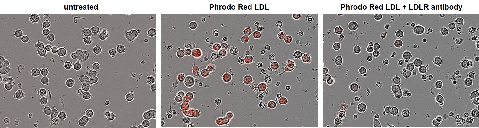
Application: Block/NeutralizeSample Tested: HEK293 human embryonic kidney cell lineSpecies: HumanVerified Customer | Posted 10/13/2020Blocking of LDL receptor in HEK293 cells by the Human LDLR Antibody (AF2148). HEK cells were incubated with Phrodo Red LDL at 10 μg/ml in the presence of 10 μg/ml of the blocking human LDLR antibody (AF2148).

-
Application: Flow CytometrySample Tested: HEK293 human embryonic kidney cell lineSpecies: HumanVerified Customer | Posted 04/21/2016
There are no reviews that match your criteria.
Protocols
Find general support by application which include: protocols, troubleshooting, illustrated assays, videos and webinars.
- Antigen Retrieval Protocol (PIER)
- Antigen Retrieval for Frozen Sections Protocol
- Appropriate Fixation of IHC/ICC Samples
- Cellular Response to Hypoxia Protocols
- Chromogenic IHC Staining of Formalin-Fixed Paraffin-Embedded (FFPE) Tissue Protocol
- Chromogenic Immunohistochemistry Staining of Frozen Tissue
- ClariTSA™ Fluorophore Kits
- Detection & Visualization of Antibody Binding
- Fluorescent IHC Staining of Frozen Tissue Protocol
- Graphic Protocol for Heat-induced Epitope Retrieval
- Graphic Protocol for the Preparation and Fluorescent IHC Staining of Frozen Tissue Sections
- Graphic Protocol for the Preparation and Fluorescent IHC Staining of Paraffin-embedded Tissue Sections
- Graphic Protocol for the Preparation of Gelatin-coated Slides for Histological Tissue Sections
- ICC Cell Smear Protocol for Suspension Cells
- ICC Immunocytochemistry Protocol Videos
- ICC for Adherent Cells
- IHC Sample Preparation (Frozen sections vs Paraffin)
- Immunocytochemistry (ICC) Protocol
- Immunocytochemistry Troubleshooting
- Immunofluorescence of Organoids Embedded in Cultrex Basement Membrane Extract
- Immunofluorescent IHC Staining of Formalin-Fixed Paraffin-Embedded (FFPE) Tissue Protocol
- Immunohistochemistry (IHC) and Immunocytochemistry (ICC) Protocols
- Immunohistochemistry Frozen Troubleshooting
- Immunohistochemistry Paraffin Troubleshooting
- Preparing Samples for IHC/ICC Experiments
- Preventing Non-Specific Staining (Non-Specific Binding)
- Primary Antibody Selection & Optimization
- Protocol for Heat-Induced Epitope Retrieval (HIER)
- Protocol for Making a 4% Formaldehyde Solution in PBS
- Protocol for VisUCyte™ HRP Polymer Detection Reagent
- Protocol for the Fluorescent ICC Staining of Cell Smears - Graphic
- Protocol for the Fluorescent ICC Staining of Cultured Cells on Coverslips - Graphic
- Protocol for the Preparation & Fixation of Cells on Coverslips
- Protocol for the Preparation and Chromogenic IHC Staining of Frozen Tissue Sections
- Protocol for the Preparation and Chromogenic IHC Staining of Frozen Tissue Sections - Graphic
- Protocol for the Preparation and Chromogenic IHC Staining of Paraffin-embedded Tissue Sections
- Protocol for the Preparation and Chromogenic IHC Staining of Paraffin-embedded Tissue Sections - Graphic
- Protocol for the Preparation and Fluorescent ICC Staining of Cells on Coverslips
- Protocol for the Preparation and Fluorescent ICC Staining of Non-adherent Cells
- Protocol for the Preparation and Fluorescent ICC Staining of Stem Cells on Coverslips
- Protocol for the Preparation and Fluorescent IHC Staining of Frozen Tissue Sections
- Protocol for the Preparation and Fluorescent IHC Staining of Paraffin-embedded Tissue Sections
- Protocol for the Preparation of Gelatin-coated Slides for Histological Tissue Sections
- Protocol for the Preparation of a Cell Smear for Non-adherent Cell ICC - Graphic
- R&D Systems Quality Control Western Blot Protocol
- TUNEL and Active Caspase-3 Detection by IHC/ICC Protocol
- The Importance of IHC/ICC Controls
- Troubleshooting Guide: Immunohistochemistry
- Troubleshooting Guide: Western Blot Figures
- Western Blot Conditions
- Western Blot Protocol
- Western Blot Protocol for Cell Lysates
- Western Blot Troubleshooting
- Western Blot Troubleshooting Guide
- View all Protocols, Troubleshooting, Illustrated assays and Webinars
Loading...
Associated Pathways
