
Key Product Details
Species Reactivity
Validated:
Human
Cited:
Human, Mouse, Primate - Chlorocebus pygerythrus (Vervet Monkey)
Applications
Validated:
Western Blot, Immunocytochemistry, Simple Western
Cited:
Western Blot, Immunoprecipitation, Co-Immunoprecipitation
Label
Unconjugated
Antibody Source
Polyclonal Goat IgG
Loading...
Product Specifications
Immunogen
E. coli-derived recombinant human Lyp
Ser306-Ser684
Accession # Q9Y2R2
Ser306-Ser684
Accession # Q9Y2R2
Specificity
Detects human Lyp in Western blots.
Clonality
Polyclonal
Host
Goat
Isotype
IgG
Scientific Data Images for Human Lyp Antibody
Detection of Human Lyp by Western Blot.
Western blot shows lysates of Daudi human Burkitt's lymphoma cell line and Ramos human Burkitt's lymphoma cell lines. PVDF membrane was probed with 0.3 µg/mL of Goat Anti-Human Lyp Antigen Affinity-purified Polyclonal Antibody (Catalog # AF3428) followed by HRP-conjugated Anti-Goat IgG Secondary Antibody (Catalog # HAF109). A specific band was detected for Lyp at approximately 108 kDa (as indicated). This experiment was conducted under reducing conditions and using Immunoblot Buffer Group 1.
Lyp in Human PBMCs.
Lyp was detected in immersion fixed human peripheral blood mononuclear cells (PBMCs) using Goat Anti-Human Lyp Antigen Affinity-purified Polyclonal Antibody (Catalog # AF3428) at 15 µg/mL for 3 hours at room temperature. Cells were stained using the NorthernLights™ 557-conjugated Anti-Goat IgG Secondary Antibody (red; Catalog # NL001) and counterstained with DAPI (blue). Specific staining was localized to cytoplasmic. View our protocol for Fluorescent ICC Staining of Non-adherent Cells.
Detection of Human Lyp by Simple WesternTM.
Simple Western lane view shows lysates of Daudi human Burkitt's lymphoma cell line and Ramos human Burkitt's lymphoma cell line, loaded at 0.2 mg/mL. A specific band was detected for Lyp at approximately 116-118 kDa (as indicated) using 3 µg/mL of Goat Anti-Human Lyp Antigen Affinity-purified Polyclonal Antibody (Catalog # AF3428) followed by 1:50 dilution of HRP-conjugated Anti-Goat IgG Secondary Antibody (Catalog # HAF109). This experiment was conducted under reducing conditions and using the 12-230 kDa separation system.
Detection of Lyp by Western Blot
Alternatively spliced forms of human protein tyrosine phosphatase, non-receptor type 22 (PTPN22). (D) Jurkat cells were transfected with indicated siRNA. Cell extract of the transfect cells was then analyzed on Western blotting using anti-PTPN22 and anti-Hsp90 (the left panel). Extract from 293 T cells transfected with an expression vector of Lyp2 was included in the Western blotting (Lyp2 tx 293 T). The levels of the dominant 110 kD PTPN22 protein bands were quantified with a densitometer and normalized against the level of Hsp90 from the corresponding samples, and are shown in the right panel. The normalized level of the mock-transfected cells was arbitrarily set as 100%. Statistical analysis of three independent experiments was performed with one-way ANOVA followed by Tukey’s test. Image collected and cropped by CiteAb from the following open publication (https://pubmed.ncbi.nlm.nih.gov/24433447), licensed under a CC-BY license. Not internally tested by R&D Systems.
Detection of Lyp by Western Blot
Alternatively spliced forms of human protein tyrosine phosphatase, non-receptor type 22 (PTPN22). (B) 293 T cells were transfected with 1 μg of an expression vector expressing indicated FLAG-PTPN22 isoforms. The protein levels of FLAG-PTPN22 isoforms and Hsp90 in the transfected cells were determined with Western blotting. Image collected and cropped by CiteAb from the following open publication (https://pubmed.ncbi.nlm.nih.gov/24433447), licensed under a CC-BY license. Not internally tested by R&D Systems.
Detection of Human Lyp by Immunocytochemistry/ Immunofluorescence
LYP co-localizes with SLP76 in T cell microclusters. Jurkat cells (A) or PBL (B) were plated on coverslips covered with antibody for CD3 for the indicated periods of time. Then, cells were fixed and stained with specific antibodies for SLP76 and LYP, as indicated. Images were taken with a confocal microscope and representative images are shown. C, Wild type and 494 Jurkat cell lines were plated on coverslips covered with CD3 antibody for the indicated periods of time. Then, cells were fixed and stained with specific antibodies for SLP76. D, PBL cells were left untreated or stimulated with PHA for 72 h to induce the expression of LYP. As before, they were plated on stimulatory coverslips and processed to detect LYP and SLP76 by immunofluorescence with a confocal microscope. E, LYP expression in PLB treated with PHA. Scale bar represents 5 μm Image collected and cropped by CiteAb from the following open publication (https://pubmed.ncbi.nlm.nih.gov/39342392), licensed under a CC-BY license. Not internally tested by R&D Systems.
Detection of Human Lyp by Western Blot
Regulation of TCR signaling by LYP downstream of ZAP70. A Activation of a luciferase reporter gene driven by the IL-2 minimal promoter in Jurkat cells transfected with Vav and stimulated with CD3 plus CD28 antibodies for 6 h, as indicated. The insert shows the IB of the Vav and LYP proteins expressed. *P < 0.05 and **P < 0.01 for comparison of cells transfected with different plasmids and cells transfected with empty vector (pEF). B Activation of a luciferase reporter gene driven by the IL-2 minimal promoter in Jurkat cells transfected with rac-QL, a dominant active mutant of rac, and LYP and and stimulated with CD3 plus CD28 antibodies for 6 h. The insert shows the IB of LYP. *P < 0.05 and **P < 0.01 for comparison of cells transfected with different plasmids and cells transfected with empty vector (pEF) C Activation of a luciferase reporter gene driven by the IL-2 minimal promoter in wild type and 494 Jurkat cells transfected with LYP and SLP76 as indicated. The cells were left untreated or stimulated with CD3/CD28 antibodies for for 6 h. The insert shows the IB of the SLP76 and LYP proteins expressed. *P < 0.05 **P < 0.01 and ***P < 0.001 for comparison of cells transfected with different plasmids and cells transfected with empty vector (pEF). D Wild type and 494 JK cells were stimulated with CD3/CD28 antibodies during the indicated periods of time. Phosphorylation of Y128 of SLP76 in each condition was measured in cell lysates by IB. Similarly, phosphorylation of LCK in Y394 and Y493 are shown. E, Wild type Jurkat cells and cells deficient in LYP (JK 494) transfected with LYP were stimulated with CD3/CD28 antibodies for 15 min and the phosphorylation of Y128 of SLP76, and Y191 of LAT were detected in each condition by IB Image collected and cropped by CiteAb from the following open publication (https://pubmed.ncbi.nlm.nih.gov/39342392), licensed under a CC-BY license. Not internally tested by R&D Systems.Applications for Human Lyp Antibody
Application
Recommended Usage
Immunocytochemistry
5-15 µg/mL
Sample: Immersion fixed human peripheral blood mononuclear cells (PBMCs)
Sample: Immersion fixed human peripheral blood mononuclear cells (PBMCs)
Simple Western
3 µg/mL
Sample: Daudi human Burkitt's lymphoma cell line and Ramos human Burkitt's lymphoma cell line
Sample: Daudi human Burkitt's lymphoma cell line and Ramos human Burkitt's lymphoma cell line
Western Blot
0.3 µg/mL
Sample: Daudi human Burkitt's lymphoma cell line and Ramos human Burkitt's lymphoma cell lines
Sample: Daudi human Burkitt's lymphoma cell line and Ramos human Burkitt's lymphoma cell lines
Reviewed Applications
Read 2 reviews rated 4 using AF3428 in the following applications:
Formulation, Preparation, and Storage
Purification
Antigen Affinity-purified
Reconstitution
Reconstitute at 0.2 mg/mL in sterile PBS. For liquid material, refer to CoA for concentration.
Loading...
Formulation
Lyophilized from a 0.2 μm filtered solution in PBS with Trehalose. *Small pack size (SP) is supplied either lyophilized or as a 0.2 µm filtered solution in PBS.
Shipping
Lyophilized product is shipped at ambient temperature. Liquid small pack size (-SP) is shipped with polar packs. Upon receipt, store immediately at the temperature recommended below.
Stability & Storage
Use a manual defrost freezer and avoid repeated freeze-thaw cycles.
- 12 months from date of receipt, -20 to -70 °C as supplied.
- 1 month, 2 to 8 °C under sterile conditions after reconstitution.
- 6 months, -20 to -70 °C under sterile conditions after reconstitution.
Calculators
Background: Lyp
Long Name
Lymphoid Phosphatase
Alternate Names
LyPTP, PEP, PTPN22, PTPN8 (former)
Gene Symbol
PTPN22
UniProt
Additional Lyp Products
Product Documents for Human Lyp Antibody
Certificate of Analysis
To download a Certificate of Analysis, please enter a lot or batch number in the search box below.
Note: Certificate of Analysis not available for kit components.
Product Specific Notices for Human Lyp Antibody
For research use only
Related Research Areas
Citations for Human Lyp Antibody
 Powered by Bioz
Customer Reviews for Human Lyp Antibody (2)
4 out of 5
2 Customer Ratings
Have you used Human Lyp Antibody?
Submit a review and receive an Amazon gift card!
$25/€18/£15/$25CAN/¥2500 Yen for a review with an image
$10/€7/£6/$10CAN/¥1110 Yen for a review without an image
Submit a review
Customer Images

Showing
1
-
2 of
2 reviews
Showing All
Filter By:
-
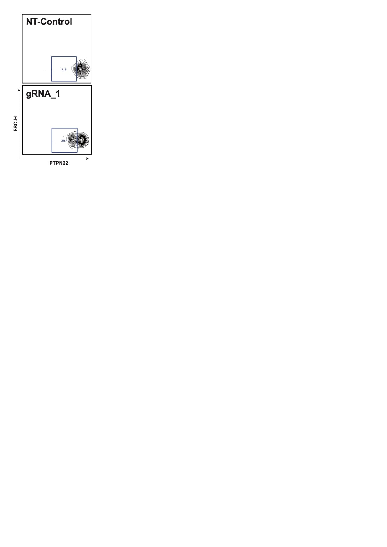
Application: Flow CytometrySample Tested: Peripheral blood mononuclear cells (PBMCs)Species: HumanVerified Customer | Posted 12/14/2021Goat anti-PTPN22 from R&D used in flow cytometry, in combination with a secondary anti-goat conjugated to AF488. Samples: PBMCs with non-targeting control and PBMCs with gRNA targeted to PTPN22/LYP. gRNAs from IDT.

-
Application: Western BlotSample Tested: Peripheral blood mononuclear cells (PBMCs)Species: HumanVerified Customer | Posted 11/03/2020Used to detect PTPN22/LYP in primary immune cells. Although a few bands can be observed per lane, the one running at 100 kDa corresponds to PTPN22/LYP (confirmed by siRNA).
There are no reviews that match your criteria.
Protocols
Find general support by application which include: protocols, troubleshooting, illustrated assays, videos and webinars.
- Appropriate Fixation of IHC/ICC Samples
- Cellular Response to Hypoxia Protocols
- ClariTSA™ Fluorophore Kits
- Detection & Visualization of Antibody Binding
- ICC Cell Smear Protocol for Suspension Cells
- ICC Immunocytochemistry Protocol Videos
- ICC for Adherent Cells
- Immunocytochemistry (ICC) Protocol
- Immunocytochemistry Troubleshooting
- Immunofluorescence of Organoids Embedded in Cultrex Basement Membrane Extract
- Immunohistochemistry (IHC) and Immunocytochemistry (ICC) Protocols
- Preparing Samples for IHC/ICC Experiments
- Preventing Non-Specific Staining (Non-Specific Binding)
- Primary Antibody Selection & Optimization
- Protocol for VisUCyte™ HRP Polymer Detection Reagent
- Protocol for the Fluorescent ICC Staining of Cell Smears - Graphic
- Protocol for the Fluorescent ICC Staining of Cultured Cells on Coverslips - Graphic
- Protocol for the Preparation and Fluorescent ICC Staining of Cells on Coverslips
- Protocol for the Preparation and Fluorescent ICC Staining of Non-adherent Cells
- Protocol for the Preparation and Fluorescent ICC Staining of Stem Cells on Coverslips
- Protocol for the Preparation of a Cell Smear for Non-adherent Cell ICC - Graphic
- R&D Systems Quality Control Western Blot Protocol
- TUNEL and Active Caspase-3 Detection by IHC/ICC Protocol
- The Importance of IHC/ICC Controls
- Troubleshooting Guide: Western Blot Figures
- Western Blot Conditions
- Western Blot Protocol
- Western Blot Protocol for Cell Lysates
- Western Blot Troubleshooting
- Western Blot Troubleshooting Guide
- View all Protocols, Troubleshooting, Illustrated assays and Webinars
Loading...