M-CSF, also known as CSF-1, is a four-alpha -helical-bundle cytokine that is the primary regulator of macrophage survival, proliferation and differentiation. M-CSF is also essential for the survival and proliferation of osteoclast progenitors. M-CSF also primes and enhances macrophage killing of tumor cells and microorganisms, regulates the release of cytokines and other inflammatory modulators from macrophages, and stimulates pinocytosis. M-CSF increases during pregnancy to support implantation and growth of the decidua and placenta. Sources of M-CSF include fibroblasts, activated macrophages, endometrial secretory epithelium, bone marrow stromal cells and activated endothelial cells. The M-CSF receptor (c-fms) transduces its pleotropic effects and mediates its endocytosis. M-CSF mRNAs of various sizes occur. Full length human M-CSF transcripts encode a 522 amino acid (aa) type I transmembrane (TM) protein with a 464 aa extracellular region, a 21 aa TM domain, and a 37 aa cytoplasmic tail that forms a 140 kDa covalent dimer. Differential processing produces two proteolytically cleaved, secreted dimers. One is an N- and O- glycosylated 86 kDa dimer, while the other is modified by both glycosylation and chondroitin-sulfate proteoglycan (PG) to generate a 200 kDa subunit. Although PG-modified M-CSF can circulate, it may be immobilized by attachment to type V collagen. Shorter transcripts encode M-CSF that lacks cleavage and PG sites and produces an N-glycosylated 68 kDa TM dimer and a slowly produced 44 kDa secreted dimer. Although forms may vary in activity and half-life, all contain the N-terminal 150 aa portion that is necessary and sufficient for interaction with the M-CSF receptor. The first 223 aa of mature human M-CSF shares 88%, 86%, 81% and 74% aa identity with corresponding regions of dog, cow, mouse and rat M-CSF, respectively. Human M-CSF is active in the mouse, but mouse M-CSF is reported to be species-specific.

Key Product Details
Species Reactivity
Validated:
Human
Cited:
Human
Applications
Validated:
Western Blot, Neutralization
Cited:
Western Blot, Neutralization, Immunocytochemistry, Cell Culture
Label
Unconjugated
Antibody Source
Polyclonal Goat IgG
Loading...
Product Specifications
Immunogen
E. coli-derived recombinant human M-CSF
Glu33-Ser190
Accession # NP_757350
Glu33-Ser190
Accession # NP_757350
Specificity
Detects human M-CSF in direct ELISAs and Western blots. In direct ELISAs, approximately 40% cross-reactivity with recombinant mouse (rm) M‑CSF is observed, and less than 1% cross-reactivity with recombinant rat M-CSF, rmSCF, and recombinant human SCF is observed.
Clonality
Polyclonal
Host
Goat
Isotype
IgG
Endotoxin Level
<0.10 EU per 1 μg of the antibody by the LAL method.
Scientific Data Images for Human M-CSF Antibody
Cell Proliferation Induced by M‑CSF and Neutralization by Human M‑CSF Antibody.
Recombinant Human M-CSF (Catalog # 216-MC) stimulates proliferation in the M-NFS-60 mouse myelogenous leukemia lymphoblast cell line in a dose-dependent manner (orange line). Proliferation elicited by Recombinant Human M-CSF (2.5 ng/mL) is neutralized (green line) by increasing concentrations of Goat Anti-Human M-CSF Antigen Affinity-purified Polyclonal Antibody (Catalog # AF216). The ND50 is typically 0.01-0.03 µg/mL.
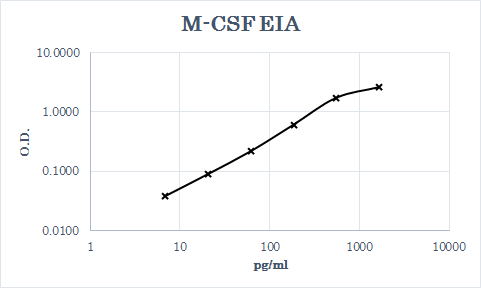
Human M-CSF ELISA Standard Curve
Recombinant Human M‑CSF (Catalog # 216-MC) was serially diluted and captured by Mouse Anti-Human M‑CSF Monoclonal Antibody (Catalog # MAB616) coated on a Clear Polystyrene Microplate (Catalog # DY990). Goat Anti-Human M‑CSF Antigen Affinity-purified Polyclonal Antibody (Catalog # AF216) was biotinylated and incubated with the protein captured on the plate. Detection of the standard curve was achieved by incubating Streptavidin-HRP (Catalog # DY998)Applications for Human M-CSF Antibody
Application
Recommended Usage
Western Blot
0.1 µg/mL
Sample: Recombinant Human M-CSF (Catalog # 216-MC)
Sample: Recombinant Human M-CSF (Catalog # 216-MC)
Neutralization
Measured by its ability to neutralize M‑CSF-induced proliferation in the M‑NFS‑60 mouse myelogenous leukemia lymphoblast cell line. Halenbeck, R. et al. (1989) Biotechnology 7:710. The Neutralization Dose (ND50) is typically 0.01-0.03 µg/mL in the presence of 2.5 ng/mL Recombinant Human M‑CSF.
Reviewed Applications
Read 2 reviews rated 5 using AF216 in the following applications:
Formulation, Preparation, and Storage
Purification
Antigen Affinity-purified
Reconstitution
Reconstitute at 0.2 mg/mL in sterile PBS. For liquid material, refer to CoA for concentration.
Loading...
Formulation
Lyophilized from a 0.2 μm filtered solution in PBS with Trehalose. *Small pack size (SP) is supplied either lyophilized or as a 0.2 µm filtered solution in PBS.
Shipping
Lyophilized product is shipped at ambient temperature. Liquid small pack size (-SP) is shipped with polar packs. Upon receipt, store immediately at the temperature recommended below.
Stability & Storage
Use a manual defrost freezer and avoid repeated freeze-thaw cycles.
- 12 months from date of receipt, -20 to -70 °C as supplied.
- 1 month, 2 to 8 °C under sterile conditions after reconstitution.
- 6 months, -20 to -70 °C under sterile conditions after reconstitution.
Calculators
Background: M-CSF
Long Name
Macrophage Colony Stimulating Factor
Alternate Names
CSF-1, CSF1, Lanimostim, MCSF
Gene Symbol
CSF1
UniProt
Additional M-CSF Products
Product Documents for Human M-CSF Antibody
Certificate of Analysis
To download a Certificate of Analysis, please enter a lot or batch number in the search box below.
Note: Certificate of Analysis not available for kit components.
Product Specific Notices for Human M-CSF Antibody
For research use only
Citations for Human M-CSF Antibody
 Powered by Bioz
Customer Reviews for Human M-CSF Antibody (2)
5 out of 5
2 Customer Ratings
Have you used Human M-CSF Antibody?
Submit a review and receive an Amazon gift card!
$25/€18/£15/$25CAN/¥2500 Yen for a review with an image
$10/€7/£6/$10CAN/¥1110 Yen for a review without an image
Submit a review
Customer Images
Showing
1
-
2 of
2 reviews
Showing All
Filter By:
-
Application: ELISASample Tested: EDTA PlasmaSpecies: HumanVerified Customer | Posted 07/23/2019

-
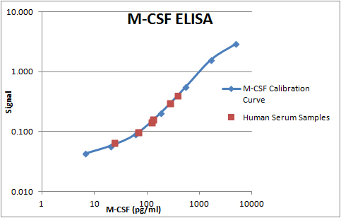
Application: ELISASample Tested: Serum and PlasmaSpecies: HumanVerified Customer | Posted 11/03/2017This goat poly clonal was biotinylated upon receipt. It was then subsequently used as a detection antibody in a sandwich ELISA with the R&D Systems monoclonal MAB616. This assay was used to measure human M-CSF in serum samples.
There are no reviews that match your criteria.
Protocols
Find general support by application which include: protocols, troubleshooting, illustrated assays, videos and webinars.
- Cellular Response to Hypoxia Protocols
- R&D Systems Quality Control Western Blot Protocol
- Troubleshooting Guide: Western Blot Figures
- Western Blot Conditions
- Western Blot Protocol
- Western Blot Protocol for Cell Lysates
- Western Blot Troubleshooting
- Western Blot Troubleshooting Guide
- View all Protocols, Troubleshooting, Illustrated assays and Webinars
Loading...
Associated Pathways

