Meprins are multimeric proteases composed of alpha and beta subunits, which are members of the astacin family of zinc endopeptidases (1, 2). Both subunits form disulfide‑linked homo- or heterooligomers, which are also referred to as Meprin A (composed of alpha subunits with or without beta subunits) and Meprin B (composed of beta subunits only) (3). Although the two subunits share 42% identity in their amino acid sequence, they differ significantly in their oligomeric structure, post-translational processing and subsequently cellular location, and substrate and peptide bond specificity (4). The 746 amino acid sequence of human meprin alpha subunit precursor consists of a signal peptide (residues 1 to 21), a pro region (residues 22 to 65), and a mature chain (residues 66 to 746) containing following domains, catalytic (residues 62 to 263), MAM (residues 264 to 433), MATH (residues 434 to 593), EGF-like (residues 670 to 710), transmembrane (residues 713 to 740), and cytoplasmic (residues 741 to 746). The pro enzyme terminating at residue 601 was expressed and the secreted protein purified from conditioned medium. The molecular masses of rhMEP1A are similar to those observed for the alpha subunit of rat Meprin A (5).
Human Meprin alpha Subunit/MEP1A Antibody
R&D Systems | Catalog # AF3220
Key Product Details
Species Reactivity
Validated:
Human
Cited:
Human, Mouse
Applications
Validated:
Immunohistochemistry, Western Blot, Intracellular Staining by Flow Cytometry, Immunoprecipitation, CyTOF-ready
Cited:
Immunohistochemistry, Immunohistochemistry-Paraffin, Western Blot
Label
Unconjugated
Antibody Source
Polyclonal Goat IgG
Loading...
Product Specifications
Immunogen
Mouse myeloma cell line NS0-derived recombinant human Meprin alpha Subunit/MEP1A
Val22-Gln601
Accession # AAA21338
Val22-Gln601
Accession # AAA21338
Specificity
Detects human Meprin alpha Subunit/MEP1A in direct ELISAs and Western blots. In direct ELISAs, approximately 10% cross‑reactivity with recombinant mouse MEP1A and recombinant human MEP1B is observed.
Clonality
Polyclonal
Host
Goat
Isotype
IgG
Applications for Human Meprin alpha Subunit/MEP1A Antibody
Application
Recommended Usage
CyTOF-ready
Ready to be labeled using established conjugation methods. No BSA or other carrier proteins that could interfere with conjugation.
Immunohistochemistry
5-15 µg/mL
Sample: Immersion fixed paraffin-embedded sections of human intestine (jejunum)
Sample: Immersion fixed paraffin-embedded sections of human intestine (jejunum)
Immunoprecipitation
25 µg/mL
Sample: Conditioned cell culture medium spiked with Recombinant Human Meprin alpha Subunit/MEP1A (Catalog # 3220-ZN), see our available Western blot detection antibodies
Sample: Conditioned cell culture medium spiked with Recombinant Human Meprin alpha Subunit/MEP1A (Catalog # 3220-ZN), see our available Western blot detection antibodies
Intracellular Staining by Flow Cytometry
0.25 µg/106 cells
Sample: HEK293 human embryonic kidney cell line fixed with paraformaldehyde and permeabilized with saponin
Sample: HEK293 human embryonic kidney cell line fixed with paraformaldehyde and permeabilized with saponin
Western Blot
0.1 µg/mL
Sample: Recombinant Human Meprin alpha Subunit/MEP1A (Catalog # 3220-ZN)
Sample: Recombinant Human Meprin alpha Subunit/MEP1A (Catalog # 3220-ZN)
Reviewed Applications
Read 1 review rated 5 using AF3220 in the following applications:
Flow Cytometry Panel Builder
Bio-Techne Knows Flow Cytometry
Save time and reduce costly mistakes by quickly finding compatible reagents using the Panel Builder Tool.
Advanced Features
- Spectra Viewer - Custom analysis of spectra from multiple fluorochromes
- Spillover Popups - Visualize the spectra of individual fluorochromes
- Antigen Density Selector - Match fluorochrome brightness with antigen density

Formulation, Preparation, and Storage
Purification
Antigen Affinity-purified
Reconstitution
Reconstitute at 0.2 mg/mL in sterile PBS. For liquid material, refer to CoA for concentration.
Loading...
Formulation
Lyophilized from a 0.2 μm filtered solution in PBS with Trehalose. *Small pack size (SP) is supplied either lyophilized or as a 0.2 µm filtered solution in PBS.
Shipping
Lyophilized product is shipped at ambient temperature. Liquid small pack size (-SP) is shipped with polar packs. Upon receipt, store immediately at the temperature recommended below.
Stability & Storage
Use a manual defrost freezer and avoid repeated freeze-thaw cycles.
- 12 months from date of receipt, -20 to -70 °C as supplied.
- 1 month, 2 to 8 °C under sterile conditions after reconstitution.
- 6 months, -20 to -70 °C under sterile conditions after reconstitution.
Calculators
Background: Meprin alpha Subunit/MEP1A
References
- Bond, J.S. and Beynon, R.J. (1995) Protein Sci. 4:1247.
- Stocker, W. et al. (1995) Protein Sci. 4:823.
- Bertenshaw, G.P. et al. (2001) J. Biol. Chem. 276:13248.
- Ishmael, F.T. et al. (2005) J. Biol. Chem. 280:13895.
- Bertenshaw, G.P. et al. (2003) J. Biol. Chem. 278:2522.
Alternate Names
MEP1A, PPHA
Gene Symbol
MEP1A
UniProt
Additional Meprin alpha Subunit/MEP1A Products
Product Documents for Human Meprin alpha Subunit/MEP1A Antibody
Certificate of Analysis
To download a Certificate of Analysis, please enter a lot or batch number in the search box below.
Note: Certificate of Analysis not available for kit components.
Product Specific Notices for Human Meprin alpha Subunit/MEP1A Antibody
For research use only
Related Research Areas
Citations for Human Meprin alpha Subunit/MEP1A Antibody
 Powered by Bioz
Customer Reviews for Human Meprin alpha Subunit/MEP1A Antibody (1)
5 out of 5
1 Customer Rating
Have you used Human Meprin alpha Subunit/MEP1A Antibody?
Submit a review and receive an Amazon gift card!
$25/€18/£15/$25CAN/¥2500 Yen for a review with an image
$10/€7/£6/$10CAN/¥1110 Yen for a review without an image
Submit a review
Customer Images
Showing
1
-
1 of
1 review
Showing All
Filter By:
-
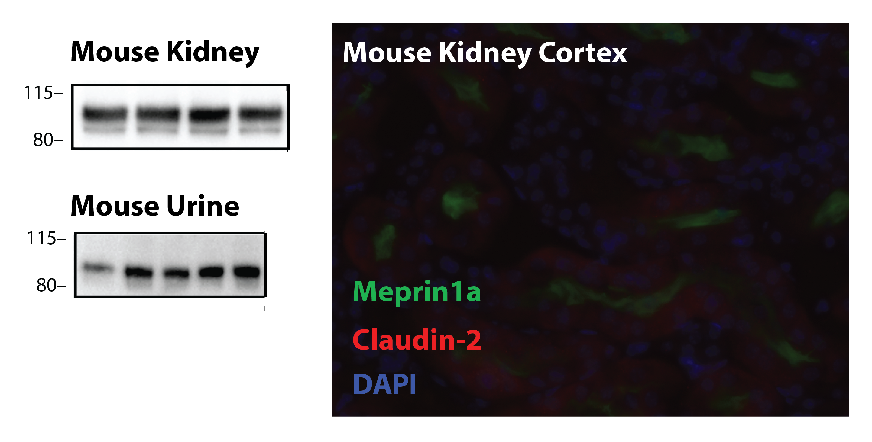
Application: Western blot and immunofluorescenceSample Tested: C57BL/6J urine and C57BL/6J kidneySpecies: MouseVerified Customer | Posted 08/20/2019Mouse kidney tissue and mouse urine western blot. Immunofluorescence of frozen mouse tissue 1:40Western blot 1:1000, IF 1:40

There are no reviews that match your criteria.
Protocols
Find general support by application which include: protocols, troubleshooting, illustrated assays, videos and webinars.
- 7-Amino Actinomycin D (7-AAD) Cell Viability Flow Cytometry Protocol
- Antigen Retrieval Protocol (PIER)
- Antigen Retrieval for Frozen Sections Protocol
- Appropriate Fixation of IHC/ICC Samples
- Cellular Response to Hypoxia Protocols
- Chromogenic IHC Staining of Formalin-Fixed Paraffin-Embedded (FFPE) Tissue Protocol
- Chromogenic Immunohistochemistry Staining of Frozen Tissue
- ClariTSA™ Fluorophore Kits
- Detection & Visualization of Antibody Binding
- Extracellular Membrane Flow Cytometry Protocol
- Flow Cytometry Protocol for Cell Surface Markers
- Flow Cytometry Protocol for Staining Membrane Associated Proteins
- Flow Cytometry Staining Protocols
- Flow Cytometry Troubleshooting Guide
- Fluorescent IHC Staining of Frozen Tissue Protocol
- Graphic Protocol for Heat-induced Epitope Retrieval
- Graphic Protocol for the Preparation and Fluorescent IHC Staining of Frozen Tissue Sections
- Graphic Protocol for the Preparation and Fluorescent IHC Staining of Paraffin-embedded Tissue Sections
- Graphic Protocol for the Preparation of Gelatin-coated Slides for Histological Tissue Sections
- IHC Sample Preparation (Frozen sections vs Paraffin)
- Immunofluorescent IHC Staining of Formalin-Fixed Paraffin-Embedded (FFPE) Tissue Protocol
- Immunohistochemistry (IHC) and Immunocytochemistry (ICC) Protocols
- Immunohistochemistry Frozen Troubleshooting
- Immunohistochemistry Paraffin Troubleshooting
- Immunoprecipitation Protocol
- Intracellular Flow Cytometry Protocol Using Alcohol (Methanol)
- Intracellular Flow Cytometry Protocol Using Detergents
- Intracellular Nuclear Staining Flow Cytometry Protocol Using Detergents
- Intracellular Staining Flow Cytometry Protocol Using Alcohol Permeabilization
- Intracellular Staining Flow Cytometry Protocol Using Detergents to Permeabilize Cells
- Preparing Samples for IHC/ICC Experiments
- Preventing Non-Specific Staining (Non-Specific Binding)
- Primary Antibody Selection & Optimization
- Propidium Iodide Cell Viability Flow Cytometry Protocol
- Protocol for Heat-Induced Epitope Retrieval (HIER)
- Protocol for Liperfluo
- Protocol for Making a 4% Formaldehyde Solution in PBS
- Protocol for VisUCyte™ HRP Polymer Detection Reagent
- Protocol for the Characterization of Human Th22 Cells
- Protocol for the Characterization of Human Th9 Cells
- Protocol for the Preparation & Fixation of Cells on Coverslips
- Protocol for the Preparation and Chromogenic IHC Staining of Frozen Tissue Sections
- Protocol for the Preparation and Chromogenic IHC Staining of Frozen Tissue Sections - Graphic
- Protocol for the Preparation and Chromogenic IHC Staining of Paraffin-embedded Tissue Sections
- Protocol for the Preparation and Chromogenic IHC Staining of Paraffin-embedded Tissue Sections - Graphic
- Protocol for the Preparation and Fluorescent IHC Staining of Frozen Tissue Sections
- Protocol for the Preparation and Fluorescent IHC Staining of Paraffin-embedded Tissue Sections
- Protocol for the Preparation of Gelatin-coated Slides for Histological Tissue Sections
- Protocol: Annexin V and PI Staining by Flow Cytometry
- Protocol: Annexin V and PI Staining for Apoptosis by Flow Cytometry
- R&D Systems Quality Control Western Blot Protocol
- TUNEL and Active Caspase-3 Detection by IHC/ICC Protocol
- The Importance of IHC/ICC Controls
- Troubleshooting Guide: Fluorokine Flow Cytometry Kits
- Troubleshooting Guide: Immunohistochemistry
- Troubleshooting Guide: Western Blot Figures
- Western Blot Conditions
- Western Blot Protocol
- Western Blot Protocol for Cell Lysates
- Western Blot Troubleshooting
- Western Blot Troubleshooting Guide
- View all Protocols, Troubleshooting, Illustrated assays and Webinars
Loading...