Matrix metalloproteinases are a family of zinc and calcium dependent endopeptidases with the combined ability to degrade all the components of the extracellular matrix. MMP-2 (gelatinase A), a type IV collagenase, can degrade a broad range of substrates including type IV, V, VII and X collagens as well as elastin and fibronectin. It is believed to act synergistically with interstitial collagenase (MMP-1) in the degradation of fibrillar collagens as it degrades their denatured gelatin forms. MMP-2 has been shown to be associated with many connective tissue cells as well as neutrophils, macrophages and monocytes. Structurally, MMP-2 may be divided into several distinct domains: a pro-domain which is cleaved upon activation; a catalytic domain containing the zinc binding site; a fibronectin-like domain thought to play a role in substrate targeting; and a carboxyl terminal (hemopexin-like) domain containing 2 N-linked glycosylation sites.

Key Product Details
Species Reactivity
Validated:
Human
Cited:
Human, Canine, Fish - Danio rerio (Zebrafish), Rabbit
Applications
Validated:
Immunohistochemistry, Western Blot, Simple Western, Immunoprecipitation
Cited:
Immunohistochemistry, Immunohistochemistry-Paraffin, Western Blot, Immunocytochemistry, ELISA Microarray Development
Label
Unconjugated
Antibody Source
Polyclonal Goat IgG
Loading...
Product Specifications
Immunogen
Chinese hamster ovary cell line CHO-derived recombinant human MMP‑2
Ile34-Cys660
Accession # P08253
Ile34-Cys660
Accession # P08253
Specificity
Detects human MMP-2 in direct ELISAs and Western blots.
Clonality
Polyclonal
Host
Goat
Isotype
IgG
Scientific Data Images for Human MMP-2 Antibody
Detection of Human MMP‑2 by Western Blot.
Western blot shows lysate of U-118-MG human glioblastoma/astrocytoma cell line. PVDF membrane was probed with 1 µg/mL of Goat Anti-Human MMP-2 Antigen Affinity-purified Polyclonal Antibody (Catalog # AF902) followed by HRP-conjugated Anti-Goat IgG Secondary Antibody (HAF017). A specific band was detected for MMP-2 at approximately 72 kDa (as indicated). This experiment was conducted under reducing conditions and using Immunoblot Buffer Group 1.
MMP‑2 in Human Ovarian Cancer Tissue.
MMP-2 was detected in immersion fixed paraffin-embedded sections of human ovarian cancer tissue using Goat Anti-Human MMP-2 Antigen Affinity-purified Poly-clonal Antibody (Catalog # AF902) at 10 µg/mL overnight at 4 °C. Tissue was stained using the Anti-Goat HRP-DAB Cell & Tissue Staining Kit (brown; CTS008) and counter-stained with hematoxylin (blue). View our protocol for Chromogenic IHC Staining of Paraffin-embedded Tissue Sections.
MMP‑2 in Human Ovary.
MMP-2 was detected in immersion fixed paraffin-embedded sections of human ovarian array using Goat Anti-Human MMP-2 Antigen Affinity-purified Polyclonal Antibody (Catalog # AF902) at 10 µg/mL overnight at 4 °C. Tissue was stained using the Anti-Goat HRP-DAB Cell & Tissue Staining Kit (brown; CTS008) and counterstained with hematoxylin (blue). Lower panel shows a lack of labeling if primary antibodies are omitted and tissue is stained only with secondary antibody followed by incubation with detection reagents. View our protocol for Chromogenic IHC Staining of Paraffin-embedded Tissue Sections.
Detection of Human MMP‑2 by Simple WesternTM.
Simple Western lane view shows lysates of U-118-MG human glioblastoma/astrocytoma cell line, loaded at 0.2 mg/mL. A specific band was detected for MMP-2 at approximately 78 kDa (as indicated) using 10 µg/mL of Goat Anti-Human MMP-2 Antigen Affinity-purified Polyclonal Antibody (Catalog # AF902) followed by 1:50 dilution of HRP-conjugated Anti-Goat IgG Secondary Antibody (HAF109). This experiment was conducted under reducing conditions and using the 12-230 kDa separation system.
Detection of Human MMP-2 by Western Blot
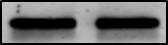
Conditioned media from HCV-expressing cells enhance invasive potential of HSC.(A) Effect of different CM on HSC invasion through transwell inserts precoated with collagen. Same amount of serum starved HSC (5×10⁴ cells/100 µL) were seeded at the upper chambers of transwells and exposed during 24 h to different CM (hepatic-derived CM, 0% FBS DMEM or 10% FBS DMEM) dispensed at the lower compartments. At the end of migration, the upper surface of the membrane was washed and HSC adhered to the lower surface were fixed in methanol, stained with DAPI and counted in 5 randomly chosen microscopic fields (400x) in an epifluorescence microscope (Leica, Wetzlar, Germany). Data from 4 independent experiments are shown as mean +SD. #p<0.05, HSC cultured with CM from HCV replicons or DMEM 10% FBS versus HSC cultured in DMEM 0% FBS; *p<0.05, HSC cultured with CM from HCV-expressing cells or 10% FBS versus HSC cultured with CM from Huh7. (B) ProMMP-2 and MMP-2 expression (AF902, 0.2 µg/mL) and activity were respectively examined by western blotting (WB) and zymography and further quantified in protein extracts (20 µL) of HSC cultured during 24 h with CM from Huh7 or HCV-expressing cells. Bars represent the mean of densitometric analysis from two independent experiments. Image collected and cropped by CiteAb from the following open publication (https://pubmed.ncbi.nlm.nih.gov/25302785), licensed under a CC-BY license. Not internally tested by R&D Systems.
Detection of MMP-2 by Western Blot
Conditioned media from G17-stimulated melanoma cells exhibit upregulation of MMP-2 and downregulation of TIMP-3 expression. Representative demonstration of the confirmation of proteomic data using Western blot analysis, with prosaposin serving as a benchmark for gastrin responsiveness (A). Mean and standard deviation (±SD) of the densitometric analysis results from three independent experiments (B). Statistical difference (* p < 0.05) between treated and untreated control groups (considered as 100%) are indicated as follows: prosaposin, TIMP-3 and TIMP-1 levels in G361 cells and MMP2 expressions in the SK-MEL-2 cell line. Image collected and cropped by CiteAb from the following open publication (https://www.mdpi.com/1422-0067/24/23/16851), licensed under a CC-BY license. Not internally tested by R&D Systems.Applications for Human MMP-2 Antibody
Application
Recommended Usage
Immunohistochemistry
5-15 µg/mL
Sample: Immersion fixed paraffin-embedded sections of human ovarian cancer tissue and normal human ovarian array
Sample: Immersion fixed paraffin-embedded sections of human ovarian cancer tissue and normal human ovarian array
Immunoprecipitation
25 µg/mL
Sample: Conditioned cell culture medium spiked with Recombinant Human MMP‑2 (Catalog # 902-MP), see our available Western blot detection antibodies
Sample: Conditioned cell culture medium spiked with Recombinant Human MMP‑2 (Catalog # 902-MP), see our available Western blot detection antibodies
Simple Western
10 µg/mL
Sample: U‑118‑MG human glioblastoma/astrocytoma cell line
Sample: U‑118‑MG human glioblastoma/astrocytoma cell line
Western Blot
1 µg/mL
Sample: U‑118‑MG human glioblastoma/astrocytoma cell line
Sample: U‑118‑MG human glioblastoma/astrocytoma cell line
Reviewed Applications
Read 4 reviews rated 4.3 using AF902 in the following applications:
Formulation, Preparation, and Storage
Purification
Antigen Affinity-purified
Reconstitution
Reconstitute at 0.2 mg/mL in sterile PBS. For liquid material, refer to CoA for concentration.
Loading...
Formulation
Lyophilized from a 0.2 μm filtered solution in PBS with Trehalose. *Small pack size (SP) is supplied either lyophilized or as a 0.2 µm filtered solution in PBS.
Shipping
Lyophilized product is shipped at ambient temperature. Liquid small pack size (-SP) is shipped with polar packs. Upon receipt, store immediately at the temperature recommended below.
Stability & Storage
Use a manual defrost freezer and avoid repeated freeze-thaw cycles.
- 12 months from date of receipt, -20 to -70 °C as supplied.
- 1 month, 2 to 8 °C under sterile conditions after reconstitution.
- 6 months, -20 to -70 °C under sterile conditions after reconstitution.
Calculators
Background: MMP-2
Long Name
Matrix Metalloproteinase 2
Alternate Names
Gelatinase A, MMP2
Gene Symbol
MMP2
UniProt
Additional MMP-2 Products
Product Documents for Human MMP-2 Antibody
Certificate of Analysis
To download a Certificate of Analysis, please enter a lot or batch number in the search box below.
Note: Certificate of Analysis not available for kit components.
Product Specific Notices for Human MMP-2 Antibody
For research use only
Citations for Human MMP-2 Antibody
 Powered by Bioz
Customer Reviews for Human MMP-2 Antibody (4)
4.3 out of 5
4 Customer Ratings
Have you used Human MMP-2 Antibody?
Submit a review and receive an Amazon gift card!
$25/€18/£15/$25CAN/¥2500 Yen for a review with an image
$10/€7/£6/$10CAN/¥1110 Yen for a review without an image
Submit a review
Customer Images
Showing
1
-
4 of
4 reviews
Showing All
Filter By:
-
Application: Western BlotSample Tested: HeLa cellsSpecies: HumanVerified Customer | Posted 07/13/2018
-
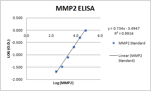
Application: ELISASample Tested: Serum and PlasmaSpecies: HumanVerified Customer | Posted 11/09/2017The polyclonal goat antibody AF902 was used as both capture and detection to build and ELISA measuring MMP-2 in human serum and plasma samples.

-
Application: Western BlotSample Tested: Human cell conditioned mediumSpecies: HumanVerified Customer | Posted 02/27/2016
-
Application: Western BlotSample Tested: Trabecular meshwork endothelialSpecies: Human and input species hereVerified Customer | Posted 11/23/2015
There are no reviews that match your criteria.
Protocols
Find general support by application which include: protocols, troubleshooting, illustrated assays, videos and webinars.
- Antigen Retrieval Protocol (PIER)
- Antigen Retrieval for Frozen Sections Protocol
- Appropriate Fixation of IHC/ICC Samples
- Cellular Response to Hypoxia Protocols
- Chromogenic IHC Staining of Formalin-Fixed Paraffin-Embedded (FFPE) Tissue Protocol
- Chromogenic Immunohistochemistry Staining of Frozen Tissue
- ClariTSA™ Fluorophore Kits
- Detection & Visualization of Antibody Binding
- Fluorescent IHC Staining of Frozen Tissue Protocol
- Graphic Protocol for Heat-induced Epitope Retrieval
- Graphic Protocol for the Preparation and Fluorescent IHC Staining of Frozen Tissue Sections
- Graphic Protocol for the Preparation and Fluorescent IHC Staining of Paraffin-embedded Tissue Sections
- Graphic Protocol for the Preparation of Gelatin-coated Slides for Histological Tissue Sections
- IHC Sample Preparation (Frozen sections vs Paraffin)
- Immunofluorescent IHC Staining of Formalin-Fixed Paraffin-Embedded (FFPE) Tissue Protocol
- Immunohistochemistry (IHC) and Immunocytochemistry (ICC) Protocols
- Immunohistochemistry Frozen Troubleshooting
- Immunohistochemistry Paraffin Troubleshooting
- Immunoprecipitation Protocol
- Preparing Samples for IHC/ICC Experiments
- Preventing Non-Specific Staining (Non-Specific Binding)
- Primary Antibody Selection & Optimization
- Protocol for Heat-Induced Epitope Retrieval (HIER)
- Protocol for Making a 4% Formaldehyde Solution in PBS
- Protocol for VisUCyte™ HRP Polymer Detection Reagent
- Protocol for the Preparation & Fixation of Cells on Coverslips
- Protocol for the Preparation and Chromogenic IHC Staining of Frozen Tissue Sections
- Protocol for the Preparation and Chromogenic IHC Staining of Frozen Tissue Sections - Graphic
- Protocol for the Preparation and Chromogenic IHC Staining of Paraffin-embedded Tissue Sections
- Protocol for the Preparation and Chromogenic IHC Staining of Paraffin-embedded Tissue Sections - Graphic
- Protocol for the Preparation and Fluorescent IHC Staining of Frozen Tissue Sections
- Protocol for the Preparation and Fluorescent IHC Staining of Paraffin-embedded Tissue Sections
- Protocol for the Preparation of Gelatin-coated Slides for Histological Tissue Sections
- R&D Systems Quality Control Western Blot Protocol
- TUNEL and Active Caspase-3 Detection by IHC/ICC Protocol
- The Importance of IHC/ICC Controls
- Troubleshooting Guide: Immunohistochemistry
- Troubleshooting Guide: Western Blot Figures
- Western Blot Conditions
- Western Blot Protocol
- Western Blot Protocol for Cell Lysates
- Western Blot Troubleshooting
- Western Blot Troubleshooting Guide
- View all Protocols, Troubleshooting, Illustrated assays and Webinars


