
Human BMP-9, also known as growth and differentiation factor 2 (GDF-2), is a member of the BMP subgroup of the TGF-beta superfamily proteins that signal through heterodimeric complexes composed of type I and type II BMP receptors. BMP-9 regulates the development and function of a variety of embryonal and adult tissues (1, 2). The human BMP-9 cDNA encodes a 429 amino acid (aa) precursor that includes a 22 aa signal sequence, a 298 aa propeptide, and a 111 aa mature protein (3). Unlike with other BMP family proteins, the propeptide does not interfere with the biological activity of BMP-9 and remains associated with the mature peptide after proteolytic cleavage (4). Human and mouse BMP-9 share 96% aa sequence identity. Within the mature protein, human BMP-9 shares 64% aa sequence identity with human BMP-10 and less than 50% aa sequence identity with other BMPs. BMP-9 is expressed by non-parenchymal cells in the liver, (5, 6) where it promotes lipid metabolism and inhibits glucose production (7). BMP-9 exerts a prolonged hypoglycemic effect which may be due to an enhancement of insulin release (7). BMP-9 interacts with a high affinity specific heteromeric receptor expressed on liver endothelial cells that has been identified as ALK-1 (4-6). In the embryonal CNS, BMP-9 functions in the development and maintenance of the cholinergic neuronal phenotype (8-10). BMP-9 also induces the differentiation of mesenchymal stem cells into the chondrogenic lineage (11, 12). At low concentrations, BMP-9 is a proliferative factor for hematopoietic progenitor cells, but at higher concentrations, it enhances TGF-beta 1 production and inhibits hematopoietic progenitor colony formation (13).

Scientific images may be resized, cropped, or adjusted for brightness or contrast for presentation purposes. These updates are limited to presentation and do not affect the underlying scientific interpretation of the data. Where available, additional source data may be provided upon request.
Key Product Details
Species Reactivity
Validated:
Cited:
Applications
Validated:
Cited:
Label
Antibody Source
Product Specifications
Immunogen
Ser320-Arg429
Accession # Q9UK05
Specificity
Clonality
Host
Isotype
Endotoxin Level
Scientific Data Images for BMP-9 Antibody
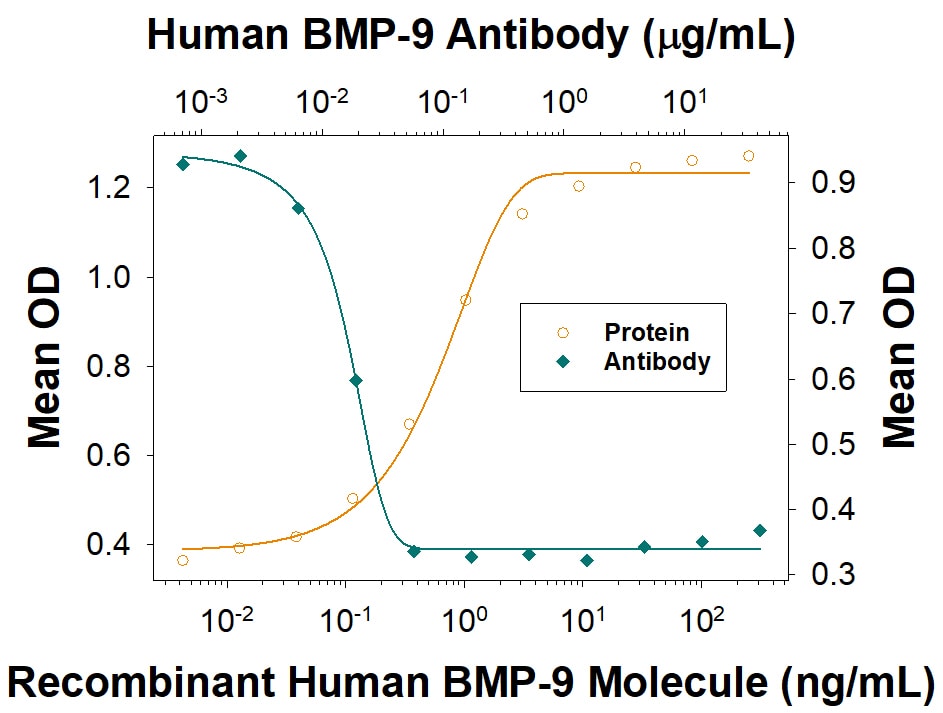
Alkaline Phosphatase Production Induced by BMP‑9 and Neutralization by Human BMP‑9 Antibody.
Recombinant Human BMP-9 (Catalog # 3209-BP) induces alkaline phosphatase production in the ATDC5 mouse chondrogenic cell line in a dose-dependent manner (orange line). Alkaline phospha-tase production elicited by Recombinant Human BMP-9 (2 ng/mL) is neutralized (green line) by increasing concen-trations of Goat Anti-Human BMP-9 Antigen Affinity-purified Polyclonal Antibody (Catalog # AF3209). The ND50 is typically 0.005-0.1 µg/mL.
Human BMP-9 ELISA Standard Curve
Recombinant Human BMP‑9 (Catalog # 3209-BP) was serially diluted and captured by Mouse Anti-Human/Mouse/Primate BMP‑9 Monoclonal Antibody (Catalog # MAB3209) coated on a Clear Polystyrene Microplate (Catalog # DY990). Goat Anti-Human/Mouse BMP‑9 Antigen Affinity-purified Polyclonal Antibody (Catalog # AF3209) was biotinylated and incubated with the protein captured on the plate. Detection of the standard curve was achieved by incubating Streptavidin-HRP (Catalog # DY998)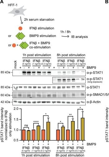
Detection of BMP-9 by Western Blot
BMP9 enhances STAT1 activation upon type I IFN, but not type II IFN stimulation.(A) After 2 h of serum starvation, HFF-1 were stimulated for either one or 8 h with IFN beta (5 ng/ml) or BMP9 (3 nM) alone, or were co-stimulated with IFN beta and BMP9, followed by cell lysis and immunoblot analysis with antibodies for phospho-STAT1, STAT1, phospho-SMAD1/5/9, and beta -Actin. Phospho-STAT1 band intensities were first normalized to corresponding total STAT1 levels, then to IFN beta stimulation only, and are shown below. (B) After 2 h of serum starvation, HFF-1 were stimulated for 1 h with either IFN alpha 2 (5 ng/ml), IFN gamma (5 ng/ml), or BMP9 (3 nM) alone, or co-stimulated with IFN alpha 2 or IFN gamma and BMP9, followed by cell lysis, immunoblot analysis and quantification as in (A). (C) After 2 h of serum starvation, HFF-1 were stimulated for 6 h with either IFN alpha 2 (5 ng/ml), IFN beta (5 ng/ml), IFN gamma (5 ng/ml), or BMP9 (3 nM) alone, or co-stimulated with IFN alpha 2, IFN beta, IFN gamma, and BMP9, followed by RNA extraction from cell lysates and RT-qPCR for Irf9, Stat2, Irf1, and Id3 transcripts. Data information: (A, B) Experiment was performed three independent times, one representative immunoblot is shown. Quantified data for the STAT1 phosphorylation levels are combined from three independent experiments. (C) Data are combined from three independent experiments. Student’s t test (unpaired, two-tailed), n.s. not significant, *P < 0.05, **P < 0.01, ****P < 0.0001. Data are shown as mean ± SD. Source data are available online for this figure. Image collected and cropped by CiteAb from the following open publication (https://pubmed.ncbi.nlm.nih.gov/38308064), licensed under a CC-BY license. Not internally tested by R&D Systems.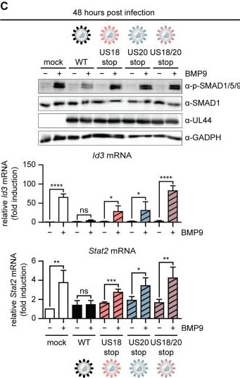
Detection of BMP-9 by Western Blot
HCMV US18 and US20 inhibit BMP9-mediated signaling during infection.(A) Schematic representation of the workflow. Recombinant HCMV US18stop, HCMV US20stop, and HCMV US18/20stop were constructed by introducing a 16 base pair (bp) stop cassette within the respective coding region. HFF-1 were infected by centrifugal enhancement with HCMV WT, HCMV US18stop, HCMV US20stop, or HCMV US18/20stop (MOI 4 for the 3 h time point, MOI 0.5 for the 48 h time point). Three or 48 h post infection, cells were stimulated with BMP9 (3 nM) for (1) 2 h, followed by cell lysis and immunoblot analysis, or (2) 6 h, followed by RNA extraction from cell lysates and RT-qPCR. (B, C) Cell lysates from (A) were subjected to immunoblot analysis with p-SMAD1/5/9, SMAD1, HCMV UL35, and GAPDH-specific antibodies (top panel), and transcript levels of Id3 and Stat2 are shown below. Data information: (B, C) Data are combined from three independent experiments. Student’s t test (unpaired, two-tailed), n.s. not significant, *P < 0.05, **P < 0.01, ***P < 0.001, ****P < 0.0001. Data are shown as mean ± SD. Source data are available online for this figure. Image collected and cropped by CiteAb from the following open publication (https://pubmed.ncbi.nlm.nih.gov/38308064), licensed under a CC-BY license. Not internally tested by R&D Systems.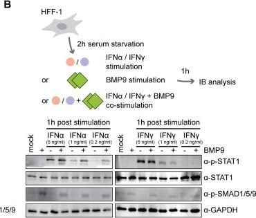
Detection of BMP-9 by Western Blot
BMP9 enhances STAT1 activation upon type I IFN, but not type II IFN stimulation.(A) After 2 h of serum starvation, HFF-1 were stimulated for either one or 8 h with IFN beta (5 ng/ml) or BMP9 (3 nM) alone, or were co-stimulated with IFN beta and BMP9, followed by cell lysis and immunoblot analysis with antibodies for phospho-STAT1, STAT1, phospho-SMAD1/5/9, and beta -Actin. Phospho-STAT1 band intensities were first normalized to corresponding total STAT1 levels, then to IFN beta stimulation only, and are shown below. (B) After 2 h of serum starvation, HFF-1 were stimulated for 1 h with either IFN alpha 2 (5 ng/ml), IFN gamma (5 ng/ml), or BMP9 (3 nM) alone, or co-stimulated with IFN alpha 2 or IFN gamma and BMP9, followed by cell lysis, immunoblot analysis and quantification as in (A). (C) After 2 h of serum starvation, HFF-1 were stimulated for 6 h with either IFN alpha 2 (5 ng/ml), IFN beta (5 ng/ml), IFN gamma (5 ng/ml), or BMP9 (3 nM) alone, or co-stimulated with IFN alpha 2, IFN beta, IFN gamma, and BMP9, followed by RNA extraction from cell lysates and RT-qPCR for Irf9, Stat2, Irf1, and Id3 transcripts. Data information: (A, B) Experiment was performed three independent times, one representative immunoblot is shown. Quantified data for the STAT1 phosphorylation levels are combined from three independent experiments. (C) Data are combined from three independent experiments. Student’s t test (unpaired, two-tailed), n.s. not significant, *P < 0.05, **P < 0.01, ****P < 0.0001. Data are shown as mean ± SD. Source data are available online for this figure. Image collected and cropped by CiteAb from the following open publication (https://pubmed.ncbi.nlm.nih.gov/38308064), licensed under a CC-BY license. Not internally tested by R&D Systems.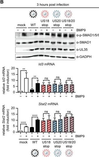
Detection of BMP-9 by Western Blot
HCMV US18 and US20 inhibit BMP9-mediated signaling during infection.(A) Schematic representation of the workflow. Recombinant HCMV US18stop, HCMV US20stop, and HCMV US18/20stop were constructed by introducing a 16 base pair (bp) stop cassette within the respective coding region. HFF-1 were infected by centrifugal enhancement with HCMV WT, HCMV US18stop, HCMV US20stop, or HCMV US18/20stop (MOI 4 for the 3 h time point, MOI 0.5 for the 48 h time point). Three or 48 h post infection, cells were stimulated with BMP9 (3 nM) for (1) 2 h, followed by cell lysis and immunoblot analysis, or (2) 6 h, followed by RNA extraction from cell lysates and RT-qPCR. (B, C) Cell lysates from (A) were subjected to immunoblot analysis with p-SMAD1/5/9, SMAD1, HCMV UL35, and GAPDH-specific antibodies (top panel), and transcript levels of Id3 and Stat2 are shown below. Data information: (B, C) Data are combined from three independent experiments. Student’s t test (unpaired, two-tailed), n.s. not significant, *P < 0.05, **P < 0.01, ***P < 0.001, ****P < 0.0001. Data are shown as mean ± SD. Source data are available online for this figure. Image collected and cropped by CiteAb from the following open publication (https://pubmed.ncbi.nlm.nih.gov/38308064), licensed under a CC-BY license. Not internally tested by R&D Systems.Applications for BMP-9 Antibody
Neutralization
Formulation, Preparation, and Storage
Purification
Reconstitution
Reconstitute at 0.2 mg/mL in sterile PBS. For liquid material, refer to CoA for concentration.
Formulation
Shipping
Stability & Storage
- 12 months from date of receipt, -20 to -70 °C as supplied.
- 1 month, 2 to 8 °C under sterile conditions after reconstitution.
- 6 months, -20 to -70 °C under sterile conditions after reconstitution.
Calculators
Background: BMP-9
References
- Chen, D. et al. (2004) Growth Factors 22:233.
- Miyazono, K. et al. (2005) Cytokine Growth Factor Rev. 16:251.
- Celeste, A.J. et al. (1994) J. Bone Miner. Res. 9:S136.
- Brown, M.A. et al. (2005) J. Biol. Chem. 280:25111.
- Song, J.J. et al. (1995) Endocrinology 136:4293.
- Miller, A.F. et al. (2000) J. Biol. Chem. 275:17937.
- Chen, C. et al. (2003) Nat. Biotechnol. 21:294.
- Lopez-Coviella, I. et al. (2000) Science 289:313.
- Lopez-Coviella, I. et al. (2005) Proc. Natl. Acad. Sci. USA 102:6984.
- Lopez-Coviella, I. et al. (2002) J. Physiol. Paris 96:53.
- Majumdar, M.K. et al. (2001) J. Cell. Physiol. 189:275.
- Hills, R.L. et al. (2005) J. Orthoped. Res. 23:611.
- Ploemacher, R.E. et al. (1999) Leukemia 13:428.
Long Name
Alternate Names
Gene Symbol
UniProt
Additional BMP-9 Products
Product Documents for BMP-9 Antibody
Certificate of Analysis
To download a Certificate of Analysis, please enter a lot or batch number in the search box below.
Note: Certificate of Analysis not available for kit components.
Product Specific Notices for BMP-9 Antibody
For research use only
Related Research Areas
Citations for BMP-9 Antibody
 Powered by Bioz
Customer Reviews for BMP-9 Antibody
There are currently no reviews for this product. Be the first to review BMP-9 Antibody and earn rewards!
Have you used BMP-9 Antibody?
Submit a review and receive an Amazon gift card!
$25/€18/£15/$25CAN/¥2500 for a review with an image
$10/€7/£6/$10CAN/¥1110 for a review without an image
Submit a review