Tie-2 (also known as TEK) is an angiogenic receptor tyrosine kinase required for the later stage of blood vessel maturation. Ligand binding induces receptor dimerization and autophosphorylation on multiple tyrosine residues. Phosphorylation of human Tie-2 at Y1102 and mouse Tie-2 at Y1100 results in the activation of PI 3‑kinase (1, 2).
phospho-Tie-2 (Y1102/Y1100) Antibody
R&D Systems | Catalog # AF3909
 by Western Blot.")
Key Product Details
Validated by
Biological Validation
Species Reactivity
Validated:
Human, Mouse
Cited:
Human, Mouse
Applications
Validated:
Immunohistochemistry, Western Blot
Cited:
Immunohistochemistry, Western Blot
Label
Unconjugated
Antibody Source
Polyclonal Rabbit IgG
Loading...
Product Specifications
Immunogen
Phosphopeptide containing human Tie-2 Y1102 site
Specificity
Detects human and mouse Tie-2 when phosphorylated at Y1102 and Y1100, respectively.
Clonality
Polyclonal
Host
Rabbit
Isotype
IgG
Scientific Data Images for phospho-Tie-2 (Y1102/Y1100) Antibody
Detection of Mouse Phospho-Tie‑2 (Y1102/Y1100) by Western Blot.
Western blot shows lysates of NIH-3T3 mouse embryonic fibroblast cell line transfected with mouse Tie-2 untreated (-) or treated (+) with 600 ng/mL Recombinant Human Angiopoietin-1 (Catalog # 923-AN) for 5 minutes. PVDF membrane was probed with 1 µg/mL of Rabbit Anti-Human/Mouse Phospho-Tie-2 (Y1102/Y1100) Antigen Affinity-purified Polyclonal Antibody (Catalog # AF3909) followed by HRP-conjugated Anti-Rabbit IgG Secondary Antibody (Catalog # HAF008). A specific band was detected for Phospho-Tie-2 (Y1102/1100) at approximately 150 kDa (as indicated). This experiment was conducted under reducing conditions and using Immunoblot Buffer Group 1.
Tie‑2 in Human Placenta.
Tie-2 phosphorylated at Y1102 was detected in immersion fixed paraffin-embedded sections of human placenta using Rabbit Anti-Human/Mouse Phospho-Tie-2 (Y1102/Y1100) Antigen Affinity-purified Polyclonal Antibody (Catalog # AF3909) at 15 µg/mL overnight at 4 °C. Tissue was stained using the Anti-Rabbit HRP-DAB Cell & Tissue Staining Kit (brown; Catalog # CTS005) and counterstained with hematoxylin (blue). Lower panel shows a lack of labeling if primary antibodies are omitted and tissue is stained only with secondary antibody followed by incubation with detection reagents. View our protocol for Chromogenic IHC Staining of Paraffin-embedded Tissue Sections.Applications for phospho-Tie-2 (Y1102/Y1100) Antibody
Application
Recommended Usage
Immunohistochemistry
5-15 µg/mL
Sample: Immersion fixed paraffin-embedded sections of mouse embryonic brain (15 d.p.c.) and human placenta
Sample: Immersion fixed paraffin-embedded sections of mouse embryonic brain (15 d.p.c.) and human placenta
Western Blot
1 µg/mL
Sample: NIH‑3T3 mouse embryonic fibroblast cell line transfected with mouse Tie-2 treated with Recombinant Human Angiopoietin-1 (Catalog # 923-AN)
Sample: NIH‑3T3 mouse embryonic fibroblast cell line transfected with mouse Tie-2 treated with Recombinant Human Angiopoietin-1 (Catalog # 923-AN)
Reviewed Applications
Read 2 reviews rated 3.5 using AF3909 in the following applications:
Formulation, Preparation, and Storage
Purification
Antigen Affinity-purified
Reconstitution
Reconstitute at 0.2 mg/mL in sterile PBS. For liquid material, refer to CoA for concentration.
Loading...
Formulation
Lyophilized from a 0.2 μm filtered solution in PBS with Trehalose. *Small pack size (SP) is supplied either lyophilized or as a 0.2 µm filtered solution in PBS.
Shipping
Lyophilized product is shipped at ambient temperature. Liquid small pack size (-SP) is shipped with polar packs. Upon receipt, store immediately at the temperature recommended below.
Stability & Storage
Use a manual defrost freezer and avoid repeated freeze-thaw cycles.
- 12 months from date of receipt, -20 to -70 °C as supplied.
- 1 month, 2 to 8 °C under sterile conditions after reconstitution.
- 6 months, -20 to -70 °C under sterile conditions after reconstitution.
Calculators
Background: Tie-2
References
- Kontos, C.D. et al. (1998) Mol. Cell Biol. 18:4131.
- Jones, N. et al. (1999) J. Biol. Chem. 274:30896.
Long Name
Tyrosine Kinase with Immunoglobulin and Epidermal Growth Factor Homology Domains 2
Alternate Names
CD202b, TEK, Tie2
Entrez Gene IDs
Gene Symbol
TEK
Additional Tie-2 Products
Product Documents for phospho-Tie-2 (Y1102/Y1100) Antibody
Certificate of Analysis
To download a Certificate of Analysis, please enter a lot or batch number in the search box below.
Note: Certificate of Analysis not available for kit components.
Product Specific Notices for phospho-Tie-2 (Y1102/Y1100) Antibody
For research use only
Citations for phospho-Tie-2 (Y1102/Y1100) Antibody
 Powered by Bioz
Customer Reviews for phospho-Tie-2 (Y1102/Y1100) Antibody (2)
3.5 out of 5
2 Customer Ratings
Have you used phospho-Tie-2 (Y1102/Y1100) Antibody?
Submit a review and receive an Amazon gift card!
$25/€18/£15/$25CAN/¥2500 Yen for a review with an image
$10/€7/£6/$10CAN/¥1110 Yen for a review without an image
Submit a review
Customer Images
Showing
1
-
2 of
2 reviews
Showing All
Filter By:
-
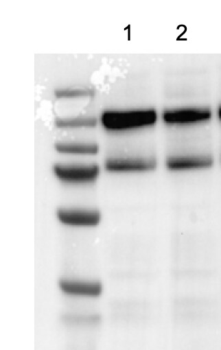
Application: Western BlotSample Tested: HUVEC human umbilical vein endothelial cellsSpecies: HumanVerified Customer | Posted 02/04/2025Seems not to work for me. It's been difficult getting a p-TIE2 antibody that works. Lane 1 is control while lane 2 is ANGPT1 treated.
 Bio-Techne ResponseThank you for reviewing our product. We are sorry to hear that this product did not perform as expected. We have been in touch with the customer to resolve this issue according to our Product Guarantee and to the customer’s satisfaction. The customer retested AF3909 on a variant of HUVEC expressing Tie-2 with the mutation L914F, which leads to increased Tie-2 activation and phosphorylation. In the HUVEC cell line carrying the Tie-2 L914F mutation, the antibody was able to differentiate between potential inhibitor treated vs control samples.
 -
Application: Western BlotSample Tested: huvecsSpecies: HumanVerified Customer | Posted 05/24/2019
There are no reviews that match your criteria.
Protocols
Find general support by application which include: protocols, troubleshooting, illustrated assays, videos and webinars.
- Antigen Retrieval Protocol (PIER)
- Antigen Retrieval for Frozen Sections Protocol
- Appropriate Fixation of IHC/ICC Samples
- Cellular Response to Hypoxia Protocols
- Chromogenic IHC Staining of Formalin-Fixed Paraffin-Embedded (FFPE) Tissue Protocol
- Chromogenic Immunohistochemistry Staining of Frozen Tissue
- ClariTSA™ Fluorophore Kits
- Detection & Visualization of Antibody Binding
- Fluorescent IHC Staining of Frozen Tissue Protocol
- Graphic Protocol for Heat-induced Epitope Retrieval
- Graphic Protocol for the Preparation and Fluorescent IHC Staining of Frozen Tissue Sections
- Graphic Protocol for the Preparation and Fluorescent IHC Staining of Paraffin-embedded Tissue Sections
- Graphic Protocol for the Preparation of Gelatin-coated Slides for Histological Tissue Sections
- IHC Sample Preparation (Frozen sections vs Paraffin)
- Immunofluorescent IHC Staining of Formalin-Fixed Paraffin-Embedded (FFPE) Tissue Protocol
- Immunohistochemistry (IHC) and Immunocytochemistry (ICC) Protocols
- Immunohistochemistry Frozen Troubleshooting
- Immunohistochemistry Paraffin Troubleshooting
- Preparing Samples for IHC/ICC Experiments
- Preventing Non-Specific Staining (Non-Specific Binding)
- Primary Antibody Selection & Optimization
- Protocol for Heat-Induced Epitope Retrieval (HIER)
- Protocol for Making a 4% Formaldehyde Solution in PBS
- Protocol for VisUCyte™ HRP Polymer Detection Reagent
- Protocol for the Preparation & Fixation of Cells on Coverslips
- Protocol for the Preparation and Chromogenic IHC Staining of Frozen Tissue Sections
- Protocol for the Preparation and Chromogenic IHC Staining of Frozen Tissue Sections - Graphic
- Protocol for the Preparation and Chromogenic IHC Staining of Paraffin-embedded Tissue Sections
- Protocol for the Preparation and Chromogenic IHC Staining of Paraffin-embedded Tissue Sections - Graphic
- Protocol for the Preparation and Fluorescent IHC Staining of Frozen Tissue Sections
- Protocol for the Preparation and Fluorescent IHC Staining of Paraffin-embedded Tissue Sections
- Protocol for the Preparation of Gelatin-coated Slides for Histological Tissue Sections
- R&D Systems Quality Control Western Blot Protocol
- TUNEL and Active Caspase-3 Detection by IHC/ICC Protocol
- The Importance of IHC/ICC Controls
- Troubleshooting Guide: Immunohistochemistry
- Troubleshooting Guide: Western Blot Figures
- Western Blot Conditions
- Western Blot Protocol
- Western Blot Protocol for Cell Lysates
- Western Blot Troubleshooting
- Western Blot Troubleshooting Guide
- View all Protocols, Troubleshooting, Illustrated assays and Webinars
Loading...
Associated Pathways

