FK506 Binding Proteins (FKBPs) are intracellular receptors for the immuno-suppressive drug FK506. The FKBP/FK506 complex exerts its immunosuppressive effects by inhibiting calcineurin, a calcium- and calmodulin-dependent serine/threonine phosphatase that functions as a critical signaling molecule during T cell activation. FKBP38, also known as FKBP8, is a 355 amino acid (aa) protein with a calculated molecular weight of 38.7 kDa and an apparent molecular mass of ~60-64 kDa in SDS-PAGE. FKBP38 binds to and inhibits calcineurin even in the absence of FK506, indicating that FKBP38 is a constitutively active inhibitor of calcineurin. Additionally, FKBP38 immunoprecipitates with Bcl-2 and Bcl-xL, suggesting that FKBP38 may regulate apoptosis by anchoring Bcl-2 and Bcl-xL to mitochondrial membranes. Mouse FKBP38 shares 94% and 87% sequence identity with human and rat FKBP38, respectively.

Key Product Details
Species Reactivity
Validated:
Human, Mouse, Rat
Cited:
Human, Mouse, Transgenic Mouse
Applications
Validated:
Immunohistochemistry, Western Blot, Simple Western
Cited:
Immunohistochemistry, Western Blot
Label
Unconjugated
Antibody Source
Polyclonal Goat IgG
Loading...
Product Specifications
Immunogen
E. coli derived recombinant mouse FKBP38
Met1-Gly326
Accession # AAH03739
Met1-Gly326
Accession # AAH03739
Specificity
Detects human, mouse, and rat FKBP38 in Western blots. In Western blots, less than 1% cross-reactivity with recombinant human FKBP-12, -12.6, -13, -25, -51, or -52 is observed.
Clonality
Polyclonal
Host
Goat
Isotype
IgG
Scientific Data Images for FKBP38 Antibody
Detection of Human/Mouse/Rat FKBP38 by Western Blot.
Western blot shows lysates of HepG2 human hepatocellular carcinoma cell line, MDA-MB-468 human breast cancer cell line, A549 human lung carcinoma cell line, C2C12 mouse myoblast cell line, and L6 rat myoblast cell line. PVDF membrane was probed with 0.5 µg/mL of Goat Anti-Human/Mouse/Rat FKBP38 Antigen Affinity-purified Polyclonal Antibody (Catalog # AF3580) followed by HRP-conjugated Anti-Goat IgG Secondary Antibody (Catalog # HAF109). A specific band was detected for FKBP38 at approximately 60 - 64 kDa (as indicated). This experiment was conducted under reducing conditions and using Immunoblot Buffer Group 2.
FKBP38 in Human Intestine.
FKBP38 was detected in immersion fixed paraffin-embedded sections of human intestine using 15 µg/mL Goat Anti-Human/Mouse/Rat FKBP38 Antigen Affinity-purified Polyclonal Antibody (Catalog # AF3580) overnight at 4 °C. Tissue was stained with the Anti-Goat HRP-DAB Cell & Tissue Staining Kit (brown; Catalog # CTS008) and counterstained with hematoxylin (blue). Specific labeling was localized to the nucleus of epithelial cells in intestinal glands. View our protocol for Chromogenic IHC Staining of Paraffin-embedded Tissue Sections. This application has not been tested in rat or mouse tissue.
Detection of Human FKBP38 by Simple WesternTM.
Simple Western lane view shows lysates of HepG2 human hepatocellular carcinoma cell line and MDA-MB-468 human breast cancer cell line, loaded at 0.2 mg/mL. A specific band was detected for FKBP38 at approximately 58 kDa (as indicated) using 5 µg/mL of Goat Anti-Human/Mouse/Rat FKBP38 Antigen Affinity-purified Polyclonal Antibody (Catalog # AF3580) followed by 1:50 dilution of HRP-conjugated Anti-Goat IgG Secondary Antibody (Catalog # HAF109). This experiment was conducted under reducing conditions and using the 12-230 kDa separation system.Applications for FKBP38 Antibody
Application
Recommended Usage
Immunohistochemistry
5-15 µg/mL
Sample: Immersion fixed paraffin-embedded sections of human intestine
Sample: Immersion fixed paraffin-embedded sections of human intestine
Simple Western
5 µg/mL
Sample: HepG2 human hepatocellular carcinoma cell line and MDA‑MB‑468 human breast cancer cell line
Sample: HepG2 human hepatocellular carcinoma cell line and MDA‑MB‑468 human breast cancer cell line
Western Blot
0.5 µg/mL
Sample: HepG2 human hepatocellular carcinoma cell line, MDA-MB-468 human breast cancer cell line, A549 human lung carcinoma cell line, C2C12 mouse myoblast cell line, and L6 rat myoblast cell line
Sample: HepG2 human hepatocellular carcinoma cell line, MDA-MB-468 human breast cancer cell line, A549 human lung carcinoma cell line, C2C12 mouse myoblast cell line, and L6 rat myoblast cell line
Reviewed Applications
Read 1 review rated 4 using AF3580 in the following applications:
Formulation, Preparation, and Storage
Purification
Antigen Affinity-purified
Reconstitution
Reconstitute at 0.2 mg/mL in sterile PBS. For liquid material, refer to CoA for concentration.
Loading...
Formulation
Lyophilized from a 0.2 μm filtered solution in PBS with Trehalose. *Small pack size (SP) is supplied either lyophilized or as a 0.2 µm filtered solution in PBS.
Shipping
Lyophilized product is shipped at ambient temperature. Liquid small pack size (-SP) is shipped with polar packs. Upon receipt, store immediately at the temperature recommended below.
Stability & Storage
Use a manual defrost freezer and avoid repeated freeze-thaw cycles.
- 12 months from date of receipt, -20 to -70 °C as supplied.
- 1 month, 2 to 8 °C under sterile conditions after reconstitution.
- 6 months, -20 to -70 °C under sterile conditions after reconstitution.
Calculators
Background: FKBP38
Long Name
38 kDa FK506 Binding Protein
Alternate Names
FKBP8
Gene Symbol
FKBP8
UniProt
Additional FKBP38 Products
Product Documents for FKBP38 Antibody
Certificate of Analysis
To download a Certificate of Analysis, please enter a lot or batch number in the search box below.
Note: Certificate of Analysis not available for kit components.
Product Specific Notices for FKBP38 Antibody
For research use only
Related Research Areas
Citations for FKBP38 Antibody
 Powered by Bioz
Customer Reviews for FKBP38 Antibody (1)
4 out of 5
1 Customer Rating
Have you used FKBP38 Antibody?
Submit a review and receive an Amazon gift card!
$25/€18/£15/$25CAN/¥2500 Yen for a review with an image
$10/€7/£6/$10CAN/¥1110 Yen for a review without an image
Submit a review
Customer Images
Showing
1
-
1 of
1 review
Showing All
Filter By:
-
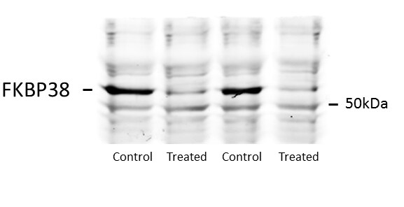
Application: Western BlotSample Tested: HEK293 human embryonic kidney cell lineSpecies: HumanVerified Customer | Posted 08/17/2016

There are no reviews that match your criteria.
Protocols
Find general support by application which include: protocols, troubleshooting, illustrated assays, videos and webinars.
- Antigen Retrieval Protocol (PIER)
- Antigen Retrieval for Frozen Sections Protocol
- Appropriate Fixation of IHC/ICC Samples
- Cellular Response to Hypoxia Protocols
- Chromogenic IHC Staining of Formalin-Fixed Paraffin-Embedded (FFPE) Tissue Protocol
- Chromogenic Immunohistochemistry Staining of Frozen Tissue
- ClariTSA™ Fluorophore Kits
- Detection & Visualization of Antibody Binding
- Fluorescent IHC Staining of Frozen Tissue Protocol
- Graphic Protocol for Heat-induced Epitope Retrieval
- Graphic Protocol for the Preparation and Fluorescent IHC Staining of Frozen Tissue Sections
- Graphic Protocol for the Preparation and Fluorescent IHC Staining of Paraffin-embedded Tissue Sections
- Graphic Protocol for the Preparation of Gelatin-coated Slides for Histological Tissue Sections
- IHC Sample Preparation (Frozen sections vs Paraffin)
- Immunofluorescent IHC Staining of Formalin-Fixed Paraffin-Embedded (FFPE) Tissue Protocol
- Immunohistochemistry (IHC) and Immunocytochemistry (ICC) Protocols
- Immunohistochemistry Frozen Troubleshooting
- Immunohistochemistry Paraffin Troubleshooting
- Preparing Samples for IHC/ICC Experiments
- Preventing Non-Specific Staining (Non-Specific Binding)
- Primary Antibody Selection & Optimization
- Protocol for Heat-Induced Epitope Retrieval (HIER)
- Protocol for Making a 4% Formaldehyde Solution in PBS
- Protocol for VisUCyte™ HRP Polymer Detection Reagent
- Protocol for the Preparation & Fixation of Cells on Coverslips
- Protocol for the Preparation and Chromogenic IHC Staining of Frozen Tissue Sections
- Protocol for the Preparation and Chromogenic IHC Staining of Frozen Tissue Sections - Graphic
- Protocol for the Preparation and Chromogenic IHC Staining of Paraffin-embedded Tissue Sections
- Protocol for the Preparation and Chromogenic IHC Staining of Paraffin-embedded Tissue Sections - Graphic
- Protocol for the Preparation and Fluorescent IHC Staining of Frozen Tissue Sections
- Protocol for the Preparation and Fluorescent IHC Staining of Paraffin-embedded Tissue Sections
- Protocol for the Preparation of Gelatin-coated Slides for Histological Tissue Sections
- R&D Systems Quality Control Western Blot Protocol
- TUNEL and Active Caspase-3 Detection by IHC/ICC Protocol
- The Importance of IHC/ICC Controls
- Troubleshooting Guide: Immunohistochemistry
- Troubleshooting Guide: Western Blot Figures
- Western Blot Conditions
- Western Blot Protocol
- Western Blot Protocol for Cell Lysates
- Western Blot Troubleshooting
- Western Blot Troubleshooting Guide
- View all Protocols, Troubleshooting, Illustrated assays and Webinars
Loading...