GSK-3 is a Ser/Thr kinase first identified as an inactivator of Glycogen Synthase. GSK-3 acts as a multifunctional downstream switch that determines the output of numerous signaling pathways. There are two mammalian GSK-3 isoforms encoded by distinct genes, GSK-3 alpha and GSK-3 beta, which are structurally similar, but functionally non-identical. GSK-3a is inhibited by phosphorylation at S21 by Akt and other kinases. GSK-3 alpha and GSK-3 beta share 85% amino acid identity. Dysregulated GSK-3 has been implicated in several diseases including type II diabetes, Alzheimer's disease, bipolar disorder, and cancer.
phospho-GSK-3 alpha/beta (S21/S9) Antibody
R&D Systems | Catalog # AF1590
 and GSK-3 beta (S9) by Western Blot.")
Key Product Details
Validated by
Biological Validation
Species Reactivity
Validated:
Human, Mouse, Rat
Cited:
Human, Mouse, Rat, Transgenic Mouse
Applications
Validated:
Western Blot, Immunocytochemistry
Cited:
Immunohistochemistry, Western Blot, Flow Cytometry
Label
Unconjugated
Antibody Source
Polyclonal Rabbit IgG
Loading...
Product Specifications
Immunogen
Phosphopeptide containing GSK-3 beta S9 site
Specificity
Detects human, mouse, and rat GSK-3 beta when phosphorylated at S9, and human, mouse, and rat GSK‑3 alpha when phosphorylated at S21.
Clonality
Polyclonal
Host
Rabbit
Isotype
IgG
Scientific Data Images for phospho-GSK-3 alpha/beta (S21/S9) Antibody
Detection of Human Phospho-GSK‑3 alpha (S21) and GSK-3 beta (S9) by Western Blot.
Western blot shows lysates of HeLa human cervical epithelial carcinoma cell line untreated (-) or treated (+) with 200 nM PMA for 20 minutes and MCF-7 human breast cancer cell line untreated or treated with 100 ng/mL Recombinant Human IGF-1 (291-G1) for 15 minutes. PVDF membrane was probed with 0.2 µg/mL of Human/Mouse/Rat Phospho-GSK-3a/ beta (S21/S9) Antigen Affinity-purified Polyclonal Antibody (Catalog # AF1590), followed by HRP-conjugated Anti-Rabbit IgG Secondary Antibody (HAF008). Specific bands were detected for Phospho-GSK-3a (S21) and GSK-3 beta (S9) at approximately 51 and 46 kDa (as indicated). This experiment was conducted under reducing conditions and using Immunoblot Buffer Group 1. in HeLa Human Cell Line.")
Phospho-GSK‑3 alpha / beta (S21/S9) in HeLa Human Cell Line.
Phospho-GSK‑3 alpha / beta (S21/S9) was detected in immersion fixed HeLa human cervical epithelial carcinoma cell line treated with PMA (left panel; positive staining) and untreated HeLa cell line (right panel; negative control) using Rabbit Anti-Human/Mouse/Rat Phospho-GSK‑3 alpha / beta (S21/S9) Antigen Affinity-purified Polyclonal Antibody (Catalog # AF1590) at 8 µg/mL for 3 hours at room temperature. Cells were stained using the NorthernLights™ 557-conjugated Anti-Rabbit IgG Secondary Antibody (red; NL004) and counterstained with DAPI (blue). Specific staining was localized to nuclei. Staining was performed using our protocol for Fluorescent ICC Staining of Non-adherent Cells.
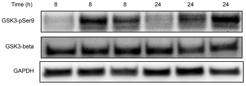
Detection of Rat GSK-3 alpha/beta by Western Blot
HYS-32 modulates GSK3 beta phosphorylation.(A) Cell lysates from control astrocytes (Con) or astrocytes treated for 2, 6, 12, or 24 h with 5 μM HYS-32 were subjected to 10% SDS-PAGE, and analyzed by immunoblotting with antibodies against GSK3 beta -pTyr216, GSK3 beta -pSer9, or GAPDH. (B) Densitometric analyses of GSK3 beta -pTyr216 and GSK3 beta -pSer9 expressed as the density of the bands in the treated groups relative to the control. *p<0.05 compared to control using one-way ANOVA with Dunnett’s post-hoc test. The results were collected from five independent experiments. (C) According to the data in (B), the stacked bar graph showing the relative percentage of phospho-GSK3 beta at indicated time. Image collected and cropped by CiteAb from the following publication (https://dx.plos.org/10.1371/journal.pone.0126217), licensed under a CC-BY license. Not internally tested by R&D Systems.
Detection of Rat GSK-3 alpha/beta by Western Blot
LY294002 inhibits the HYS-32-induced phosphorylation of GSK3 beta -pS9 and GSK3 beta -pY216.(A) Control astrocytes (Con) or astrocytes treated for 24 h with 5 μM HYS-32 (HYS), co-treated for 24 h with 5 μM HYS-32 and 20 μM LY294002 (HYS+LY), or treated with 20 μM LY294002 (LY) were subjected to 10% SDS-PAGE, and analyzed by immunoblotting with antibodies against GSK3 beta -pSer9, GSK3 beta -pY216, totalGSK3 beta, or GAPDH. (B) Densitometric analyses of GSK3 beta -pSer9 and GSK3 beta -pY216 expressed as the density of the bands in the treated groups relative to the control. (C) According to the data in (B), the stacked bar graph showing relative percentage ofphospho-GSK3 beta in various treatment. (D)Astrocytes treated as in (A) were fixed in cold acetone and triple-stained for beta -tubulin, N-cadherin, and F-actin. Quantitative analysis of the straight distance between microtubule tips and cell border were performed as described in Materials and Methods. The results were collected from three independent experiments.*p<0.01 compared to HYS using one-way ANOVA with Dunnett’s post-hoctest. Image collected and cropped by CiteAb from the following publication (https://dx.plos.org/10.1371/journal.pone.0126217), licensed under a CC-BY license. Not internally tested by R&D Systems. by Western Blot")
Detection of Phospho-GSK-3 alpha/ beta (S21/S9) by Western Blot
Galectin-3 expression induces activation of PYK2, STAT1 and GSK3 alpha / beta signalling. Expression of 37 protein kinases in SW620 cells in response to 10 µg/ml galectin-3 or BSA for 0.5 h was assessed by Proteome Profiler Human Phospho-Kinase Array (A, Percentage changes of the kinases in cell response to galectin-3 in comparison to control are shown at the bottom panel). The presence of galectin-3 increases the phosphorylation of PYK2, GSK3 alpha / beta, and STAT1 and decreases phosphorylation of STAT3. SW620 cells treated with 10 µg/ml galectin-3 for different times were assessed by immunoblotting using antibodies against p-PYK2, p-STAT-1, p-GSK3 alpha / beta or p-STAT-3 (B). The blots were striped and reprobed with antibodies against PYK2, STAT-1, GSK3 alpha / beta or STAT-3. The band density was quantified and expressed as percentages of phospho-/non-phosphorylated proteins (C). In D and E, SW620 cells were treated with 10 µg/ml galectin-3 or BSA followed by introduction of GSK3 alpha / beta inhibitor SB 216763 (SB) or PKY2 inhibitor PF-431396 (PF) for 15 min and the levels of phosphorylated PYK2, STAT-1, GSK3 alpha / beta or STAT-3 were analysed by immunoblotting. The blots were striped and reprobed with antibodies against PYK2, STAT-1, GSK3 alpha / beta or STAT-3. The densities of the blots from three independent experiments were quantified and are expressed as the percentage of phosphorylated/non-phosphorylated levels of each protein. ***P < 0.001, **P < 0.01, *P < 0.05 (ANOVA). Image collected and cropped by CiteAb from the following open publication (https://pubmed.ncbi.nlm.nih.gov/37055381), licensed under a CC-BY license. Not internally tested by R&D Systems. by Western Blot")
Detection of Phospho-GSK-3 alpha/ beta (S21/S9) by Western Blot
Galectin-3 expression induces activation of PYK2, STAT1 and GSK3 alpha / beta signalling. Expression of 37 protein kinases in SW620 cells in response to 10 µg/ml galectin-3 or BSA for 0.5 h was assessed by Proteome Profiler Human Phospho-Kinase Array (A, Percentage changes of the kinases in cell response to galectin-3 in comparison to control are shown at the bottom panel). The presence of galectin-3 increases the phosphorylation of PYK2, GSK3 alpha / beta, and STAT1 and decreases phosphorylation of STAT3. SW620 cells treated with 10 µg/ml galectin-3 for different times were assessed by immunoblotting using antibodies against p-PYK2, p-STAT-1, p-GSK3 alpha / beta or p-STAT-3 (B). The blots were striped and reprobed with antibodies against PYK2, STAT-1, GSK3 alpha / beta or STAT-3. The band density was quantified and expressed as percentages of phospho-/non-phosphorylated proteins (C). In D and E, SW620 cells were treated with 10 µg/ml galectin-3 or BSA followed by introduction of GSK3 alpha / beta inhibitor SB 216763 (SB) or PKY2 inhibitor PF-431396 (PF) for 15 min and the levels of phosphorylated PYK2, STAT-1, GSK3 alpha / beta or STAT-3 were analysed by immunoblotting. The blots were striped and reprobed with antibodies against PYK2, STAT-1, GSK3 alpha / beta or STAT-3. The densities of the blots from three independent experiments were quantified and are expressed as the percentage of phosphorylated/non-phosphorylated levels of each protein. ***P < 0.001, **P < 0.01, *P < 0.05 (ANOVA). Image collected and cropped by CiteAb from the following open publication (https://pubmed.ncbi.nlm.nih.gov/37055381), licensed under a CC-BY license. Not internally tested by R&D Systems.Applications for phospho-GSK-3 alpha/beta (S21/S9) Antibody
Application
Recommended Usage
Immunocytochemistry
5-15 µg/mL
Sample: Immersion fixed HeLa human cervical epithelial carcinoma cell treated with PMA
Sample: Immersion fixed HeLa human cervical epithelial carcinoma cell treated with PMA
Western Blot
0.2 µg/mL
Sample: PMA-treated HeLa human cervical epithelial carcinoma cell line and MCF‑7 human breast cancer cell line treated with Recombinant Human IGF-1 (Catalog # 291-G1)
Sample: PMA-treated HeLa human cervical epithelial carcinoma cell line and MCF‑7 human breast cancer cell line treated with Recombinant Human IGF-1 (Catalog # 291-G1)
Reviewed Applications
Read 1 review rated 5 using AF1590 in the following applications:
Formulation, Preparation, and Storage
Purification
Antigen and protein A Affinity-purified
Reconstitution
Reconstitute at 0.2 mg/mL in sterile PBS. For liquid material, refer to CoA for concentration.
Loading...
Formulation
Lyophilized from a 0.2 μm filtered solution in PBS with Trehalose. See Certificate of Analysis for details.
*Small pack size (-SP) is supplied either lyophilized or as a 0.2 µm filtered solution in PBS.
*Small pack size (-SP) is supplied either lyophilized or as a 0.2 µm filtered solution in PBS.
Shipping
Lyophilized product is shipped at ambient temperature. Liquid small pack size (-SP) is shipped with polar packs. Upon receipt, store immediately at the temperature recommended below.
Stability & Storage
Use a manual defrost freezer and avoid repeated freeze-thaw cycles.
- 12 months from date of receipt, -20 to -70 °C as supplied.
- 1 month, 2 to 8 °C under sterile conditions after reconstitution.
- 6 months, -20 to -70 °C under sterile conditions after reconstitution.
Calculators
Background: GSK-3 alpha/beta
Long Name
Glycogen Synthase Kinase 3
Alternate Names
DKFZp686D0638, EC 2.7.11, EC 2.7.11.26, glycogen synthase kinase 3 alpha, glycogen synthase kinase-3 alpha, GSK-3 alpha
Additional GSK-3 alpha/beta Products
Product Documents for phospho-GSK-3 alpha/beta (S21/S9) Antibody
Certificate of Analysis
To download a Certificate of Analysis, please enter a lot or batch number in the search box below.
Note: Certificate of Analysis not available for kit components.
Product Specific Notices for phospho-GSK-3 alpha/beta (S21/S9) Antibody
For research use only
Related Research Areas
Citations for phospho-GSK-3 alpha/beta (S21/S9) Antibody
 Powered by Bioz
Customer Reviews for phospho-GSK-3 alpha/beta (S21/S9) Antibody (1)
5 out of 5
1 Customer Rating
Have you used phospho-GSK-3 alpha/beta (S21/S9) Antibody?
Submit a review and receive an Amazon gift card!
$25/€18/£15/$25CAN/¥2500 Yen for a review with an image
$10/€7/£6/$10CAN/¥1110 Yen for a review without an image
Submit a review
Customer Images
Showing
1
-
1 of
1 review
Showing All
Filter By:
-
Application: Western BlotSample Tested: Colon cancer cell lineSpecies: HumanVerified Customer | Posted 09/14/2018

There are no reviews that match your criteria.
Protocols
Find general support by application which include: protocols, troubleshooting, illustrated assays, videos and webinars.
- Appropriate Fixation of IHC/ICC Samples
- Cellular Response to Hypoxia Protocols
- ClariTSA™ Fluorophore Kits
- Detection & Visualization of Antibody Binding
- ICC Cell Smear Protocol for Suspension Cells
- ICC Immunocytochemistry Protocol Videos
- ICC for Adherent Cells
- Immunocytochemistry (ICC) Protocol
- Immunocytochemistry Troubleshooting
- Immunofluorescence of Organoids Embedded in Cultrex Basement Membrane Extract
- Immunohistochemistry (IHC) and Immunocytochemistry (ICC) Protocols
- Preparing Samples for IHC/ICC Experiments
- Preventing Non-Specific Staining (Non-Specific Binding)
- Primary Antibody Selection & Optimization
- Protocol for VisUCyte™ HRP Polymer Detection Reagent
- Protocol for the Fluorescent ICC Staining of Cell Smears - Graphic
- Protocol for the Fluorescent ICC Staining of Cultured Cells on Coverslips - Graphic
- Protocol for the Preparation and Fluorescent ICC Staining of Cells on Coverslips
- Protocol for the Preparation and Fluorescent ICC Staining of Non-adherent Cells
- Protocol for the Preparation and Fluorescent ICC Staining of Stem Cells on Coverslips
- Protocol for the Preparation of a Cell Smear for Non-adherent Cell ICC - Graphic
- R&D Systems Quality Control Western Blot Protocol
- TUNEL and Active Caspase-3 Detection by IHC/ICC Protocol
- The Importance of IHC/ICC Controls
- Troubleshooting Guide: Western Blot Figures
- Western Blot Conditions
- Western Blot Protocol
- Western Blot Protocol for Cell Lysates
- Western Blot Troubleshooting
- Western Blot Troubleshooting Guide
- View all Protocols, Troubleshooting, Illustrated assays and Webinars
Loading...