VAMP-1 (vesicle-associated membrane protein 1; also synaptobrevin-1/SYB1) is an 18 kDa member of the synaptobrevin family of proteins. It is expressed in neurons, neutrophils, and skeletal muscle cells, and participates in vesicle fusion with the plasma membrane. Human VAMP-1 is 118 amino acids (aa) in length. It is a type IV transmembrane protein that contains an N-terminal cytoplasmic region (aa 1-96) and a 22 aa transmembrane domain (aa 97-118). There is one coiled-coil region between aa 33-93. Multiple splice variants are known that show two, three, four and 81 aa substitutions for the C-terminal five amino acids. One three aa variant creates a mitochondrial targeting motif. Over aa 1-96, human VAMP-1 is 98% aa identical to mouse VAMP-1.

Key Product Details
Species Reactivity
Validated:
Human, Mouse
Cited:
Human, Rat
Applications
Validated:
Immunohistochemistry, Western Blot
Cited:
Immunohistochemistry, Western Blot
Label
Unconjugated
Antibody Source
Polyclonal Goat IgG
Loading...
Product Specifications
Immunogen
E. coli-derived recombinant human VAMP-1
Met1-Lys96
Accession # P23763
Met1-Lys96
Accession # P23763
Specificity
Detects human and mouse VAMP-1 in direct ELISAs and Western blots. In direct ELISAs, approximately 20% cross-reactivity with recombinant human (rh) VAMP-2 and less than 1% cross-reactivity with rhVAMP-5 is observed.
Clonality
Polyclonal
Host
Goat
Isotype
IgG
Scientific Data Images for VAMP-1 Antibody
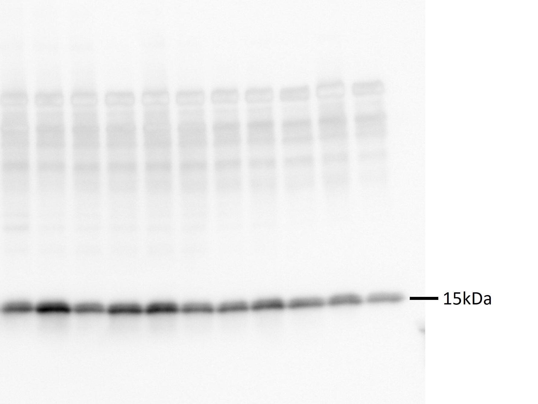
Detection of Human/Mouse VAMP‑1 by Western Blot.
Western blot shows lysates of human heart and brain tissue. PVDF membrane was probed with 1 µg/mL of Goat Anti-Human/Mouse VAMP-1 Antigen Affinity-purified Polyclonal Antibody (Catalog # AF4828) followed by HRP-conjugated Anti-Goat IgG Secondary Antibody (Catalog # HAF019). A specific band was detected for VAMP-1 at approximately 17 kDa (as indicated). This experiment was conducted under reducing conditions and using Immunoblot Buffer Group 8.
VAMP‑1 in Mouse Spinal Cord.
VAMP-1 was detected in perfusion fixed frozen sections of mouse spinal cord using 1.7 µg/mL Goat Anti-Human/Mouse VAMP-1 Antigen Affinity-purified Polyclonal Antibody (Catalog # AF4828) overnight at 4 °C. Tissue was stained with the NorthernLights™ 557-conjugated Anti-Goat IgG Secondary Antibody (red; Catalog # NL001) and counterstained (green). View our protocol for Fluorescent IHC Staining of Frozen Tissue Sections.Applications for VAMP-1 Antibody
Application
Recommended Usage
Immunohistochemistry
5-15 µg/mL
Sample: Perfusion fixed frozen sections of mouse spinal cord
Sample: Perfusion fixed frozen sections of mouse spinal cord
Western Blot
1 µg/mL
Sample: Human heart and brain tissue
Sample: Human heart and brain tissue
Reviewed Applications
Read 2 reviews rated 5 using AF4828 in the following applications:
Formulation, Preparation, and Storage
Purification
Antigen Affinity-purified
Reconstitution
Reconstitute at 0.2 mg/mL in sterile PBS. For liquid material, refer to CoA for concentration.
Loading...
Formulation
Lyophilized from a 0.2 μm filtered solution in PBS with Trehalose. *Small pack size (SP) is supplied either lyophilized or as a 0.2 µm filtered solution in PBS.
Shipping
Lyophilized product is shipped at ambient temperature. Liquid small pack size (-SP) is shipped with polar packs. Upon receipt, store immediately at the temperature recommended below.
Stability & Storage
Use a manual defrost freezer and avoid repeated freeze-thaw cycles.
- 12 months from date of receipt, -20 to -70 °C as supplied.
- 1 month, 2 to 8 °C under sterile conditions after reconstitution.
- 6 months, -20 to -70 °C under sterile conditions after reconstitution.
Calculators
Background: VAMP-1
Long Name
Vesicle-Associated Membrane Protein 1
Alternate Names
SYB1, Synaptobrevin-1, VAMP1
Gene Symbol
VAMP1
UniProt
Additional VAMP-1 Products
Product Documents for VAMP-1 Antibody
Certificate of Analysis
To download a Certificate of Analysis, please enter a lot or batch number in the search box below.
Note: Certificate of Analysis not available for kit components.
Product Specific Notices for VAMP-1 Antibody
For research use only
Related Research Areas
Citations for VAMP-1 Antibody
 Powered by Bioz
Customer Reviews for VAMP-1 Antibody (2)
5 out of 5
2 Customer Ratings
Have you used VAMP-1 Antibody?
Submit a review and receive an Amazon gift card!
$25/€18/£15/$25CAN/¥2500 Yen for a review with an image
$10/€7/£6/$10CAN/¥1110 Yen for a review without an image
Submit a review
Customer Images
Showing
1
-
2 of
2 reviews
Showing All
Filter By:
-
Application: Western BlotSample Tested: Brain (cerebral cortex)Species: MouseVerified Customer | Posted 07/02/2022Used at 1:500 in 5% non-fat milk overnight at 4 degree

-
Application: Western BlotSample Tested: See PMID 23206873Species: RatVerified Customer | Posted 01/08/2015
There are no reviews that match your criteria.
Protocols
Find general support by application which include: protocols, troubleshooting, illustrated assays, videos and webinars.
- Antigen Retrieval Protocol (PIER)
- Antigen Retrieval for Frozen Sections Protocol
- Appropriate Fixation of IHC/ICC Samples
- Cellular Response to Hypoxia Protocols
- Chromogenic IHC Staining of Formalin-Fixed Paraffin-Embedded (FFPE) Tissue Protocol
- Chromogenic Immunohistochemistry Staining of Frozen Tissue
- ClariTSA™ Fluorophore Kits
- Detection & Visualization of Antibody Binding
- Fluorescent IHC Staining of Frozen Tissue Protocol
- Graphic Protocol for Heat-induced Epitope Retrieval
- Graphic Protocol for the Preparation and Fluorescent IHC Staining of Frozen Tissue Sections
- Graphic Protocol for the Preparation and Fluorescent IHC Staining of Paraffin-embedded Tissue Sections
- Graphic Protocol for the Preparation of Gelatin-coated Slides for Histological Tissue Sections
- IHC Sample Preparation (Frozen sections vs Paraffin)
- Immunofluorescent IHC Staining of Formalin-Fixed Paraffin-Embedded (FFPE) Tissue Protocol
- Immunohistochemistry (IHC) and Immunocytochemistry (ICC) Protocols
- Immunohistochemistry Frozen Troubleshooting
- Immunohistochemistry Paraffin Troubleshooting
- Preparing Samples for IHC/ICC Experiments
- Preventing Non-Specific Staining (Non-Specific Binding)
- Primary Antibody Selection & Optimization
- Protocol for Heat-Induced Epitope Retrieval (HIER)
- Protocol for Making a 4% Formaldehyde Solution in PBS
- Protocol for VisUCyte™ HRP Polymer Detection Reagent
- Protocol for the Preparation & Fixation of Cells on Coverslips
- Protocol for the Preparation and Chromogenic IHC Staining of Frozen Tissue Sections
- Protocol for the Preparation and Chromogenic IHC Staining of Frozen Tissue Sections - Graphic
- Protocol for the Preparation and Chromogenic IHC Staining of Paraffin-embedded Tissue Sections
- Protocol for the Preparation and Chromogenic IHC Staining of Paraffin-embedded Tissue Sections - Graphic
- Protocol for the Preparation and Fluorescent IHC Staining of Frozen Tissue Sections
- Protocol for the Preparation and Fluorescent IHC Staining of Paraffin-embedded Tissue Sections
- Protocol for the Preparation of Gelatin-coated Slides for Histological Tissue Sections
- R&D Systems Quality Control Western Blot Protocol
- TUNEL and Active Caspase-3 Detection by IHC/ICC Protocol
- The Importance of IHC/ICC Controls
- Troubleshooting Guide: Immunohistochemistry
- Troubleshooting Guide: Western Blot Figures
- Western Blot Conditions
- Western Blot Protocol
- Western Blot Protocol for Cell Lysates
- Western Blot Troubleshooting
- Western Blot Troubleshooting Guide
- View all Protocols, Troubleshooting, Illustrated assays and Webinars
Loading...