Nectin-1 (designated CD111), also called PRR-1 (poliovirus receptor-related protein 1) or HVEC (herpesvirus entry mediator C), is a widely expressed 110 kDa type I transmembrane glycoprotein important in formation of adherens junctions and synapses. It is a member of the nectin family within the Ig superfamily (1, 2). The Latin word necto means “to connect”, indicating the role of nectins in Ca2+-independent cell-cell adhesion (2). Nectin-1 forms homodimers in cis, followed by interactions in trans with Nectin-1, -3 or -4 (2). The 517 amino acid (aa) human Nectin-1 isoform 1 contains a 30 aa signal sequence, a 325 aa extracellular domain (ECD), a 21 aa transmembrane segment (TM), and a 141 aa cytoplasmic region. Nectin ECDs contain three Ig-like domains: an N-terminal V-type that mediates ligand binding and two C2-type (3). Nectin-1, like other nectins, has a splice form (isoform 2 or HigR, 458 aa) with alternate TM and cytoplasmic sequences. Another, isoform 3, is a 352 aa secreted protein (4). The common region of mature human Nectin-1 (aa 31-334) shares 93%, 94%, 96% and 96% aa identity with mouse, rat, bovine and porcine Nectin-1, respectively. Nectin-1 binds viral glycoprotein D to mediate herpesvirus (but not poxvirus) entry into vaginal mucosa, sensory neurons and fibroblasts (4 - 7). In forming adherens junctions and synapses, nectins 1 and 3 initiate cell-cell interactions, recruiting alpha v beta 3 integrin extracellularly and cadherins intracellularly through afadin and other junctional proteins (2, 8 - 11). These interactions organize the cytoskeleton, strengthen attachment to basement membrane and promote further cell-cell connections. Nectin-1 also recognizes CD96 on NK cells (12). Deficiency of Nectin-1 can result in cleft lip/palate ectodermal dysplasia (13). Nectin-1 downregulation in epithelial cancers, mediated in part by ectodomain shedding, may contribute to invasiveness (14).
Human Nectin-1 Antibody (610835)
R&D Systems | Catalog # MAB2880

Key Product Details
Species Reactivity
Validated:
Human
Cited:
Human
Applications
Validated:
Flow Cytometry, CyTOF-ready
Cited:
Flow Cytometry
Label
Unconjugated
Antibody Source
Monoclonal Mouse IgG2A Clone # 610835
Loading...
Product Specifications
Immunogen
Mouse myeloma cell line NS0-derived recombinant human Nectin-1
Gln31-Thr334
Accession # Q15223
Gln31-Thr334
Accession # Q15223
Specificity
Detects human Nectin-1 in direct ELISAs. In direct ELISAs, no cross-reactivity with recombinant human Nectin-2, 3, 4, or recombinant mouse Nectin-1 is observed.
Clonality
Monoclonal
Host
Mouse
Isotype
IgG2A
Scientific Data Images for Human Nectin-1 Antibody (610835)
Detection of Nectin‑1 in U937 Human Cell Line by Flow Cytometry.
U937 human histiocytic lymphoma cell line was stained with Human Nectin-1 Monoclonal Antibody (Catalog # MAB2880, filled histogram) or isotype control antibody (Catalog # MAB003, open histogram), followed by Allophycocyanin-conjugated Anti-Mouse IgG F(ab')2Secondary Antibody (Catalog # F0101B).Applications for Human Nectin-1 Antibody (610835)
Application
Recommended Usage
CyTOF-ready
Ready to be labeled using established conjugation methods. No BSA or other carrier proteins that could interfere with conjugation.
Flow Cytometry
2.5 µg/106 cells
Sample: U937 human histiocytic lymphoma cell line
Sample: U937 human histiocytic lymphoma cell line
Reviewed Applications
Read 1 review rated 5 using MAB2880 in the following applications:
Flow Cytometry Panel Builder
Bio-Techne Knows Flow Cytometry
Save time and reduce costly mistakes by quickly finding compatible reagents using the Panel Builder Tool.
Advanced Features
- Spectra Viewer - Custom analysis of spectra from multiple fluorochromes
- Spillover Popups - Visualize the spectra of individual fluorochromes
- Antigen Density Selector - Match fluorochrome brightness with antigen density

Formulation, Preparation, and Storage
Purification
Protein A or G purified from hybridoma culture supernatant
Reconstitution
Sterile PBS to a final concentration of 0.5 mg/mL. For liquid material, refer to CoA for concentration.
Loading...
Formulation
Lyophilized from a 0.2 μm filtered solution in PBS with Trehalose. *Small pack size (SP) is supplied either lyophilized or as a 0.2 µm filtered solution in PBS.
Shipping
Lyophilized product is shipped at ambient temperature. Liquid small pack size (-SP) is shipped with polar packs. Upon receipt, store immediately at the temperature recommended below.
Stability & Storage
Use a manual defrost freezer and avoid repeated freeze-thaw cycles.
- 12 months from date of receipt, -20 to -70 °C as supplied.
- 1 month, 2 to 8 °C under sterile conditions after reconstitution.
- 6 months, -20 to -70 °C under sterile conditions after reconstitution.
Calculators
Background: Nectin-1
References
- Lopez, M. et al. (1995) Gene 155:261.
- Takai, Y. et al. (2008) Nat. Rev. Mol. Cell Biol. 9:603.
- Fabre, S. et al. (2002) J. Biol. Chem. 277:27006.
- Lopez, M. et al. (2001) J. Virol. 75:5684.
- Cocchi, F. et al. (1998) Proc. Natl. Acad. Sci. USA 95:15700.
- Linehan, M. M. et al. (2004) J. Virol. 78:2530.
- Simpson, S. A. et al. (2005) J. Neurovirol. 11:208.
- Mizoguchi, A. et al. (2002) J. Cell Biol. 156:555.
- Togashi, H. et al. (2006) J. Cell Biol. 174:141.
- Tachibana, K. et al. (2000) J. Cell Biol. 150:1161.
- Takai, Y. and H. Nakanishi (2003) J. Cell Science 116:17.
- Seth, S. et al. (2007) Biochem. Biophys. Res. Commun. 364:959.
- Suzuki, K. et al. (2000) Nat. Genet. 25:427.
- Tanaka, Y. et al. (2002) Biochem. Biophys. Res. Commun. 299:472.
Long Name
Poliovirus Receptor Related 1
Alternate Names
CD111, HVEC, Nectin1, PRR1, PVRL1, PVRR1
Gene Symbol
NECTIN1
UniProt
Additional Nectin-1 Products
Product Documents for Human Nectin-1 Antibody (610835)
Certificate of Analysis
To download a Certificate of Analysis, please enter a lot or batch number in the search box below.
Note: Certificate of Analysis not available for kit components.
Product Specific Notices for Human Nectin-1 Antibody (610835)
For research use only
Citations for Human Nectin-1 Antibody (610835)
 Powered by Bioz
Customer Reviews for Human Nectin-1 Antibody (610835) (1)
5 out of 5
1 Customer Rating
Have you used Human Nectin-1 Antibody (610835)?
Submit a review and receive an Amazon gift card!
$25/€18/£15/$25CAN/¥2500 Yen for a review with an image
$10/€7/£6/$10CAN/¥1110 Yen for a review without an image
Submit a review
Customer Images
Showing
1
-
1 of
1 review
Showing All
Filter By:
-
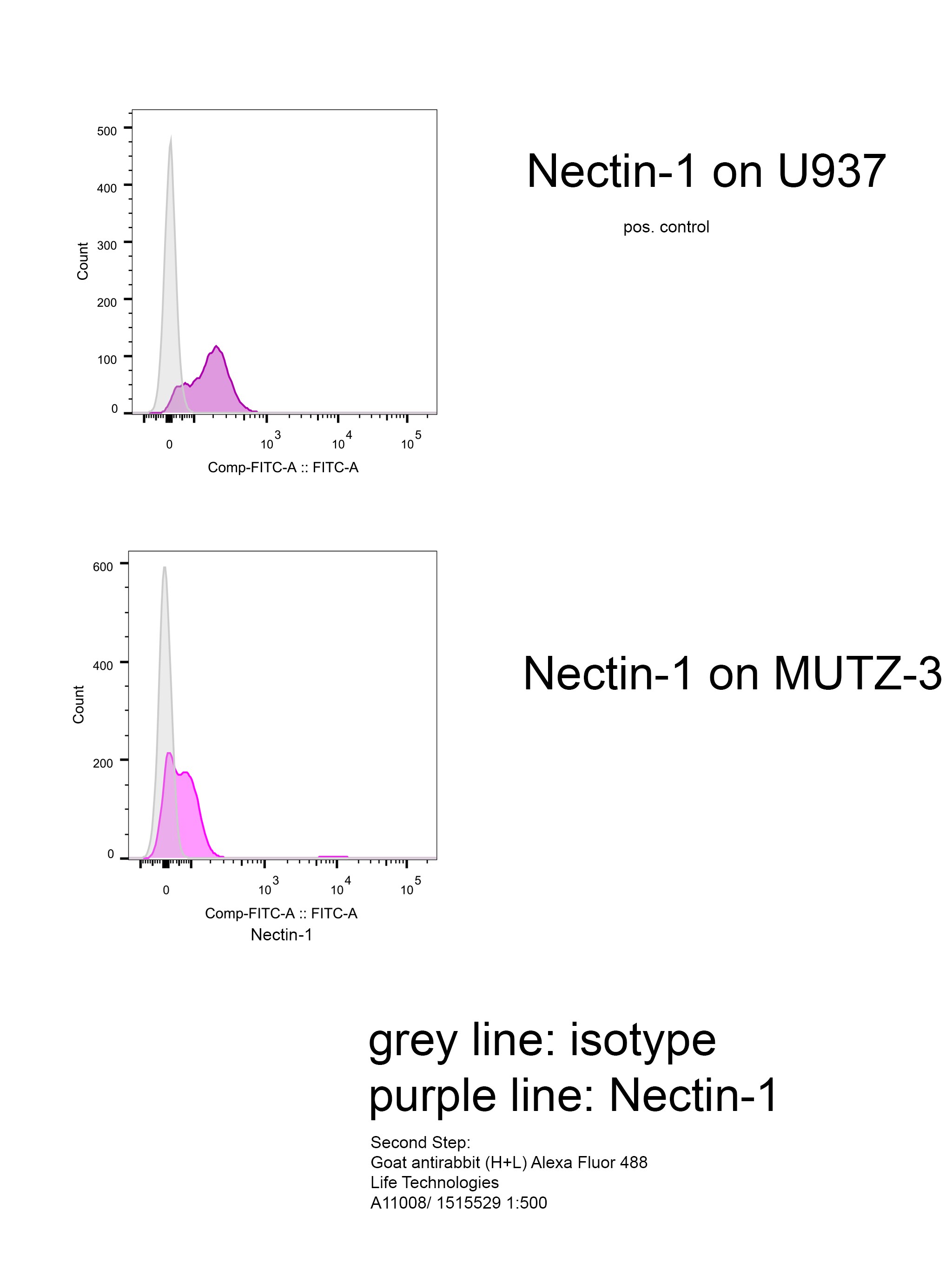
Application: Flow CytometrySample Tested: MUTZ-3 cell line and U937 human histiocytic lymphoma cell lineSpecies: HumanVerified Customer | Posted 12/08/2016second step antibody: Goat antirabbit (H+L) Alexa Fluor 488 A11008/ 1515529 1:500

There are no reviews that match your criteria.
Protocols
Find general support by application which include: protocols, troubleshooting, illustrated assays, videos and webinars.
- 7-Amino Actinomycin D (7-AAD) Cell Viability Flow Cytometry Protocol
- Extracellular Membrane Flow Cytometry Protocol
- Flow Cytometry Protocol for Cell Surface Markers
- Flow Cytometry Protocol for Staining Membrane Associated Proteins
- Flow Cytometry Staining Protocols
- Flow Cytometry Troubleshooting Guide
- Intracellular Flow Cytometry Protocol Using Alcohol (Methanol)
- Intracellular Flow Cytometry Protocol Using Detergents
- Intracellular Nuclear Staining Flow Cytometry Protocol Using Detergents
- Intracellular Staining Flow Cytometry Protocol Using Alcohol Permeabilization
- Intracellular Staining Flow Cytometry Protocol Using Detergents to Permeabilize Cells
- Propidium Iodide Cell Viability Flow Cytometry Protocol
- Protocol for Liperfluo
- Protocol for the Characterization of Human Th22 Cells
- Protocol for the Characterization of Human Th9 Cells
- Protocol: Annexin V and PI Staining by Flow Cytometry
- Protocol: Annexin V and PI Staining for Apoptosis by Flow Cytometry
- Troubleshooting Guide: Fluorokine Flow Cytometry Kits
- View all Protocols, Troubleshooting, Illustrated assays and Webinars
Loading...