Signal transduction and activator of transcription 5 (STAT5) is a member of the Jak/STAT signal transduction pathway and is activated by a variety of cytokines (IL‑22, IL-6, IFN-alpha ). STAT5 has two isoforms (A and B) that share 93% amino acid identity and bind the DNA consensus site TTCN3GAA. STAT5 mediates cytokine signaling by acting as a signal transducer in the cytoplasm and, upon phosphorylation, translocates to the nucleus and activates transcription of specific genes. STAT5 is involved in a wide array of biological processes ranging from regulating apoptosis to adult mammary gland proliferation, differentiation, and survival.
Human phospho-STAT5a/b (Y694/Y699) Antibody (1247C)
R&D Systems | Catalog # MAB41901
Recombinant Monoclonal Antibody.
 by Western Blot.")
Key Product Details
Validated by
Biological Validation
Species Reactivity
Validated:
Human
Cited:
Human
Applications
Validated:
Western Blot, Intracellular Staining by Flow Cytometry, CyTOF-ready
Cited:
Western Blot
Label
Unconjugated
Antibody Source
Recombinant Monoclonal Rabbit IgG Clone # 1247C
Loading...
Product Specifications
Immunogen
Phosphopeptide containing human STAT5b Y699 site (amino acid sequence of
this peptide is identical to a corresponding region of human STAT5a
containing Y694)
Accession # P51692
Accession # P51692
Specificity
Detects human STAT5a/b when phosphorylated at Y964/Y699 in direct ELISAs and Western blots.
Clonality
Monoclonal
Host
Rabbit
Isotype
IgG
Scientific Data Images for Human phospho-STAT5a/b (Y694/Y699) Antibody (1247C)
Detection of Human Phospho-STAT5a/b (Y694/Y699) by Western Blot.
Western blot shows lysates of Daudi human Burkitt's lymphoma cell line and HeLa human cervical epithelial carcinoma cell line untreated (-) or treated (+) with 500 U/mL Recombinant Human IFN-aA (Catalog # 11100-1) for 20 minutes. PVDF membrane was probed with 0.1 µg/mL of Rabbit Anti-Human Phospho-STAT5a/b (Y694/Y699) Monoclonal Antibody (Catalog # MAB41901) followed by HRP-conjugated Anti-Rabbit IgG Secondary Antibody (Catalog # HAF008). A specific band was detected for STAT5a/b at approximately 95 kDa (as indicated). This experiment was conducted under reducing conditions and using Immunoblot Buffer Group 1. in IFN alpha-treated Daudi Human Cell Line by Flow Cytometry.")
Detection of Phospho-STAT5a/b (Y694/Y699) in IFN alpha-treated Daudi Human Cell Line by Flow Cytometry.
Daudi human Burkitt's lymphoma cell line was unstimulated (open histogram) or treated with 500 U/mL rhIFN-alpha for 20 minutes (filled histogram) and stained with Rabbit anti-Human Phospho-STAT5a/b (Y694/Y699) Molyclonal Antibody (Catalog # MAB41901) followed by APC-conjugated Anti-Rabbit IgG Secondary Antibody (Catalog # F0111). To facilitate intracellular staining, cells were fixed with paraformaldehyde and permeabilized with methanol. View our protocol for Staining Intracellular Molecules.Applications for Human phospho-STAT5a/b (Y694/Y699) Antibody (1247C)
Application
Recommended Usage
CyTOF-ready
Ready to be labeled using established conjugation methods. No BSA or other carrier proteins that could interfere with conjugation.
Intracellular Staining by Flow Cytometry
0.25 µg/106 cells
Sample: IFN alpha-treated Daudi Human Cell Line fixed with paraformaldehyde and permeabilized with methanol
Sample: IFN alpha-treated Daudi Human Cell Line fixed with paraformaldehyde and permeabilized with methanol
Western Blot
0.1 µg/mL
Sample: Daudi human Burkitt's lymphoma cell line and HeLa human cervical epithelial carcinoma cell line
Sample: Daudi human Burkitt's lymphoma cell line and HeLa human cervical epithelial carcinoma cell line
Reviewed Applications
Read 3 reviews rated 5 using MAB41901 in the following applications:
Flow Cytometry Panel Builder
Bio-Techne Knows Flow Cytometry
Save time and reduce costly mistakes by quickly finding compatible reagents using the Panel Builder Tool.
Advanced Features
- Spectra Viewer - Custom analysis of spectra from multiple fluorochromes
- Spillover Popups - Visualize the spectra of individual fluorochromes
- Antigen Density Selector - Match fluorochrome brightness with antigen density

Formulation, Preparation, and Storage
Purification
Protein A or G purified from cell culture supernatant
Reconstitution
Reconstitute at 0.5 mg/mL in sterile PBS. For liquid material, refer to CoA for concentration.
Loading...
Formulation
Lyophilized from a 0.2 μm filtered solution in PBS with Trehalose. *Small pack size (SP) is supplied either lyophilized or as a 0.2 µm filtered solution in PBS.
Shipping
Lyophilized product is shipped at ambient temperature. Liquid small pack size (-SP) is shipped with polar packs. Upon receipt, store immediately at the temperature recommended below.
Stability & Storage
Use a manual defrost freezer and avoid repeated freeze-thaw cycles.
- 12 months from date of receipt, -20 to -70 °C as supplied.
- 1 month, 2 to 8 °C under sterile conditions after reconstitution.
- 6 months, -20 to -70 °C under sterile conditions after reconstitution.
Calculators
Background: STAT5a/b
Long Name
Signal Transducer and Activator of Transcription 5b
Alternate Names
MGF, STAT5, STAT5A signal transducer and activator of transcription 5A
UniProt
Additional STAT5a/b Products
Product Documents for Human phospho-STAT5a/b (Y694/Y699) Antibody (1247C)
Certificate of Analysis
To download a Certificate of Analysis, please enter a lot or batch number in the search box below.
Note: Certificate of Analysis not available for kit components.
Product Specific Notices for Human phospho-STAT5a/b (Y694/Y699) Antibody (1247C)
For research use only
Citations for Human phospho-STAT5a/b (Y694/Y699) Antibody (1247C)
 Powered by Bioz
Customer Reviews for Human phospho-STAT5a/b (Y694/Y699) Antibody (1247C) (3)
5 out of 5
3 Customer Ratings
Have you used Human phospho-STAT5a/b (Y694/Y699) Antibody (1247C)?
Submit a review and receive an Amazon gift card!
$25/€18/£15/$25CAN/¥2500 Yen for a review with an image
$10/€7/£6/$10CAN/¥1110 Yen for a review without an image
Submit a review
Customer Images
Showing
1
-
3 of
3 reviews
Showing All
Filter By:
-
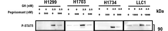
Application: Western BlotSample Tested: LLC1, H1703, H1299 and H1734 LUNG CANCER CELL LINE, H1703 and H1299 and H1734 LUNG CANCER CELL LINESpecies: Human and mouse lung cancer cellsVerified Customer | Posted 05/21/2026Four Lung cancer cell lines used. Three are human and one mouse lung cancer cell line.in vitro

-
Application: Western BlotSample Tested: SK-Mel-28 human malignant melanoma cell line, SK-Mel-30 human malignant melanoma cell line and human IM9 lymphoblastoid cellsSpecies: HumanVerified Customer | Posted 04/16/2018
-
Application: Western BlotSample Tested: MDA-MB-231 human breast cancer cell line and MCF 10A human breast epithelial cell lineSpecies: HumanVerified Customer | Posted 07/24/2017
There are no reviews that match your criteria.
Protocols
Find general support by application which include: protocols, troubleshooting, illustrated assays, videos and webinars.
- 7-Amino Actinomycin D (7-AAD) Cell Viability Flow Cytometry Protocol
- Cellular Response to Hypoxia Protocols
- Extracellular Membrane Flow Cytometry Protocol
- Flow Cytometry Protocol for Cell Surface Markers
- Flow Cytometry Protocol for Staining Membrane Associated Proteins
- Flow Cytometry Staining Protocols
- Flow Cytometry Troubleshooting Guide
- Intracellular Flow Cytometry Protocol Using Alcohol (Methanol)
- Intracellular Flow Cytometry Protocol Using Detergents
- Intracellular Nuclear Staining Flow Cytometry Protocol Using Detergents
- Intracellular Staining Flow Cytometry Protocol Using Alcohol Permeabilization
- Intracellular Staining Flow Cytometry Protocol Using Detergents to Permeabilize Cells
- Propidium Iodide Cell Viability Flow Cytometry Protocol
- Protocol for Liperfluo
- Protocol for the Characterization of Human Th22 Cells
- Protocol for the Characterization of Human Th9 Cells
- Protocol: Annexin V and PI Staining by Flow Cytometry
- Protocol: Annexin V and PI Staining for Apoptosis by Flow Cytometry
- R&D Systems Quality Control Western Blot Protocol
- Troubleshooting Guide: Fluorokine Flow Cytometry Kits
- Troubleshooting Guide: Western Blot Figures
- Western Blot Conditions
- Western Blot Protocol
- Western Blot Protocol for Cell Lysates
- Western Blot Troubleshooting
- Western Blot Troubleshooting Guide
- View all Protocols, Troubleshooting, Illustrated assays and Webinars
Loading...
Associated Pathways


