PIK3C2 beta (Phosphadidylinositol-4-phosphate 3 kinase C2 domain-containing beta subunit; also HsC2-PI3K) is a 175-185 kDa member of the Class II PI3/PI4 kinase family of enzymes. It is widely expressed, being found in neurons, urinary transitional and columnar epithelium, and fibroblasts. PIK3C2 beta likely participates in growth factor, integrin and chemokine signaling by interacting with select cell membrane receptors, and appears to phosphorylate both phosphatidylinositol (PI) and PI 4‑monophosphate upon activation. Human PIK3C2 beta is 1634 amino acids (aa) in length. It contains an N-terminal Pro-rich region (aa 156-174), two PI3K Class II domains (aa 622-794 and 1505-1627), a PI3K catalytic region (aa 987-1340), one PX domain (aa 1365-1481) and an NLS (aa 1555-1565). There are two potential C2 beta isoform variants. One shows a deletion of aa 850-877, while another contains a three aa insertion after Gln1016. Over aa 2-134, human PIK3C2 beta shares 91% aa identity with mouse PIK3C2 beta.
Human PI 3-Kinase C2 beta Antibody
R&D Systems | Catalog # AF7249

Key Product Details
Species Reactivity
Validated:
Human
Cited:
Human
Applications
Validated:
Western Blot, Immunocytochemistry
Cited:
Western Blot
Label
Unconjugated
Antibody Source
Polyclonal Goat IgG
Loading...
Product Specifications
Immunogen
E. coli-derived recombinant human PI 3‑Kinase C2 beta
Ser2-Ser134
Accession # O00750
Ser2-Ser134
Accession # O00750
Specificity
Detects human PI 3‑Kinase C2 beta in direct ELISAs and Western blots.
Clonality
Polyclonal
Host
Goat
Isotype
IgG
Scientific Data Images for Human PI 3-Kinase C2 beta Antibody
Detection of Human PI 3‑Kinase C2 beta by Western Blot.
Western blot shows lysates of HepG2 human hepatocellular carcinoma cell line and U2OS human osteosarcoma cell line. PVDF membrane was probed with 2 µg/mL of Goat Anti-Human PI 3-Kinase C2 beta Antigen Affinity-purified Polyclonal Antibody (Catalog # AF7249) followed by HRP-conjugated Anti-Goat IgG Secondary Antibody (Catalog # HAF017). A specific band was detected for PI 3-Kinase C2 beta at approximately 185 kDa (as indicated). This experiment was conducted under reducing conditions and using Immunoblot Buffer Group 1.
PI 3‑Kinase C2 beta in HEK293 Human Cell Line.
PI 3-Kinase C2 beta was detected in immersion fixed HEK293 human embryonic kidney cell line using Goat Anti-Human PI 3-Kinase C2 beta Antigen Affinity-purified Polyclonal Antibody (Catalog # AF7249) at 10 µg/mL for 3 hours at room temperature. Cells were stained using the NorthernLights™ 557-conjugated Anti-Goat IgG Secondary Antibody (red; Catalog # NL001) and counterstained with DAPI (blue). Specific staining was localized to cytoplasm. View our protocol for Fluorescent ICC Staining of Cells on Coverslips.
Detection of Human PI 3-Kinase C2 beta by Western Blot
Predator cell PIK3C2B colocalizes with PI(3)P during engulfment and is required for complete internalization.(A) CRISPR-Cas9 single-cell clone PIK3C2B knockouts of MCF-7 were screened by immunoblot. Clones indicated by asterisk were chosen for further testing. (B) Predator cell engulfment rates (left) and confluency as a measure of viability (right) for senescent MCF-7 parental cells and 3 PIK3C2B knockout clones were determined by time course imaging in a DOXO-NT culture. Underlying data can be found at S1 Data. (C) Time course live-cell imaging of a senescent MCF-7 cell expressing 2xFYVE-mCherry and PIK3C2B-GFP throughout the entire process of engulfing an NIR-MCF-7 cell, scale bar = 100 μm. (D) Left: Merged (top) and separate (middle, lower) color channels of axial planes from bottom to top of a senescent MCF-7 cell expressing 2xFYVE-mCherry and PIK3C2B-GFP, engulfing NIR-MCF-7 cells. Right, volume view reconstruction of the same image, scale bar = 10 μm. Image collected and cropped by CiteAb from the following open publication (https://pubmed.ncbi.nlm.nih.gov/36279312), licensed under a CC-BY license. Not internally tested by R&D Systems.Applications for Human PI 3-Kinase C2 beta Antibody
Application
Recommended Usage
Immunocytochemistry
5-15 µg/mL
Sample: Immersion fixed HEK293 human embryonic kidney cell line
Sample: Immersion fixed HEK293 human embryonic kidney cell line
Western Blot
2 µg/mL
Sample: HepG2 human hepatocellular carcinoma cell line and U2OS human osteosarcoma cell line
Sample: HepG2 human hepatocellular carcinoma cell line and U2OS human osteosarcoma cell line
Reviewed Applications
Read 1 review rated 5 using AF7249 in the following applications:
Formulation, Preparation, and Storage
Purification
Antigen Affinity-purified
Reconstitution
Sterile PBS to a final concentration of 0.2 mg/mL. For liquid material, refer to CoA for concentration.
Loading...
Formulation
Lyophilized from a 0.2 μm filtered solution in PBS with Trehalose. *Small pack size (SP) is supplied either lyophilized or as a 0.2 µm filtered solution in PBS.
Shipping
Lyophilized product is shipped at ambient temperature. Liquid small pack size (-SP) is shipped with polar packs. Upon receipt, store immediately at the temperature recommended below.
Stability & Storage
Use a manual defrost freezer and avoid repeated freeze-thaw cycles.
- 12 months from date of receipt, -20 to -70 °C as supplied.
- 1 month, 2 to 8 °C under sterile conditions after reconstitution.
- 6 months, -20 to -70 °C under sterile conditions after reconstitution.
Calculators
Background: PI 3-Kinase C2 beta
Long Name
Phosphatidylinositol-4-Phosphate 3-Kinase C2 Domain-containing Subunit beta
Alternate Names
C2-PI3K, Phosphoinositide 3-kinase-C2-beta, PI 3Kinase C2 beta, PIK3C2B, PTDINS-3-kinase C2 beta
Gene Symbol
PIK3C2B
UniProt
Additional PI 3-Kinase C2 beta Products
Product Documents for Human PI 3-Kinase C2 beta Antibody
Certificate of Analysis
To download a Certificate of Analysis, please enter a lot or batch number in the search box below.
Note: Certificate of Analysis not available for kit components.
Product Specific Notices for Human PI 3-Kinase C2 beta Antibody
For research use only
Citations for Human PI 3-Kinase C2 beta Antibody
 Powered by Bioz
Customer Reviews for Human PI 3-Kinase C2 beta Antibody (1)
5 out of 5
1 Customer Rating
Have you used Human PI 3-Kinase C2 beta Antibody?
Submit a review and receive an Amazon gift card!
$25/€18/£15/$25CAN/¥2500 Yen for a review with an image
$10/€7/£6/$10CAN/¥1110 Yen for a review without an image
Submit a review
Customer Images
Showing
1
-
1 of
1 review
Showing All
Filter By:
-
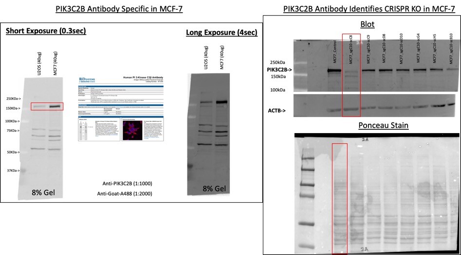
Application: Western BlotSample Tested: MCF-7 human breast cancer cell line, MCF-7 PIK3C2B CRISPR single cell clones, U2OS human osteosarcoma cell line and MCF-7 sgPIK3C2B CRISPR Single-cell clonesSpecies: HumanVerified Customer | Posted 09/28/2020

There are no reviews that match your criteria.
Protocols
Find general support by application which include: protocols, troubleshooting, illustrated assays, videos and webinars.
- Appropriate Fixation of IHC/ICC Samples
- Cellular Response to Hypoxia Protocols
- ClariTSA™ Fluorophore Kits
- Detection & Visualization of Antibody Binding
- ICC Cell Smear Protocol for Suspension Cells
- ICC Immunocytochemistry Protocol Videos
- ICC for Adherent Cells
- Immunocytochemistry (ICC) Protocol
- Immunocytochemistry Troubleshooting
- Immunofluorescence of Organoids Embedded in Cultrex Basement Membrane Extract
- Immunohistochemistry (IHC) and Immunocytochemistry (ICC) Protocols
- Preparing Samples for IHC/ICC Experiments
- Preventing Non-Specific Staining (Non-Specific Binding)
- Primary Antibody Selection & Optimization
- Protocol for VisUCyte™ HRP Polymer Detection Reagent
- Protocol for the Fluorescent ICC Staining of Cell Smears - Graphic
- Protocol for the Fluorescent ICC Staining of Cultured Cells on Coverslips - Graphic
- Protocol for the Preparation and Fluorescent ICC Staining of Cells on Coverslips
- Protocol for the Preparation and Fluorescent ICC Staining of Non-adherent Cells
- Protocol for the Preparation and Fluorescent ICC Staining of Stem Cells on Coverslips
- Protocol for the Preparation of a Cell Smear for Non-adherent Cell ICC - Graphic
- R&D Systems Quality Control Western Blot Protocol
- TUNEL and Active Caspase-3 Detection by IHC/ICC Protocol
- The Importance of IHC/ICC Controls
- Troubleshooting Guide: Western Blot Figures
- Western Blot Conditions
- Western Blot Protocol
- Western Blot Protocol for Cell Lysates
- Western Blot Troubleshooting
- Western Blot Troubleshooting Guide
- View all Protocols, Troubleshooting, Illustrated assays and Webinars
Loading...
Associated Pathways
