Prosaposin behaves as a myelinotrophic and neurotrophic factor, whose effects are
mediated by its G-protein-coupled receptors, GPR37 and GPR37L1,
undergoing ligand-mediated internalization followed by ERK
phosphorylation signaling.By similarity Saposins
are specific low-molecular mass non-enzymic proteins and participate
in the lysosomal degradation of sphingolipids, which takes place by the
sequential action of specific hydrolases. The Prosaposin gets cleaved into 5 chains, Saposin A, B, B-Val, C and D. It also contains several disulfide bonds as well as glycosylation sites.

Loading...
Key Product Details
Validated by
Knockout/Knockdown
Species Reactivity
Validated:
Human
Cited:
Human
Applications
Validated:
Knockout Validated, Western Blot, Immunocytochemistry, Simple Western, Immunoprecipitation
Cited:
Immunohistochemistry-Frozen, Western Blot, Immunoprecipitation
Label
Unconjugated
Antibody Source
Polyclonal Rabbit IgG
Loading...
Product Specifications
Immunogen
S. frugiperda insect ovarian cell line Sf21-derived recombinant human PSAP
Gly17-Asn524
Accession # P07602
Gly17-Asn524
Accession # P07602
Specificity
Detects human PSAP in direct ELISAs and Western blots.
Clonality
Polyclonal
Host
Rabbit
Isotype
IgG
Scientific Data Images for Human PSAP Antibody
Detection of Human PSAP by Western Blot.
Western blot shows lysates of A431 human epithelial carcinoma cell line. PVDF membrane was probed with 1:1000 dilution of Rabbit Anti-Human PSAP Antigen Affinity-purified Polyclonal Antibody (Catalog # AF8520) followed by HRP-conjugated Anti-Rabbit IgG Secondary Antibody (Catalog # HAF008). Specific bands were detected for PSAP at approximately 60-80 kDa (as indicated). This experiment was conducted under reducing conditions and using Immunoblot Buffer Group 1.
PSAP in A431 Human Cell Line.
PSAP was detected in immersion fixed A431 human epithelial carcinoma cell line using Rabbit Anti-Human PSAP Antigen Affinity-purified Polyclonal Antibody (Catalog # AF8520) at 1:100 dilution for 3 hours at room temperature. Cells were stained using the NorthernLights™ 557-conjugated Anti-Rabbit IgG Secondary Antibody (red; Catalog # NL004) and counterstained with DAPI (blue). Specific staining was localized to Golgi. View our protocol for Fluorescent ICC Staining of Cells on Coverslips.
Detection of Human PSAP by Simple WesternTM.
Simple Western lane view shows lysates of A431 human epithelial carcinoma cell line, loaded at 0.2 mg/mL. A specific band was detected for PSAP at approximately 98 kDa (as indicated) using 2 µg/mL of Rabbit Anti-Human PSAP Antigen Affinity-purified Polyclonal Antibody (Catalog # AF8520). This experiment was conducted under reducing conditions and using the 12-230 kDa separation system.
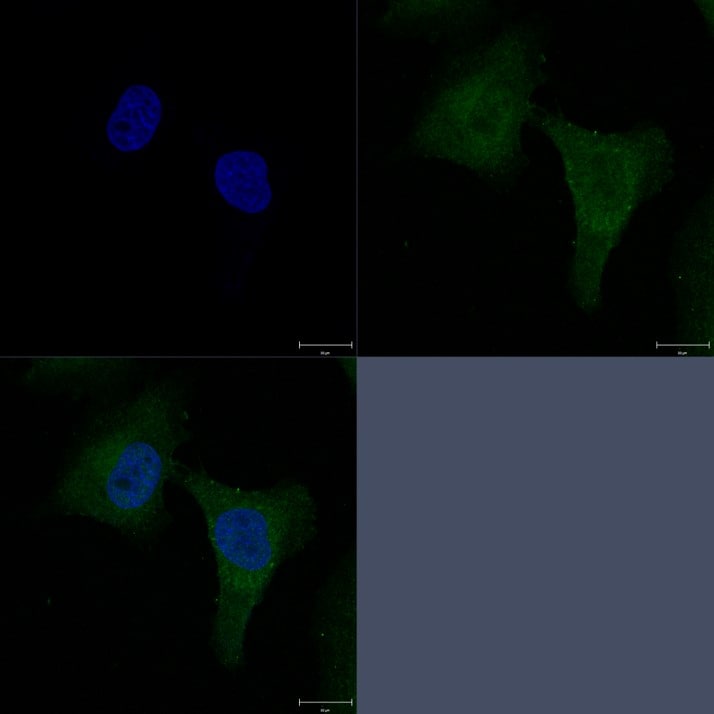
Detection of Human PSAP by Immunocytochemistry/Immunofluorescence
Confocal microscopy of prosaposin localization on plaques and different cell types. (a-c). A beta (green) (a) and PSAP (red) plaque (b) with limited colocalization (C – yellow) in an AD case. Scale bar represents 30 μm. (d-f). Comparison of colocalization in plaque of PGRN (green) and A beta (blue) (d) with PSAP (red) and A beta (blue) in triple-stained AD section. Merged images show extensive colocalization of PGRN and PSAP (yellow) but limited overlap with A beta -positive structures. Scale bar represents 30 μm. (G-I). Merged images of PSAP (red) immunoreactivity with microglial markers IBA-1 (g) and CD68 (h) (green) and astrocyte marker GFAP (green) show some expression of PSAP in both cell types (yellow). These images show that PSAP (red) is predominantly in cells with morphology of neurons. Scale bar represents 10 μm. (j-l). Merged images of CD68 (green) and PSAP (red) on plaques in low plaque case (J), high plaque case (k) and AD case (l). Significant amounts of PSAP immunoreactivity (red) can be observed on all plaques but with only limited colocalization with CD68 in infiltrating microglia. Scale bar represents 30 μm. (m-n) Merged images of AT180 (pTau) (green) and PSAP (red) on tangle in low plaque case (M), high plaque case (n) and Alzheimer’s disease case (o). Very limited amounts of PSAP immunoreactivity (yellow) can be observed on tangles. Panel M and N show intracellular tangles with DAPI-positive nuclei, while panel O shows extracellular tangle. Scale bar represents 30 μm. Image collected and cropped by CiteAb from the following publication (https://pubmed.ncbi.nlm.nih.gov/31864418), licensed under a CC-BY license. Not internally tested by R&D Systems.
Western Blot Shows PSAP Specificity Using Knockout Cell Line.
Western blot shows culture media of HeLa human cervical epithelial carcinoma parental cell line and PSAP knockout HeLa cell line (KO). Nitrocellulose membrane was probed with Rabbit Anti-Human PSAP Antigen Affinity-purified Polyclonal Antibody (Catalog # AF8520) followed by HRP-conjugated secondary antibody. A specific band was detected for PSAP at approximately 58.1 kDa (as indicated) in the parental HeLa cell line, but is not detectable in knockout HeLa cell line. Primary antibody dilution used: 1:1000. The Ponceau stained transfer of the blot is shown. This experiment was conducted under reducing conditions. Image, protocol, and testing courtesy of YCharOS Inc. See ycharos.com for additional details.
Detection of PSAP by Immunoprecipitation.
HeLa human cervical epithelial carcinoma cell line culture medium were prepared and immunoprecipitation was performed using 2.0 μg of Rabbit Anti-Human PSAP Antigen Affinity-purified Polyclonal Antibody (Catalog # AF8520) pre-coupled to Dynabeads Protein A. Immunoprecipitated PSAP was detected in Western Blot with a rabbit PSAP antibody used at 1/1000. The Ponceau stained transfer of the blot is shown. SM=4% starting material; UB=4% unbound fraction; IP=immunoprecipitate; HC=antibody heavy chain. Image, protocol and testing courtesy of YCharOS Inc. (ycharos.com).Applications for Human PSAP Antibody
Application
Recommended Usage
Immunocytochemistry
1:100 dilution
Sample: Immersion fixed A431 human epithelial carcinoma cell line
Sample: Immersion fixed A431 human epithelial carcinoma cell line
Immunoprecipitation
1 µg/mL
Sample: Culture medium of HeLa human cervical epithelial carcinoma cell line
Sample: Culture medium of HeLa human cervical epithelial carcinoma cell line
Knockout Validated
PSAP is specifically detected in HeLa human cervical epithelial carcinoma parental cell line but is not detectable in knockout HeLa human cervical epithelial carcinoma cell line.
Simple Western
2 µg/mL
Sample: A431 human epithelial carcinoma cell line
Sample: A431 human epithelial carcinoma cell line
Western Blot
1:1000 dilution
Sample: A431 human epithelial carcinoma cell line
Sample: A431 human epithelial carcinoma cell line
Reviewed Applications
Read 2 reviews rated 5 using AF8520 in the following applications:
Formulation, Preparation, and Storage
Purification
Antigen Affinity-purified
Formulation
Supplied as a solution in PBS containing BSA, Glycerol and Sodium Azide. See Certificate of Analysis for details.
Shipping
The product is shipped with polar packs. Upon receipt, store it immediately at the temperature recommended below.
Stability & Storage
Use a manual defrost freezer and avoid repeated freeze-thaw cycles.
- 12 months from date of receipt, -20 to -70 °C, as supplied.
- 1 month, 2 to 8 °C under sterile conditions after opening.
- 6 months, -20 to -70 °C under sterile conditions after opening.
Background: PSAP
Long Name
Prosaposin
Alternate Names
GLBA, SAP1
Gene Symbol
PSAP
UniProt
Additional PSAP Products
Product Documents for Human PSAP Antibody
Certificate of Analysis
To download a Certificate of Analysis, please enter a lot or batch number in the search box below.
Note: Certificate of Analysis not available for kit components.
Product Specific Notices for Human PSAP Antibody
* Contains <0.1% Sodium Azide, which is not hazardous at this concentration according to GHS classifications. Refer to SDS for additional information and handling instructions.
For research use only
Related Research Areas
Citations for Human PSAP Antibody
 Powered by Bioz
Customer Reviews for Human PSAP Antibody (2)
5 out of 5
2 Customer Ratings
Have you used Human PSAP Antibody?
Submit a review and receive an Amazon gift card!
$25/€18/£15/$25CAN/¥2500 Yen for a review with an image
$10/€7/£6/$10CAN/¥1110 Yen for a review without an image
Submit a review
Customer Images

Showing
1
-
2 of
2 reviews
Showing All
Filter By:
-
Application: Western BlotSample Tested: A549 cells and H441Species: HumanVerified Customer | Posted 04/04/2020

-
Application: Immunocytochemistry/ImmunofluorescenceSample Tested: A431 human epithelial carcinoma cell lineSpecies: HumanVerified Customer | Posted 10/28/2018
There are no reviews that match your criteria.
Protocols
Find general support by application which include: protocols, troubleshooting, illustrated assays, videos and webinars.
- Appropriate Fixation of IHC/ICC Samples
- Cellular Response to Hypoxia Protocols
- ClariTSA™ Fluorophore Kits
- Detection & Visualization of Antibody Binding
- ICC Cell Smear Protocol for Suspension Cells
- ICC Immunocytochemistry Protocol Videos
- ICC for Adherent Cells
- Immunocytochemistry (ICC) Protocol
- Immunocytochemistry Troubleshooting
- Immunofluorescence of Organoids Embedded in Cultrex Basement Membrane Extract
- Immunohistochemistry (IHC) and Immunocytochemistry (ICC) Protocols
- Immunoprecipitation Protocol
- Preparing Samples for IHC/ICC Experiments
- Preventing Non-Specific Staining (Non-Specific Binding)
- Primary Antibody Selection & Optimization
- Protocol for VisUCyte™ HRP Polymer Detection Reagent
- Protocol for the Fluorescent ICC Staining of Cell Smears - Graphic
- Protocol for the Fluorescent ICC Staining of Cultured Cells on Coverslips - Graphic
- Protocol for the Preparation and Fluorescent ICC Staining of Cells on Coverslips
- Protocol for the Preparation and Fluorescent ICC Staining of Non-adherent Cells
- Protocol for the Preparation and Fluorescent ICC Staining of Stem Cells on Coverslips
- Protocol for the Preparation of a Cell Smear for Non-adherent Cell ICC - Graphic
- R&D Systems Quality Control Western Blot Protocol
- TUNEL and Active Caspase-3 Detection by IHC/ICC Protocol
- The Importance of IHC/ICC Controls
- Troubleshooting Guide: Western Blot Figures
- Western Blot Conditions
- Western Blot Protocol
- Western Blot Protocol for Cell Lysates
- Western Blot Troubleshooting
- Western Blot Troubleshooting Guide
- View all Protocols, Troubleshooting, Illustrated assays and Webinars
Loading...