RAB27A (Ras-related protein Rab 27A; also GTP-binding protein Ram) is a 27-28 kDa member of the Rab27 subfamily, Rab family, Small GTPase superfamily of proteins. It is widely expressed, and found in cells diverse as mast cells, cytotoxic T cells, melanocytes, retinal pigment epithelium and pancreatic beta -cells. RAB27A plays a key role in the secretion of specialized lysosomes termed secretory lysosomes. In melanocytes, for example, RAB27A is incorporated into the melanosome membrane where it serves as a docking factor for melanophilin and myosin-Va, regulating melanosome transport to, and concentration at, sites of release. Human RAB27A is 221 amino acids (aa) in length. It contains multiple Rab family and subfamily motifs, and concludes with a C-terminal CXC prenylation sequence (aa 219‑221). There is one potential splice variant that shows a deletion of aa 146-153. Over aa 135-218, human RAB27A shares 92% and 94% aa sequence identity with mouse Rab27A and rat RAB27A, respectively.
Human Rab27a Antibody (2537A)
R&D Systems | Catalog # MAB7245
Recombinant Monoclonal Antibody.

Key Product Details
Validated by
Knockout/Knockdown
Species Reactivity
Validated:
Human
Cited:
Human
Applications
Validated:
Western Blot, Immunocytochemistry, Simple Western
Cited:
Western Blot
Label
Unconjugated
Antibody Source
Recombinant Monoclonal Rabbit IgG Clone # 2537A
Loading...
Product Specifications
Immunogen
E. coli-derived recombinant human RAB27a
Ser135-Ala218
Accession # P51159
Ser135-Ala218
Accession # P51159
Specificity
Detects human RAB27a in direct ELISAs.
Clonality
Monoclonal
Host
Rabbit
Isotype
IgG
Scientific Data Images for Human Rab27a Antibody (2537A)
Detection of Human Rab27a by Western Blot.
Western blot shows lysates of human prostate tissue. PVDF membrane was probed with 2 µg/mL of Rabbit Anti-Human Rab27a Monoclonal Antibody (Catalog # MAB7245) followed by HRP-conjugated Anti-Rabbit IgG Secondary Antibody (HAF008). A specific band was detected for Rab27a at approximately 26 kDa (as indicated). This experiment was conducted under reducing conditions and using Immunoblot Buffer Group 1.
Detection of Human Rab27a by Simple WesternTM.
Simple Western shows lysates of Exosome Standards (K562) (NBP2-49864) and SK‑Mel‑28 human malignant melanoma cell line, loaded at 0.5 mg/ml. A specific band was detected for Rab27a at approximately 32 kDa (as indicated) using 20 µg/mL of Rabbit Anti-Human Rab27a Monoclonal Antibody (Catalog # MAB7245). This experiment was conducted under reducing conditions and using the 12-230 kDa separation system.
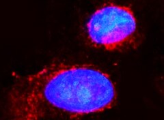
Rab27a in SK‑Mel‑28 Human Cell Line.
Rab27a was detected in immersion fixed SK-Mel-28 human malignant melanoma cell line (left panel; positive staining) and Daudi human Burkitt's lymphoma cell line (right panel; negative staining) using Rabbit Anti-Human Rab27a Monoclonal Antibody (Catalog # MAB7245) at 3 µg/mL for 3 hours at room temperature. Cells were stained using the NorthernLights™ 557-conjugated Anti-Rabbit IgG Secondary Antibody (red; NL004) and counterstained with DAPI (blue). Specific staining was localized to cytoplasm. View our protocol for Fluorescent ICC Staining of Cells on Coverslips.
Detection of Human Rab27a by Simple WesternTM.
Simple Western lane view shows lysates of K562 human chronic myelogenous leukemia cell line, loaded at 0.2 mg/mL. A specific band was detected for Rab27a at approximately 31 kDa (as indicated) using 20 µg/mL of Rabbit Anti-Human Rab27a Monoclonal Antibody (Catalog # MAB7245). This experiment was conducted under reducing conditions and using the 12-230 kDa separation system.
Western Blot Shows Rab27a Specificity Using Knockdown Cell Line.
Western blot shows lysates of U‑87 MG human glioblastoma/astrocytoma parental cell line and Rab27a knockdown U-87 MG cell line (KD). Nitrocellulose membrane was probed with 2.5 µg/mL Rabbit Anti-Human Rab27a Monoclonal Antibody (Catalog # MAB7245) followed by HRP-conjugated secondary antibody. A specific band was detected for Rab27a at approximately 26 kDa (as indicated) in the parental U-87 MG cell line, but is significantly reduced in knockdown U-87 MG cell line. The Ponceau stained transfer of the blot is shown. This experiment was conducted under reducing conditions. Image, protocol, and testing courtesy of YCharOS Inc. See ycharos.com for additional details.
Rab27a Specificity is Shown by Immunocytochemistry in Knockdown Cell Line.
U‑87 MG human glioblastoma/astrocytoma parental cell line ctrl and Rab27a U-87 MG KD cells were labelled with a green or a far-red fluorescent dye, respectively. Cells were stained with Rabbit Anti-Human Rab27a Monoclonal Antibody (Catalog # MAB7245) followed by incubation with an Alexa-fluor 555 conjugated secondary antibody (upper panel). DAPI-only counterstained cells shown on a lower panel. Acquisition of the blue (nucleus-DAPI), green (identification of ctrl cells), red (antibody staining) and far-red (identification of KD cells) channels was performed. Representative images of the blue and red (grayscale) channels are shown. Ctrl and KD cells are outlined with green and magenta dashed line, respectively. Primary antibody concentration used: 3.33 µg/mL. Image, protocol and testing courtesy of YCharOS Inc. (ycharos.com).
Detection of Human Rab27a by Western Blot
Assessment of the effect of GTP gamma S and GDP beta S loading of cell lysate before pulldown. To investigate the impact of the GTP/GDP cycle of small GTPases on their affinity to ARHGAP25, we performed GST-pulldowns on samples pretreated with GTP gamma S and GDP beta S. (a) Comparison of LFQ values of small GTPases pulled down with ARHGAP25 following GTP gamma S and GDP beta S loading of the neutrophilic cell lysate. Employed statistics: paired t-tests (n = 3). Data are presented as mean ± SD. p values are presented numerically; bold values are considered significant (p < 0.05). LFQ values RAC2 and RHOG were significantly elevated in GTP gamma S samples, implying a GTP/GDP dependent effect on their affinity towards ARHGAP25. ARF4 and RAB27A did not show any significant difference. (b) Representative Western Blot of RHOG, RAC2 and RAB27A protein levels in GST-ARHGAP25 pulldown samples following GTP gamma S- and GDP beta S-loading of the neutrophilic cell lysate. The difference in the band intensities supported our proteomic results. Image collected and cropped by CiteAb from the following open publication (https://www.nature.com/articles/s41598-024-71002-4), licensed under a CC-BY license. Not internally tested by R&D Systems.
Detection of Human Rab27a by Western Blot
Identification of ARHGAP25’s interactome in neutrophilic granulocytes. Out of the identified proteins, a list of potential candidates of interaction partners was selected and further supported by co-immunoprecipitation assays from neutrophil cell lysates. (a) Volcano plot of the GST-pulldown-derived proteomic data. Employed statistics: multiple t-tests with Benjamini–Hochberg correction (n = 6); significance thresholds are marked by dotted lines. ARHGAP25’s potential partners are colored green. Gray dots represent proteins below the threshold. Ninety proteins showed a significant preference towards GST-ARHGAP25. (b) Western Blot validation of the proteomic analysis. Six proteins were selected to check their presence in the samples. Representative images reinsured the presence of ACSL1, SYK, RAB27A, RHOG, and RAC2, and the absence of LDHA. (c) Functional enrichment of ‘GO: Biological Function’ and ‘Reactome’ gene sets of identified protein partners. Gene sets with redundant or overlapping themes are grouped and colored differently for clarity. (d) STRING (‘Search Tool for Retrieval of Interacting Genes/Proteins’) analysis between identified partners of ARHGAP25. Based on molecular function and localization, proteins are separated into different groups. Lines represent documented interactions between proteins extracted from the STRING database. Image collected and cropped by CiteAb from the following open publication (https://www.nature.com/articles/s41598-024-71002-4), licensed under a CC-BY license. Not internally tested by R&D Systems.Applications for Human Rab27a Antibody (2537A)
Application
Recommended Usage
Immunocytochemistry
3-25 µg/mL
Sample: Immersion fixed SK-Mel-28 human malignant melanoma cell line
Sample: Immersion fixed SK-Mel-28 human malignant melanoma cell line
Simple Western
20 µg/mL
Sample: Exosome Standards (K562) (Catalog # NBP2-49864), SK-Mel-28 human malignant melanoma cell line and K562 human chronic myelogenous leukemia cell line
Sample: Exosome Standards (K562) (Catalog # NBP2-49864), SK-Mel-28 human malignant melanoma cell line and K562 human chronic myelogenous leukemia cell line
Western Blot
2 µg/mL
Sample: Human prostate tissue
Sample: Human prostate tissue
Reviewed Applications
Read 1 review rated 5 using MAB7245 in the following applications:
Formulation, Preparation, and Storage
Purification
Protein A or G purified from cell culture supernatant
Reconstitution
Reconstitute at 0.5 mg/mL in sterile PBS. For liquid material, refer to CoA for concentration.
Loading...
Formulation
Lyophilized from a 0.2 μm filtered solution in PBS with Trehalose. See Certificate of Analysis for details.
*Small pack size (-SP) is supplied either lyophilized or as a 0.2 µm filtered solution in PBS.
*Small pack size (-SP) is supplied either lyophilized or as a 0.2 µm filtered solution in PBS.
Shipping
Lyophilized product is shipped at ambient temperature. Liquid small pack size (-SP) is shipped with polar packs. Upon receipt, store immediately at the temperature recommended below.
Stability & Storage
Use a manual defrost freezer and avoid repeated freeze-thaw cycles.
- 12 months from date of receipt, -20 to -70 °C as supplied.
- 1 month, 2 to 8 °C under sterile conditions after reconstitution.
- 6 months, -20 to -70 °C under sterile conditions after reconstitution.
Calculators
Background: Rab27a
Long Name
RAs Genes from Brain Protein 27A
Alternate Names
GS2, HsT18676, RAM
Entrez Gene IDs
5873 (Human)
Gene Symbol
RAB27A
UniProt
Additional Rab27a Products
Product Documents for Human Rab27a Antibody (2537A)
Certificate of Analysis
To download a Certificate of Analysis, please enter a lot or batch number in the search box below.
Note: Certificate of Analysis not available for kit components.
Product Specific Notices for Human Rab27a Antibody (2537A)
For research use only
Related Research Areas
Citations for Human Rab27a Antibody (2537A)
 Powered by Bioz
Customer Reviews for Human Rab27a Antibody (2537A) (1)
5 out of 5
1 Customer Rating
Have you used Human Rab27a Antibody (2537A)?
Submit a review and receive an Amazon gift card!
$25/€18/£15/$25CAN/¥2500 Yen for a review with an image
$10/€7/£6/$10CAN/¥1110 Yen for a review without an image
Submit a review
Customer Images
Showing
1
-
1 of
1 review
Showing All
Filter By:
-
Application: Immunocytochemistry/ImmunofluorescenceSample Tested: SK-Mel-28 human malignant melanoma cell lineSpecies: HumanVerified Customer | Posted 06/05/2022

There are no reviews that match your criteria.
Protocols
Find general support by application which include: protocols, troubleshooting, illustrated assays, videos and webinars.
- Appropriate Fixation of IHC/ICC Samples
- Cellular Response to Hypoxia Protocols
- ClariTSA™ Fluorophore Kits
- Detection & Visualization of Antibody Binding
- ICC Cell Smear Protocol for Suspension Cells
- ICC Immunocytochemistry Protocol Videos
- ICC for Adherent Cells
- Immunocytochemistry (ICC) Protocol
- Immunocytochemistry Troubleshooting
- Immunofluorescence of Organoids Embedded in Cultrex Basement Membrane Extract
- Immunohistochemistry (IHC) and Immunocytochemistry (ICC) Protocols
- Preparing Samples for IHC/ICC Experiments
- Preventing Non-Specific Staining (Non-Specific Binding)
- Primary Antibody Selection & Optimization
- Protocol for VisUCyte™ HRP Polymer Detection Reagent
- Protocol for the Fluorescent ICC Staining of Cell Smears - Graphic
- Protocol for the Fluorescent ICC Staining of Cultured Cells on Coverslips - Graphic
- Protocol for the Preparation and Fluorescent ICC Staining of Cells on Coverslips
- Protocol for the Preparation and Fluorescent ICC Staining of Non-adherent Cells
- Protocol for the Preparation and Fluorescent ICC Staining of Stem Cells on Coverslips
- Protocol for the Preparation of a Cell Smear for Non-adherent Cell ICC - Graphic
- R&D Systems Quality Control Western Blot Protocol
- TUNEL and Active Caspase-3 Detection by IHC/ICC Protocol
- The Importance of IHC/ICC Controls
- Troubleshooting Guide: Western Blot Figures
- Western Blot Conditions
- Western Blot Protocol
- Western Blot Protocol for Cell Lysates
- Western Blot Troubleshooting
- Western Blot Troubleshooting Guide
- View all Protocols, Troubleshooting, Illustrated assays and Webinars
Loading...