
Tollip (Toll-interacting protein) is a 28 kDa, cytoplasmic protein that is involved in IL-1 and Toll receptor signaling. It is classified as an adaptor protein that performs multiple functions. It binds both IRAK and Toll receptors, inhibiting constitutive IL-1 and Toll receptor signaling. It also binds to ubiquinated IL-1 RI, directing it to the endosomal degradation compartment. Human Tollip is 274 amino acids(aa) in length. It contains a C2 Ca++-binding region (aa 54‑154) and a CUE domain (aa 229‑270) that binds ubiquitin. One potential splice form exists that shows a deletion of aa 6‑33. Human Tollip is 92% identical to mouse Tollip.
Tollip Antibody (503315)
R&D Systems | Catalog # MAB4678


Scientific images may be resized, cropped, or adjusted for brightness or contrast for presentation purposes. These updates are limited to presentation and do not affect the underlying scientific interpretation of the data. Where available, additional source data may be provided upon request.
Key Product Details
Species Reactivity
Validated:
Human, Rat
Cited:
Fish - Danio rerio (Zebrafish)
Applications
Validated:
Western Blot
Cited:
Immunohistochemistry, Western Blot
Label
Unconjugated
Antibody Source
Monoclonal Mouse IgG2A Clone # 503315
Loading...
Product Specifications
Immunogen
E. coli-derived recombinant human Tollip
Met1-Pro274
Accession # Q9H0E2
Met1-Pro274
Accession # Q9H0E2
Specificity
Detects human and rat Tollip in Western blots.
Clonality
Monoclonal
Host
Mouse
Isotype
IgG2A
Scientific Data Images for Tollip Antibody (503315)
Detection of Human/Rat Tollip by Western Blot.
Western blot shows lysates of HepG2 human hepatocellular carcinoma cell line and NRK rat normal kidney cell line. PVDF membrane was probed with 1 µg/mL of Mouse Anti-Human/Rat Tollip Monoclonal Antibody (Catalog # MAB4678) followed by HRP-conjugated Anti-Mouse IgG Secondary Antibody (Catalog # HAF007). A specific band was detected for Tollip at approximately 28 kDa (as indicated). This experiment was conducted under reducing conditions and using Immunoblot Buffer Group 2.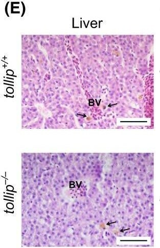
Detection of Tollip by Immunohistochemistry
Knockout of the tollip gene has no effect on the fish morphology. (A) Wild‐type (tollip+/+) and mutant (tollip−/−) embryos at 14 h, and 1 and 5 days post fertilization (hpf, dpf) show normal development and morphology. Scale bar 500 μm. Right image, wild‐type (tollip+/+) and mutant (tollip−/−) male adults at 14 mpf are without visible body morphological defects. Scale bar 1 cm. (B) Morphology of wild‐type tollip+/+ and mutant tollip−/− embryos injected with 2 ng of translation‐blocking morpholino targeting tollip (MO), observed at 48 hpf. Scale bar 1 mm. (C–H) Histology and quantitative analysis of hematoxylin and eosin stained sections of testes (C), spleen (D), liver (E, F), and anterior intestine (G, H) with specific cells and structures marked, including spermatozoa (Sm), spermatogonia (Sg), spermatocytes (Sp), red pulp (RP), white pulp (WP), blood vessels (BV), liver macrophages (arrows in E), and goblet cells (gc). For liver analyses, areas of hepatocytes and their nuclei are quantified in F. The height of intestinal folds is quantified in H. Scale bar 50 µm (C, G), 100 µm (D, E). Bars represent the means ± SEM (n = 3, each encompassing 40–60 (liver) or 25 (intestine) measurements from 5 selected areas) t‐test, ns.—nonsignificant. Image collected and cropped by CiteAb from the following open publication (https://pubmed.ncbi.nlm.nih.gov/35506194), licensed under a CC-BY license. Not internally tested by R&D Systems.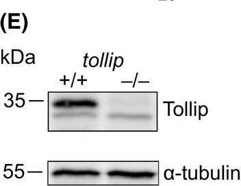
Detection of Tollip by Western Blot
CRISPR/Cas9‐based genome editing allows for the generation of a Tollip‐deficient zebrafish line. (A) Schemes of the zebrafish tollip transcript variants 1 and 2 (v1 and v2), based on the Ensembl database, showing exons (E), translated sequences (gray), and UTR regions (white). (B) Schematic illustration of the structure of Tollip protein isoforms with the C2 and CUE domains indicated. (C) Partial DNA sequence of the target site within exon 2 of the tollip gene in wild‐type tollip+/+ fish (left) and homozygous tollip−/− knockout fish (right). Deletion of eight nucleotides observed in the mutant line is shadowed in dark gray. There is an additional nucleotide change flanking the deletion (double peak marked R in the chromatogram, corresponding to A or G, with a predicted amino acid change D to G in the truncated protein product), indicating mosaicism of the generated line. (D) Schematic illustration of the predicted structure of Tollip protein isoforms synthesized from the mutated tollip gene. (E) Western blot of the 5 dpf protein lysates from the wild‐type (tollip+/+) line and tollip−/− siblings. Top panel shows Tollip (~ 35 kDa) and a bottom panel shows alpha ‐tubulin (~ 55 kDa) signal. (F) qPCR analysis of the expression of tollip transcripts during early zebrafish development (1–5 dpf). Bars represent the means ± SEM from 3–4 independent experiments (encompassing a pool of 10 larvae/condition). Mann–Whitney U test, *P < 0.05, ****P < 0.0001. Image collected and cropped by CiteAb from the following open publication (https://pubmed.ncbi.nlm.nih.gov/35506194), licensed under a CC-BY license. Not internally tested by R&D Systems.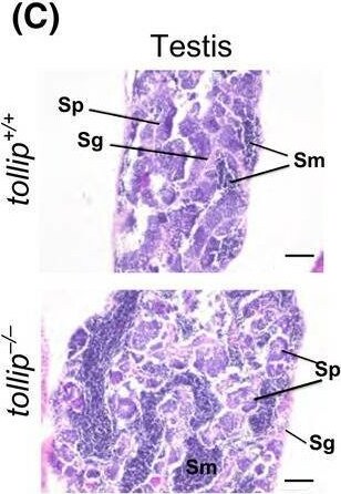
Detection of Tollip by Immunohistochemistry
Knockout of the tollip gene has no effect on the fish morphology. (A) Wild‐type (tollip+/+) and mutant (tollip−/−) embryos at 14 h, and 1 and 5 days post fertilization (hpf, dpf) show normal development and morphology. Scale bar 500 μm. Right image, wild‐type (tollip+/+) and mutant (tollip−/−) male adults at 14 mpf are without visible body morphological defects. Scale bar 1 cm. (B) Morphology of wild‐type tollip+/+ and mutant tollip−/− embryos injected with 2 ng of translation‐blocking morpholino targeting tollip (MO), observed at 48 hpf. Scale bar 1 mm. (C–H) Histology and quantitative analysis of hematoxylin and eosin stained sections of testes (C), spleen (D), liver (E, F), and anterior intestine (G, H) with specific cells and structures marked, including spermatozoa (Sm), spermatogonia (Sg), spermatocytes (Sp), red pulp (RP), white pulp (WP), blood vessels (BV), liver macrophages (arrows in E), and goblet cells (gc). For liver analyses, areas of hepatocytes and their nuclei are quantified in F. The height of intestinal folds is quantified in H. Scale bar 50 µm (C, G), 100 µm (D, E). Bars represent the means ± SEM (n = 3, each encompassing 40–60 (liver) or 25 (intestine) measurements from 5 selected areas) t‐test, ns.—nonsignificant. Image collected and cropped by CiteAb from the following open publication (https://pubmed.ncbi.nlm.nih.gov/35506194), licensed under a CC-BY license. Not internally tested by R&D Systems.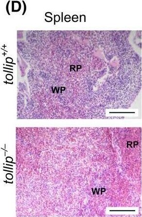
Detection of Tollip by Immunohistochemistry
Knockout of the tollip gene has no effect on the fish morphology. (A) Wild‐type (tollip+/+) and mutant (tollip−/−) embryos at 14 h, and 1 and 5 days post fertilization (hpf, dpf) show normal development and morphology. Scale bar 500 μm. Right image, wild‐type (tollip+/+) and mutant (tollip−/−) male adults at 14 mpf are without visible body morphological defects. Scale bar 1 cm. (B) Morphology of wild‐type tollip+/+ and mutant tollip−/− embryos injected with 2 ng of translation‐blocking morpholino targeting tollip (MO), observed at 48 hpf. Scale bar 1 mm. (C–H) Histology and quantitative analysis of hematoxylin and eosin stained sections of testes (C), spleen (D), liver (E, F), and anterior intestine (G, H) with specific cells and structures marked, including spermatozoa (Sm), spermatogonia (Sg), spermatocytes (Sp), red pulp (RP), white pulp (WP), blood vessels (BV), liver macrophages (arrows in E), and goblet cells (gc). For liver analyses, areas of hepatocytes and their nuclei are quantified in F. The height of intestinal folds is quantified in H. Scale bar 50 µm (C, G), 100 µm (D, E). Bars represent the means ± SEM (n = 3, each encompassing 40–60 (liver) or 25 (intestine) measurements from 5 selected areas) t‐test, ns.—nonsignificant. Image collected and cropped by CiteAb from the following open publication (https://pubmed.ncbi.nlm.nih.gov/35506194), licensed under a CC-BY license. Not internally tested by R&D Systems.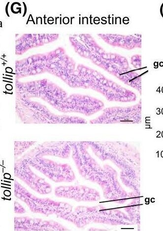
Detection of Tollip by Immunohistochemistry
Knockout of the tollip gene has no effect on the fish morphology. (A) Wild‐type (tollip+/+) and mutant (tollip−/−) embryos at 14 h, and 1 and 5 days post fertilization (hpf, dpf) show normal development and morphology. Scale bar 500 μm. Right image, wild‐type (tollip+/+) and mutant (tollip−/−) male adults at 14 mpf are without visible body morphological defects. Scale bar 1 cm. (B) Morphology of wild‐type tollip+/+ and mutant tollip−/− embryos injected with 2 ng of translation‐blocking morpholino targeting tollip (MO), observed at 48 hpf. Scale bar 1 mm. (C–H) Histology and quantitative analysis of hematoxylin and eosin stained sections of testes (C), spleen (D), liver (E, F), and anterior intestine (G, H) with specific cells and structures marked, including spermatozoa (Sm), spermatogonia (Sg), spermatocytes (Sp), red pulp (RP), white pulp (WP), blood vessels (BV), liver macrophages (arrows in E), and goblet cells (gc). For liver analyses, areas of hepatocytes and their nuclei are quantified in F. The height of intestinal folds is quantified in H. Scale bar 50 µm (C, G), 100 µm (D, E). Bars represent the means ± SEM (n = 3, each encompassing 40–60 (liver) or 25 (intestine) measurements from 5 selected areas) t‐test, ns.—nonsignificant. Image collected and cropped by CiteAb from the following open publication (https://pubmed.ncbi.nlm.nih.gov/35506194), licensed under a CC-BY license. Not internally tested by R&D Systems.Applications for Tollip Antibody (503315)
Application
Recommended Usage
Western Blot
1 µg/mL
Sample: HepG2 human hepatocellular carcinoma cell line and NRK rat normal kidney cells
Sample: HepG2 human hepatocellular carcinoma cell line and NRK rat normal kidney cells
Formulation, Preparation, and Storage
Purification
Protein A or G purified from hybridoma culture supernatant
Reconstitution
Reconstitute at 0.5 mg/mL in sterile PBS. For liquid material, refer to CoA for concentration.
Loading...
Formulation
Lyophilized from a 0.2 μm filtered solution in PBS with Trehalose. *Small pack size (SP) is supplied either lyophilized or as a 0.2 µm filtered solution in PBS.
Shipping
Lyophilized product is shipped at ambient temperature. Liquid small pack size (-SP) is shipped with polar packs. Upon receipt, store immediately at the temperature recommended below.
Stability & Storage
Use a manual defrost freezer and avoid repeated freeze-thaw cycles.
- 12 months from date of receipt, -20 to -70 °C as supplied.
- 1 month, 2 to 8 °C under sterile conditions after reconstitution.
- 6 months, -20 to -70 °C under sterile conditions after reconstitution.
Calculators
Background: Tollip
Long Name
Toll Interacting Protein
Alternate Names
IL-1RAcPIP
Gene Symbol
TOLLIP
UniProt
Additional Tollip Products
Product Documents for Tollip Antibody (503315)
Certificate of Analysis
To download a Certificate of Analysis, please enter a lot or batch number in the search box below.
Note: Certificate of Analysis not available for kit components.
Product Specific Notices for Tollip Antibody (503315)
For research use only
Citations for Tollip Antibody (503315)
 Powered by Bioz
Customer Reviews for Tollip Antibody (503315)
There are currently no reviews for this product. Be the first to review Tollip Antibody (503315) and earn rewards!
Have you used Tollip Antibody (503315)?
Submit a review and receive an Amazon gift card!
$25/€18/£15/$25CAN/¥2500 for a review with an image
$10/€7/£6/$10CAN/¥1110 for a review without an image
Submit a review
Protocols
Find general support by application which include: protocols, troubleshooting, illustrated assays, videos and webinars.
- Cellular Response to Hypoxia Protocols
- R&D Systems Quality Control Western Blot Protocol
- Troubleshooting Guide: Western Blot Figures
- Western Blot Conditions
- Western Blot Protocol
- Western Blot Protocol for Cell Lysates
- Western Blot Troubleshooting
- Western Blot Troubleshooting Guide
- View all Protocols, Troubleshooting, Illustrated assays and Webinars
Loading...