S100A8 (also known as MRP8 and calgranulin A) is a 10 kDa member of the S100 family, EF-hand superfamily of Ca-binding proteins. It is produced by neutrophils and monocytes, and forms Ca‑dependent heterodimer/heterotetramer complexes (termed calprotectin) with S100A9. It functions both intracellularly and extracellularly, where it binds to RAGE and CD36. Human S100A8 is 93 amino acids (aa) in length. It contains two EF-hand motifs (aa 12-47 and 46-81) and one high-affinity Ca‑binding site (aa 59-70). There may be one splice form that shows a 15 aa substitution for the C-terminal 14 amino acids. Although mouse S100A8 is cleaved by MMP‑2 after Asn21, it is unclear if human S100A8 is susceptible. Full-length human S100A8 is 57% and 74% aa identical to mouse and canine S100A8, respectively.

Key Product Details
Species Reactivity
Validated:
Human
Cited:
Human
Applications
Validated:
Immunohistochemistry, Western Blot
Cited:
Immunohistochemistry, Western Blot, Immunocytochemistry
Label
Unconjugated
Antibody Source
Monoclonal Mouse IgG1 Clone # 749916
Loading...
Product Specifications
Immunogen
E. coli-derived recombinant human S100A8
Met1-Glu93
Accession # P05109
Met1-Glu93
Accession # P05109
Specificity
Detects human S100A8 in direct ELISAs.
In direct ELISAs, 100% cross-reactivity
with recombinant human (rh) S100A8/A9 is observed and no cross-reactivity with
rhS100A9, recombinant mouse (rm) S100A8, rmS100A9, or rmS100A8/A9 is observed.
Clonality
Monoclonal
Host
Mouse
Isotype
IgG1
Scientific Data Images for Human S100A8 Antibody (749916)
Detection of Human S100A8 by Western Blot.
Western blot shows lysates of HL-60 human acute promyelocytic leukemia cell line. PVDF membrane was probed with 2 µg/mL of Mouse Anti-Human S100A8 Monoclonal Antibody (Catalog # MAB4570) followed by HRP-conjugated Anti-Mouse IgG Secondary Antibody (Catalog # HAF007). A specific band was detected for S100A8 at approximately 11 kDa (as indicated). This experiment was conducted under reducing conditions and using Immunoblot Buffer Group 1.
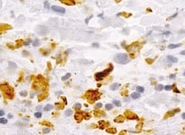
S100A8 in Human Lung Cancer Tissue.
S100A8 was detected in immersion fixed paraffin-embedded sections of human lung cancer tissue using Mouse Anti-Human S100A8 Monoclonal Antibody (Catalog # MAB4570) at 15 µg/mL overnight at 4 °C. Before incubation with the primary antibody, tissue was subjected to heat-induced epitope retrieval using Antigen Retrieval Reagent-Basic (Catalog # CTS013). Tissue was stained using the Anti-Mouse HRP-DAB Cell & Tissue Staining Kit (brown; Catalog # CTS002) and counterstained with hematoxylin (blue). Specific staining was localized to nuclei and cytoplasm of cancer cells. View our protocol for Chromogenic IHC Staining of Paraffin-embedded Tissue Sections.
Detection of Human S100A8 by Western Blot
Quantitative PCR and immunoblotting of five candidate gene products in DLBCL and LCL lines. (A) Reverse transcriptase and real-time PCR analysis of mRNAs for BMP8A, CCR6, HOXA9, NANOG and S100A8 in LCL and DLBCL (HBL-2, HT, SU-DHL-5, U2932 and U2940) cell lines. Data were normalized by the amount of GAPDH mRNA, expressed relative to the corresponding value for LCL, and are shown as means ± SD from triplicate data. (B) After quantification of Western blot intensity, the relative expression levels of each protein are shown with normalization of GAPDH. U2940 cells have the highest expression of the stem cell genes, HOXA9 and NANOG. Molecular weight listed in parenthesis. Error bars represent the standard error of the mean of three independent experiments. *p < 0.05, **p < 0.01, ***p < 0.001, Student t-test. Image collected and cropped by CiteAb from the following open publication (https://pubmed.ncbi.nlm.nih.gov/33288848), licensed under a CC-BY license. Not internally tested by R&D Systems.
Detection of Human S100A8 by Western Blot
Quantitative PCR and immunoblotting of five candidate gene products in DLBCL and LCL lines. (A) Reverse transcriptase and real-time PCR analysis of mRNAs for BMP8A, CCR6, HOXA9, NANOG and S100A8 in LCL and DLBCL (HBL-2, HT, SU-DHL-5, U2932 and U2940) cell lines. Data were normalized by the amount of GAPDH mRNA, expressed relative to the corresponding value for LCL, and are shown as means ± SD from triplicate data. (B) After quantification of Western blot intensity, the relative expression levels of each protein are shown with normalization of GAPDH. U2940 cells have the highest expression of the stem cell genes, HOXA9 and NANOG. Molecular weight listed in parenthesis. Error bars represent the standard error of the mean of three independent experiments. *p < 0.05, **p < 0.01, ***p < 0.001, Student t-test. Image collected and cropped by CiteAb from the following open publication (https://pubmed.ncbi.nlm.nih.gov/33288848), licensed under a CC-BY license. Not internally tested by R&D Systems.Applications for Human S100A8 Antibody (749916)
Application
Recommended Usage
Immunohistochemistry
8-25 µg/mL
Sample: Immersion fixed paraffin-embedded sections of human lung cancer tissue
Sample: Immersion fixed paraffin-embedded sections of human lung cancer tissue
Western Blot
2 µg/mL
Sample: HL‑60 human acute promyelocytic leukemia cell line
Sample: HL‑60 human acute promyelocytic leukemia cell line
Reviewed Applications
Read 1 review rated 5 using MAB4570 in the following applications:
Formulation, Preparation, and Storage
Purification
Protein A or G purified from hybridoma culture supernatant
Reconstitution
Sterile PBS to a final concentration of 0.5 mg/mL. For liquid material, refer to CoA for concentration.
Loading...
Formulation
Lyophilized from a 0.2 μm filtered solution in PBS with Trehalose. *Small pack size (SP) is supplied either lyophilized or as a 0.2 µm filtered solution in PBS.
Shipping
Lyophilized product is shipped at ambient temperature. Liquid small pack size (-SP) is shipped with polar packs. Upon receipt, store immediately at the temperature recommended below.
Stability & Storage
Use a manual defrost freezer and avoid repeated freeze-thaw cycles.
- 12 months from date of receipt, -20 to -70 °C as supplied.
- 1 month, 2 to 8 °C under sterile conditions after reconstitution.
- 6 months, -20 to -70 °C under sterile conditions after reconstitution.
Calculators
Background: S100A8
Long Name
S100 Calcium Binding Protein A8
Alternate Names
Calgranulin A, CFAG, MRP8
Gene Symbol
S100A8
UniProt
Additional S100A8 Products
Product Documents for Human S100A8 Antibody (749916)
Certificate of Analysis
To download a Certificate of Analysis, please enter a lot or batch number in the search box below.
Note: Certificate of Analysis not available for kit components.
Product Specific Notices for Human S100A8 Antibody (749916)
For research use only
Citations for Human S100A8 Antibody (749916)
 Powered by Bioz
Customer Reviews for Human S100A8 Antibody (749916) (1)
5 out of 5
1 Customer Rating
Have you used Human S100A8 Antibody (749916)?
Submit a review and receive an Amazon gift card!
$25/€18/£15/$25CAN/¥2500 Yen for a review with an image
$10/€7/£6/$10CAN/¥1110 Yen for a review without an image
Submit a review
Customer Images
Showing
1
-
1 of
1 review
Showing All
Filter By:
-
Application: ImmunohistochemistrySample Tested: AppendixSpecies: HumanVerified Customer | Posted 10/25/2021

There are no reviews that match your criteria.
Protocols
Find general support by application which include: protocols, troubleshooting, illustrated assays, videos and webinars.
- Antigen Retrieval Protocol (PIER)
- Antigen Retrieval for Frozen Sections Protocol
- Appropriate Fixation of IHC/ICC Samples
- Cellular Response to Hypoxia Protocols
- Chromogenic IHC Staining of Formalin-Fixed Paraffin-Embedded (FFPE) Tissue Protocol
- Chromogenic Immunohistochemistry Staining of Frozen Tissue
- ClariTSA™ Fluorophore Kits
- Detection & Visualization of Antibody Binding
- Fluorescent IHC Staining of Frozen Tissue Protocol
- Graphic Protocol for Heat-induced Epitope Retrieval
- Graphic Protocol for the Preparation and Fluorescent IHC Staining of Frozen Tissue Sections
- Graphic Protocol for the Preparation and Fluorescent IHC Staining of Paraffin-embedded Tissue Sections
- Graphic Protocol for the Preparation of Gelatin-coated Slides for Histological Tissue Sections
- IHC Sample Preparation (Frozen sections vs Paraffin)
- Immunofluorescent IHC Staining of Formalin-Fixed Paraffin-Embedded (FFPE) Tissue Protocol
- Immunohistochemistry (IHC) and Immunocytochemistry (ICC) Protocols
- Immunohistochemistry Frozen Troubleshooting
- Immunohistochemistry Paraffin Troubleshooting
- Preparing Samples for IHC/ICC Experiments
- Preventing Non-Specific Staining (Non-Specific Binding)
- Primary Antibody Selection & Optimization
- Protocol for Heat-Induced Epitope Retrieval (HIER)
- Protocol for Making a 4% Formaldehyde Solution in PBS
- Protocol for VisUCyte™ HRP Polymer Detection Reagent
- Protocol for the Preparation & Fixation of Cells on Coverslips
- Protocol for the Preparation and Chromogenic IHC Staining of Frozen Tissue Sections
- Protocol for the Preparation and Chromogenic IHC Staining of Frozen Tissue Sections - Graphic
- Protocol for the Preparation and Chromogenic IHC Staining of Paraffin-embedded Tissue Sections
- Protocol for the Preparation and Chromogenic IHC Staining of Paraffin-embedded Tissue Sections - Graphic
- Protocol for the Preparation and Fluorescent IHC Staining of Frozen Tissue Sections
- Protocol for the Preparation and Fluorescent IHC Staining of Paraffin-embedded Tissue Sections
- Protocol for the Preparation of Gelatin-coated Slides for Histological Tissue Sections
- R&D Systems Quality Control Western Blot Protocol
- TUNEL and Active Caspase-3 Detection by IHC/ICC Protocol
- The Importance of IHC/ICC Controls
- Troubleshooting Guide: Immunohistochemistry
- Troubleshooting Guide: Western Blot Figures
- Western Blot Conditions
- Western Blot Protocol
- Western Blot Protocol for Cell Lysates
- Western Blot Troubleshooting
- Western Blot Troubleshooting Guide
- View all Protocols, Troubleshooting, Illustrated assays and Webinars
Loading...