Secretagogin (SCGN) is a 32 kDa member of the EF-hand family of Ca-binding proteins. It is expressed in select neurons, as well as neuroendocrine cells and pancreatic islets. Human Secretagogin is 276 amino acids (aa) in length and contains six 35 aa EF-hand domains (aa 12‑276). There is one alternative splice variant that shows a 22 aa substitution for aa 28‑276, and which demonstrates no Ca-binding ability. Secretagogin is known to circulate, presumably released during cell apoptosis. Over aa 164‑276, human Secretagogin is more than 97% aa identical to mouse, porcine and canine Secretagogin.

Key Product Details
Species Reactivity
Human
Applications
Immunohistochemistry, Western Blot, Simple Western
Label
Unconjugated
Antibody Source
Monoclonal Mouse IgG1 Clone # 778518
Loading...
Product Specifications
Immunogen
E. coli-derived recombinant human Secretagogin
Asn164-Pro276
Accession # O76038
Asn164-Pro276
Accession # O76038
Specificity
Detects human Secretagogin in ELISAs and Western Blots. Detects mouse Secretagogin in Western Blots
Clonality
Monoclonal
Host
Mouse
Isotype
IgG1
Scientific Data Images for Human Secretagogin Antibody (778518)
Detection of Human and Mouse Secretagogin by Western Blot.
Western blot shows lysates of beta TC-6 mouse beta cell insulinoma cell line and human pancreas tissue. PVDF membrane was probed with 1 µg/mL of Mouse Anti-Human Secretagogin Monoclonal Antibody (Catalog # MAB4878) followed by HRP-conjugated Anti-Mouse IgG Secondary Antibody (Catalog # HAF018). A specific band was detected for Secretagogin at approximately 32 kDa (as indicated). This experiment was conducted under reducing conditions and using Immunoblot Buffer Group 1.
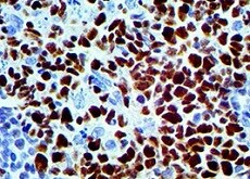
Secretagogin in Human Pancreas.
Secretagogin was detected in immersion fixed paraffin-embedded sections of human pancreas using Mouse Anti-Human Secretagogin Monoclonal Antibody (Catalog # MAB4878) at 15 µg/mL overnight at 4 °C. Before incubation with the primary antibody, tissue was subjected to heat-induced epitope retrieval using Antigen Retrieval Reagent-Basic (Catalog # CTS013). Tissue was stained using the Anti-Mouse HRP-DAB Cell & Tissue Staining Kit (brown; Catalog # CTS002) and counterstained with hematoxylin (blue). Specific staining was localized to islets. View our protocol for Chromogenic IHC Staining of Paraffin-embedded Tissue Sections.
Detection of Human and Mouse Secretagogin by Simple WesternTM.
Simple Western lane view shows lysates of beta TC‑6 mouse beta cell insulinoma cell line and human pancreas tissue, loaded at 0.5 mg/mL. A specific band was detected for Secretagogin at approximately 37 kDa (as indicated) using 10 µg/mL of Mouse Anti-Human Secretagogin Monoclonal Antibody (Catalog # MAB4878). This experiment was conducted under reducing conditions and using the 12-230 kDa separation system.Applications for Human Secretagogin Antibody (778518)
Application
Recommended Usage
Immunohistochemistry
8-25 µg/mL
Sample: Immersion fixed paraffin-embedded sections of human pancreas
Sample: Immersion fixed paraffin-embedded sections of human pancreas
Simple Western
10 µg/mL
Sample: beta TC‑6 mouse beta cell insulinoma cell line and human pancreas tissue
Sample: beta TC‑6 mouse beta cell insulinoma cell line and human pancreas tissue
Western Blot
1 µg/mL
Sample: beta TC‑6 mouse beta cell insulinoma cell line and human pancreas tissue
Sample: beta TC‑6 mouse beta cell insulinoma cell line and human pancreas tissue
Reviewed Applications
Read 1 review rated 5 using MAB4878 in the following applications:
Formulation, Preparation, and Storage
Purification
Protein A or G purified from hybridoma culture supernatant
Reconstitution
Sterile PBS to a final concentration of 0.5 mg/mL. For liquid material, refer to CoA for concentration.
Loading...
Formulation
Lyophilized from a 0.2 μm filtered solution in PBS with Trehalose. *Small pack size (SP) is supplied either lyophilized or as a 0.2 µm filtered solution in PBS.
Shipping
Lyophilized product is shipped at ambient temperature. Liquid small pack size (-SP) is shipped with polar packs. Upon receipt, store immediately at the temperature recommended below.
Stability & Storage
Use a manual defrost freezer and avoid repeated freeze-thaw cycles.
- 12 months from date of receipt, -20 to -70 °C as supplied.
- 1 month, 2 to 8 °C under sterile conditions after reconstitution.
- 6 months, -20 to -70 °C under sterile conditions after reconstitution.
Calculators
Background: Secretagogin
Alternate Names
CALBL, SCGN, SECRET, SEGN, Setagin
Gene Symbol
SCGN
UniProt
Additional Secretagogin Products
Product Documents for Human Secretagogin Antibody (778518)
Certificate of Analysis
To download a Certificate of Analysis, please enter a lot or batch number in the search box below.
Note: Certificate of Analysis not available for kit components.
Product Specific Notices for Human Secretagogin Antibody (778518)
For research use only
Related Research Areas
Customer Reviews for Human Secretagogin Antibody (778518) (1)
5 out of 5
1 Customer Rating
Have you used Human Secretagogin Antibody (778518)?
Submit a review and receive an Amazon gift card!
$25/€18/£15/$25CAN/¥2500 Yen for a review with an image
$10/€7/£6/$10CAN/¥1110 Yen for a review without an image
Submit a review
Customer Images
Showing
1
-
1 of
1 review
Showing All
Filter By:
-
Application: Immunohistochemistry-ParaffinSample Tested: Pancreas tissueSpecies: HumanVerified Customer | Posted 04/05/2022human pancreas tissue stainingIncubated tissue overnight at 4C at 15 µg/mL

There are no reviews that match your criteria.
Protocols
Find general support by application which include: protocols, troubleshooting, illustrated assays, videos and webinars.
- Antigen Retrieval Protocol (PIER)
- Antigen Retrieval for Frozen Sections Protocol
- Appropriate Fixation of IHC/ICC Samples
- Cellular Response to Hypoxia Protocols
- Chromogenic IHC Staining of Formalin-Fixed Paraffin-Embedded (FFPE) Tissue Protocol
- Chromogenic Immunohistochemistry Staining of Frozen Tissue
- ClariTSA™ Fluorophore Kits
- Detection & Visualization of Antibody Binding
- Fluorescent IHC Staining of Frozen Tissue Protocol
- Graphic Protocol for Heat-induced Epitope Retrieval
- Graphic Protocol for the Preparation and Fluorescent IHC Staining of Frozen Tissue Sections
- Graphic Protocol for the Preparation and Fluorescent IHC Staining of Paraffin-embedded Tissue Sections
- Graphic Protocol for the Preparation of Gelatin-coated Slides for Histological Tissue Sections
- IHC Sample Preparation (Frozen sections vs Paraffin)
- Immunofluorescent IHC Staining of Formalin-Fixed Paraffin-Embedded (FFPE) Tissue Protocol
- Immunohistochemistry (IHC) and Immunocytochemistry (ICC) Protocols
- Immunohistochemistry Frozen Troubleshooting
- Immunohistochemistry Paraffin Troubleshooting
- Preparing Samples for IHC/ICC Experiments
- Preventing Non-Specific Staining (Non-Specific Binding)
- Primary Antibody Selection & Optimization
- Protocol for Heat-Induced Epitope Retrieval (HIER)
- Protocol for Making a 4% Formaldehyde Solution in PBS
- Protocol for VisUCyte™ HRP Polymer Detection Reagent
- Protocol for the Preparation & Fixation of Cells on Coverslips
- Protocol for the Preparation and Chromogenic IHC Staining of Frozen Tissue Sections
- Protocol for the Preparation and Chromogenic IHC Staining of Frozen Tissue Sections - Graphic
- Protocol for the Preparation and Chromogenic IHC Staining of Paraffin-embedded Tissue Sections
- Protocol for the Preparation and Chromogenic IHC Staining of Paraffin-embedded Tissue Sections - Graphic
- Protocol for the Preparation and Fluorescent IHC Staining of Frozen Tissue Sections
- Protocol for the Preparation and Fluorescent IHC Staining of Paraffin-embedded Tissue Sections
- Protocol for the Preparation of Gelatin-coated Slides for Histological Tissue Sections
- R&D Systems Quality Control Western Blot Protocol
- TUNEL and Active Caspase-3 Detection by IHC/ICC Protocol
- The Importance of IHC/ICC Controls
- Troubleshooting Guide: Immunohistochemistry
- Troubleshooting Guide: Western Blot Figures
- Western Blot Conditions
- Western Blot Protocol
- Western Blot Protocol for Cell Lysates
- Western Blot Troubleshooting
- Western Blot Troubleshooting Guide
- View all Protocols, Troubleshooting, Illustrated assays and Webinars
Loading...