Human SHBG (also known as ABP or androgen-binding protein) is a variably glycosylated, secreted non-disulfide linked homodimer that belongs to the SHBG family. Members of this small family have tandem repeats of 170 amino acid (aa) long laminin alpha chain G-like domains. Each SHBG monomer is a 47-53 kDa, 373 aa glycoprotein that contains one steroid-binding site in its N-terminal G-like domain. Male and female sex hormones are bound with equal affinity. SHBG is synthesized by the liver and circulates in blood; ABP (SHBG in the testis) is synthesized by Sertoli cells and circulates in the male reproductive system. Human SHBG shares approximately 67% aa sequence identity with mouse and rat SHBG.

Key Product Details
Species Reactivity
Validated:
Human
Cited:
Human
Applications
Validated:
Immunohistochemistry, Western Blot
Cited:
Western Blot, Immunoprecipitation
Label
Unconjugated
Antibody Source
Polyclonal Goat IgG
Loading...
Product Specifications
Immunogen
Mouse myeloma cell line NS0-derived recombinant human SHBG
Leu30-His402
Accession # P04278
Leu30-His402
Accession # P04278
Specificity
Detects human SHBG in direct ELISAs and Western blots.
Clonality
Polyclonal
Host
Goat
Isotype
IgG
Scientific Data Images for Human SHBG Antibody
SHBG in Human Liver.
SHBG was detected in immersion fixed paraffin-embedded sections of human liver using Goat Anti-Human SHBG Antigen Affinity-purified Polyclonal Antibody (Catalog # AF2656) at 1.7 µg/mL overnight at 4 °C. Tissue was stained using the Anti-Goat HRP-DAB Cell & Tissue Staining Kit (brown; Catalog # CTS008) and counterstained with hematoxylin (blue). Specific labeling was localized to the plasma membrane of hepatocytes. View our protocol for Chromogenic IHC Staining of Paraffin-embedded Tissue Sections.
Detection of Human SHBG by Western Blot.
Western blot shows lysates of Human Liver. PVDF membrane was probed with 1 µg/mL of Goat Anti-Human SHBG Antigen Affinity-purified Polyclonal Antibody (Catalog # AF2656) followed by HRP-conjugated Anti-Goat IgG Secondary Antibody (Catalog # HAF017). A specific band was detected for SHBG at approximately ~37 kDa (as indicated). This experiment was conducted under reducing conditions and using Western Blot Buffer Group 1.
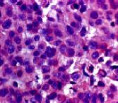
Detection of SHBG by Immunohistochemistry
Immunohistochemistry of SHBG expression in prostate cancer tissues.(A) Positive and negative control of SHBG in liver tissues. (B) Weak SHBG positivity is shown in a benign prostate tumor and strong SHBG immunoreactivity is revealed in a malignant Gleason score 8 cancer tissue. (C) Representative immunohistochemical images of different scores of prostate cancer samples are shown. Bar scale in all of the images is 150 µm. Image collected and cropped by CiteAb from the following open publication (https://pubmed.ncbi.nlm.nih.gov/23936228), licensed under a CC-BY license. Not internally tested by R&D Systems.Applications for Human SHBG Antibody
Application
Recommended Usage
Immunohistochemistry
5-15 µg/mL
Sample: Immersion fixed paraffin-embedded sections of human liver
Sample: Immersion fixed paraffin-embedded sections of human liver
Western Blot
1 µg/mL
Sample: Human Liver
Sample: Human Liver
Reviewed Applications
Read 2 reviews rated 5 using AF2656 in the following applications:
Formulation, Preparation, and Storage
Purification
Antigen Affinity-purified
Reconstitution
Reconstitute at 0.2 mg/mL in sterile PBS. For liquid material, refer to CoA for concentration.
Loading...
Formulation
Lyophilized from a 0.2 μm filtered solution in PBS with Trehalose. *Small pack size (SP) is supplied either lyophilized or as a 0.2 µm filtered solution in PBS.
Shipping
Lyophilized product is shipped at ambient temperature. Liquid small pack size (-SP) is shipped with polar packs. Upon receipt, store immediately at the temperature recommended below.
Stability & Storage
Use a manual defrost freezer and avoid repeated freeze-thaw cycles.
- 12 months from date of receipt, -20 to -70 °C as supplied.
- 1 month, 2 to 8 °C under sterile conditions after reconstitution.
- 6 months, -20 to -70 °C under sterile conditions after reconstitution.
Calculators
Background: SHBG
Long Name
Sex Hormone-binding Globulin
Alternate Names
ABP
Entrez Gene IDs
6462 (Human)
Gene Symbol
SHBG
UniProt
Additional SHBG Products
Product Documents for Human SHBG Antibody
Certificate of Analysis
To download a Certificate of Analysis, please enter a lot or batch number in the search box below.
Note: Certificate of Analysis not available for kit components.
Product Specific Notices for Human SHBG Antibody
For research use only
Citations for Human SHBG Antibody
 Powered by Bioz
Customer Reviews for Human SHBG Antibody (2)
5 out of 5
2 Customer Ratings
Have you used Human SHBG Antibody?
Submit a review and receive an Amazon gift card!
$25/€18/£15/$25CAN/¥2500 Yen for a review with an image
$10/€7/£6/$10CAN/¥1110 Yen for a review without an image
Submit a review
Customer Images

Showing
1
-
2 of
2 reviews
Showing All
Filter By:
-
Application: Immunohistochemistry-ParaffinSample Tested: Liver cancer tissueSpecies: HumanVerified Customer | Posted 04/04/2020

-
Application: Western BlotSample Tested: HepG2 human hepatocellular carcinoma cell line and Huh-7 human hepatoma cell lineSpecies: HumanVerified Customer | Posted 10/28/2018
There are no reviews that match your criteria.
Protocols
Find general support by application which include: protocols, troubleshooting, illustrated assays, videos and webinars.
- Antigen Retrieval Protocol (PIER)
- Antigen Retrieval for Frozen Sections Protocol
- Appropriate Fixation of IHC/ICC Samples
- Cellular Response to Hypoxia Protocols
- Chromogenic IHC Staining of Formalin-Fixed Paraffin-Embedded (FFPE) Tissue Protocol
- Chromogenic Immunohistochemistry Staining of Frozen Tissue
- ClariTSA™ Fluorophore Kits
- Detection & Visualization of Antibody Binding
- Fluorescent IHC Staining of Frozen Tissue Protocol
- Graphic Protocol for Heat-induced Epitope Retrieval
- Graphic Protocol for the Preparation and Fluorescent IHC Staining of Frozen Tissue Sections
- Graphic Protocol for the Preparation and Fluorescent IHC Staining of Paraffin-embedded Tissue Sections
- Graphic Protocol for the Preparation of Gelatin-coated Slides for Histological Tissue Sections
- IHC Sample Preparation (Frozen sections vs Paraffin)
- Immunofluorescent IHC Staining of Formalin-Fixed Paraffin-Embedded (FFPE) Tissue Protocol
- Immunohistochemistry (IHC) and Immunocytochemistry (ICC) Protocols
- Immunohistochemistry Frozen Troubleshooting
- Immunohistochemistry Paraffin Troubleshooting
- Preparing Samples for IHC/ICC Experiments
- Preventing Non-Specific Staining (Non-Specific Binding)
- Primary Antibody Selection & Optimization
- Protocol for Heat-Induced Epitope Retrieval (HIER)
- Protocol for Making a 4% Formaldehyde Solution in PBS
- Protocol for VisUCyte™ HRP Polymer Detection Reagent
- Protocol for the Preparation & Fixation of Cells on Coverslips
- Protocol for the Preparation and Chromogenic IHC Staining of Frozen Tissue Sections
- Protocol for the Preparation and Chromogenic IHC Staining of Frozen Tissue Sections - Graphic
- Protocol for the Preparation and Chromogenic IHC Staining of Paraffin-embedded Tissue Sections
- Protocol for the Preparation and Chromogenic IHC Staining of Paraffin-embedded Tissue Sections - Graphic
- Protocol for the Preparation and Fluorescent IHC Staining of Frozen Tissue Sections
- Protocol for the Preparation and Fluorescent IHC Staining of Paraffin-embedded Tissue Sections
- Protocol for the Preparation of Gelatin-coated Slides for Histological Tissue Sections
- R&D Systems Quality Control Western Blot Protocol
- TUNEL and Active Caspase-3 Detection by IHC/ICC Protocol
- The Importance of IHC/ICC Controls
- Troubleshooting Guide: Immunohistochemistry
- Troubleshooting Guide: Western Blot Figures
- Western Blot Conditions
- Western Blot Protocol
- Western Blot Protocol for Cell Lysates
- Western Blot Troubleshooting
- Western Blot Troubleshooting Guide
- View all Protocols, Troubleshooting, Illustrated assays and Webinars
Loading...