Siglecs (sialic acid binding Ig-like lectins) are I-type lectins that belong to the immunoglobulin superfamily. They are characterized by an N‑terminal Ig-like V-type domain which mediates sialic acid binding, followed by a varying number of Ig-like C2-type domains. Siglecs 5‑11 constitute the CD33/Siglec-3 related group, and are differentially expressed in the hematopoietic system (1‑3). Siglec-G is the apparent ortholog of human Siglec-10 (4). The human Siglec-10 cDNA encodes a 697 amino acid (aa) precursor that includes a 16 aa signal sequence, a 534 aa extracellular domain (ECD), a 21 aa transmembrane segment, and a 126 aa cytoplasmic domain. The ECD contains one Ig-like V‑type domain and four Ig-like C2-type domains, while the cytoplasmic domain contains two immunoreceptor tyrosine-based inhibitory motifs (ITIM) (5‑8). Five splice variants of human Siglec-10 differ in their deletions within the ECD. A potentially secreted sixth variant contains the Ig-like V-type domain followed by a 45 aa substitution (5‑7, 9). Within the ECD, human Siglec-10 is most closely related to Siglec-5 (42% aa sequence identity). It shares 63% aa sequence identity with mouse Siglec-G. Siglec-10 is expressed on eosinophils, neutrophils, monocytes, and B cells (5, 8) with some splice variants predominating in particular cell types and tissue locations (6, 7, 9). It is up‑regulated on eosinophils in mouse models of allergic respiratory inflammation (10). Siglec-10 binds sialated proteins and lipids in alpha 2,3 or alpha 2,6 linkage and shows a preference for GT1b gangliosides (7, 11). This binding can be modulated by cis interactions of Siglec-10 with sialated molecules expressed on the same cell (7). When tyrosine phosphorylated, the cytoplasmic ITIMs interact with phosphatases SHP-1 and SHP-2 to propagate inhibitory signals (5, 9).

Key Product Details
Species Reactivity
Validated:
Human
Cited:
Human, Porcine
Applications
Validated:
Western Blot, Blockade of Receptor-ligand Interaction, Flow Cytometry, CyTOF-ready
Cited:
Western Blot, Neutralization, Immunocytochemistry
Label
Unconjugated
Antibody Source
Polyclonal Goat IgG
Loading...
Product Specifications
Immunogen
Mouse myeloma cell line NS0-derived recombinant human Siglec-10
Met17-Thr546
Accession # Q96LC7
Met17-Thr546
Accession # Q96LC7
Specificity
Detects human Siglec‑10 in direct ELISAs and Western blots. In direct ELISAs and Western blots, approximately 5% cross‑reactivity with recombinant human (rh) Siglec-5 is observed and less than 1% cross-reactivity with rhSiglec-2, rhSiglec-3, rhSiglec-7, rhSiglec-9, and recombinant mouse Siglec-F is observed.
Clonality
Polyclonal
Host
Goat
Isotype
IgG
Endotoxin Level
<0.10 EU per 1 μg of the antibody by the LAL method.
Scientific Data Images for Human Siglec-10 Antibody
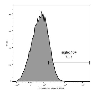
Detection of Siglec‑10 in Human B Cells by Flow Cytometry.
Human whole blood CD19+B cells were stained with Goat Anti-Human Siglec-10 Antigen Affinity-purified Polyclonal Antibody (Catalog # AF2130, filled histogram) or isotype control antibody (Catalog # AB-108-C, open histogram), followed by Phy-coerythrin-conjugated Anti-Goat IgG Secondary Anti-body (Catalog # F0107).Applications for Human Siglec-10 Antibody
Application
Recommended Usage
Blockade of Receptor-ligand Interaction
In a functional ELISA, 1-4 µg/mL of this antibody will block 50% of the binding of 1.5 μg/mL of biotinylated 6’‑Sialyllactose-Polyacrylamide to immobilized Recombinant Human Siglec-10 Fc Chimera (Catalog # 2130-SL) coated at 5 µg/mL (100 µL/well). At 30 μg/mL, this antibody will block >90% of the binding.
CyTOF-ready
Ready to be labeled using established conjugation methods. No BSA or other carrier proteins that could interfere with conjugation.
Flow Cytometry
2.5 µg/106 cells
Sample: Human whole blood CD19+ B cells
Sample: Human whole blood CD19+ B cells
Western Blot
0.1 µg/mL
Sample: Recombinant Human Siglec‑10 Fc Chimera (Catalog # 2130-SL)
Sample: Recombinant Human Siglec‑10 Fc Chimera (Catalog # 2130-SL)
Reviewed Applications
Read 3 reviews rated 4.3 using AF2130 in the following applications:
Flow Cytometry Panel Builder
Bio-Techne Knows Flow Cytometry
Save time and reduce costly mistakes by quickly finding compatible reagents using the Panel Builder Tool.
Advanced Features
- Spectra Viewer - Custom analysis of spectra from multiple fluorochromes
- Spillover Popups - Visualize the spectra of individual fluorochromes
- Antigen Density Selector - Match fluorochrome brightness with antigen density

Formulation, Preparation, and Storage
Purification
Antigen Affinity-purified
Reconstitution
Reconstitute at 0.2 mg/mL in sterile PBS. For liquid material, refer to CoA for concentration.
Loading...
Formulation
Lyophilized from a 0.2 μm filtered solution in PBS with Trehalose. *Small pack size (SP) is supplied either lyophilized or as a 0.2 µm filtered solution in PBS.
Shipping
Lyophilized product is shipped at ambient temperature. Liquid small pack size (-SP) is shipped with polar packs. Upon receipt, store immediately at the temperature recommended below.
Stability & Storage
Use a manual defrost freezer and avoid repeated freeze-thaw cycles.
- 12 months from date of receipt, -20 to -70 °C as supplied.
- 1 month, 2 to 8 °C under sterile conditions after reconstitution.
- 6 months, -20 to -70 °C under sterile conditions after reconstitution.
Calculators
Background: Siglec-10
References
- Crocker, P.R. (2005) Curr. Opin. Pharmacol. 5:431.
- Crocker, P.R. (2002) Curr. Opin. Struct. Biol. 12:609.
- Crocker, P.R. and J. Zhang (2002) Biochem. Soc. Symp. 69:83.
- Angata, T. et al. (2001) J. Biol. Chem. 276:45128.
- Whitney, G. et al. (2001) Eur. J. Biochem. 268:6083.
- Yousef, G.M. et al. (2001) Biochem. Biophys. Res. Commun. 284:900.
- Li, N. et al. (2001) J. Biol. Chem. 276:28106.
- Munday, J. et al. (2001) Biochem. J. 355:489.
- Kitzig, F. et al. (2002) Biochem. Biophys. Res. Commun. 296:355.
- Aizawa, H. et al. (2003) Genomics 82:521.
- Rapoport, E. et al. (2003) Bioorg. Med. Chem. Lett. 13:675.
Long Name
Sialic Acid Binding Ig-like Lectin 10
Alternate Names
Siglec10, SLG2
Entrez Gene IDs
89790 (Human)
Gene Symbol
SIGLEC10
UniProt
Additional Siglec-10 Products
Product Documents for Human Siglec-10 Antibody
Certificate of Analysis
To download a Certificate of Analysis, please enter a lot or batch number in the search box below.
Note: Certificate of Analysis not available for kit components.
Product Specific Notices for Human Siglec-10 Antibody
For research use only
Citations for Human Siglec-10 Antibody
 Powered by Bioz
Customer Reviews for Human Siglec-10 Antibody (3)
4.3 out of 5
3 Customer Ratings
Have you used Human Siglec-10 Antibody?
Submit a review and receive an Amazon gift card!
$25/€18/£15/$25CAN/¥2500 Yen for a review with an image
$10/€7/£6/$10CAN/¥1110 Yen for a review without an image
Submit a review
Customer Images
Showing
1
-
3 of
3 reviews
Showing All
Filter By:
-
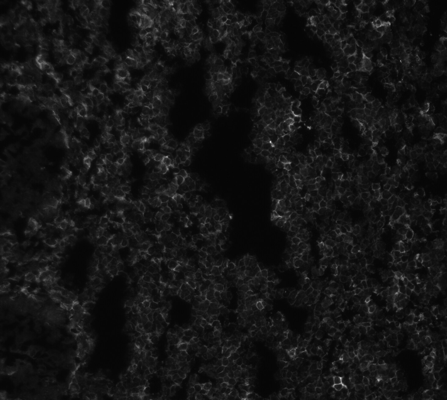
Application: Immunocytochemistry/ImmunofluorescenceSample Tested: Melanoma tissueSpecies: HumanVerified Customer | Posted 11/04/2020

-
Application: Block/NeutralizeSample Tested: NK cellSpecies: HumanVerified Customer | Posted 03/13/2020
-
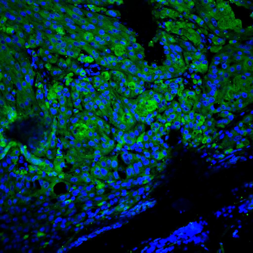
Application: Immunofluorescence - fresh-frozenSample Tested: Colon tissueSpecies: HumanVerified Customer | Posted 02/29/2020Siglec10 dilution 1:100, colon tissue
There are no reviews that match your criteria.
Protocols
Find general support by application which include: protocols, troubleshooting, illustrated assays, videos and webinars.
- 7-Amino Actinomycin D (7-AAD) Cell Viability Flow Cytometry Protocol
- Cellular Response to Hypoxia Protocols
- Extracellular Membrane Flow Cytometry Protocol
- Flow Cytometry Protocol for Cell Surface Markers
- Flow Cytometry Protocol for Staining Membrane Associated Proteins
- Flow Cytometry Staining Protocols
- Flow Cytometry Troubleshooting Guide
- Intracellular Flow Cytometry Protocol Using Alcohol (Methanol)
- Intracellular Flow Cytometry Protocol Using Detergents
- Intracellular Nuclear Staining Flow Cytometry Protocol Using Detergents
- Intracellular Staining Flow Cytometry Protocol Using Alcohol Permeabilization
- Intracellular Staining Flow Cytometry Protocol Using Detergents to Permeabilize Cells
- Propidium Iodide Cell Viability Flow Cytometry Protocol
- Protocol for Liperfluo
- Protocol for the Characterization of Human Th22 Cells
- Protocol for the Characterization of Human Th9 Cells
- Protocol: Annexin V and PI Staining by Flow Cytometry
- Protocol: Annexin V and PI Staining for Apoptosis by Flow Cytometry
- R&D Systems Quality Control Western Blot Protocol
- Troubleshooting Guide: Fluorokine Flow Cytometry Kits
- Troubleshooting Guide: Western Blot Figures
- Western Blot Conditions
- Western Blot Protocol
- Western Blot Protocol for Cell Lysates
- Western Blot Troubleshooting
- Western Blot Troubleshooting Guide
- View all Protocols, Troubleshooting, Illustrated assays and Webinars
Loading...