TBX3 belongs to the T-box family of transcription factors, which are involved in regulation of developmental processes. TBX3 acts as a transcriptional repressor regulating anterior/posterior axis of the forelimb. Mutations in TBX3 are associated with ulnar-mammary syndrome, affecting limb, apocrine gland, tooth, hair, and genital development. TBX3 has 3 splice isoforms.

Key Product Details
Species Reactivity
Validated:
Human
Cited:
Human, Mouse, Transgenic Mouse
Applications
Validated:
Western Blot, Immunocytochemistry
Cited:
Immunohistochemistry, Immunocytochemistry
Label
Unconjugated
Antibody Source
Polyclonal Goat IgG
Loading...
Product Specifications
Immunogen
E. coli-derived recombinant human TBX3
Ser321-Thr483
Accession # O15119
Ser321-Thr483
Accession # O15119
Specificity
Detects human TBX3 in direct ELISAs and Western blots. In direct ELISAs, less than 1% cross-reactivity with recombinant human (rh) TBX2, rhTBX6, and rhTBX20 is observed.
Clonality
Polyclonal
Host
Goat
Isotype
IgG
Scientific Data Images for Human TBX3 Antibody
Detection of Human TBX3 by Western Blot.
Western blot shows lysates of HeLa human cervical epithelial carcinoma cell line and Daudi human Burkitt's lymphoma cell line. PVDF membrane was probed with 1 µg/mL of Goat Anti-Human TBX3 Antigen Affinity-purified Polyclonal Antibody (Catalog # AF4509) followed by HRP-conjugated Anti-Goat IgG Secondary Antibody (HAF109). A specific band was detected for TBX3 at approximately 80 kDa (as indicated). This experiment was conducted under reducing conditions and using Immunoblot Buffer Group 3.
Detection of Human TBX3 by Western Blot.
Western blot shows lysates of LNCaP human prostate cancer cell line and HepG2 human hepatocellular carcinoma cell line. PVDF membrane was probed with 2 µg/mL of Goat Anti-Human TBX3 Antigen Affinity-purified Polyclonal Antibody (Catalog # AF4509) followed by HRP-conjugated Anti-Goat IgG Secondary Antibody (HAF017). A specific band was detected for TBX3 at approximately 80 kDa (as indicated). This experiment was conducted under reducing conditions and using Western Blot Buffer Group 1.
TBX3 in LNCaP Human Cell Line.
TBX3 was detected in immersion fixed LNCaP human prostate cancer cell line using Goat Anti-Human TBX3 Antigen Affinity-purified Polyclonal Antibody (Catalog # AF4509) at 5 µg/mL for 3 hours at room temperature. Cells were stained using the NorthernLights™ 557-conjugated Anti-Goat IgG Secondary Antibody (red; NL001) and counterstained with DAPI (blue). Specific staining was localized to nuclei. View our protocol for Fluorescent ICC Staining of Cells on Coverslips.Applications for Human TBX3 Antibody
Application
Recommended Usage
Immunocytochemistry
5-15 µg/mL
Sample: Immersion fixed ZR-75-1 human mammary ductal carcinoma cell line and LNCaP human prostate cancer cell line
Sample: Immersion fixed ZR-75-1 human mammary ductal carcinoma cell line and LNCaP human prostate cancer cell line
Western Blot
1-2 µg/mL
Sample: HeLa human cervical epithelial carcinoma cell line, Daudi human Burkitt's lymphoma cell line, LNCaP human prostate cancer cell line and HepG2 human hepatocellular carcinoma cell line
Sample: HeLa human cervical epithelial carcinoma cell line, Daudi human Burkitt's lymphoma cell line, LNCaP human prostate cancer cell line and HepG2 human hepatocellular carcinoma cell line
Reviewed Applications
Read 1 review rated 5 using AF4509 in the following applications:
Formulation, Preparation, and Storage
Purification
Antigen Affinity-purified
Reconstitution
Reconstitute at 0.2 mg/mL in sterile PBS. For liquid material, refer to CoA for concentration.
Loading...
Formulation
Lyophilized from a 0.2 μm filtered solution in PBS with Trehalose. *Small pack size (SP) is supplied either lyophilized or as a 0.2 µm filtered solution in PBS.
Shipping
Lyophilized product is shipped at ambient temperature. Liquid small pack size (-SP) is shipped with polar packs. Upon receipt, store immediately at the temperature recommended below.
Stability & Storage
Use a manual defrost freezer and avoid repeated freeze-thaw cycles.
- 12 months from date of receipt, -20 to -70 °C as supplied.
- 1 month, 2 to 8 °C under sterile conditions after reconstitution.
- 6 months, -20 to -70 °C under sterile conditions after reconstitution.
Calculators
Background: TBX3
Long Name
T-box Transcription Factor 3
Alternate Names
UMS, XHL
Gene Symbol
TBX3
UniProt
Additional TBX3 Products
Product Documents for Human TBX3 Antibody
Certificate of Analysis
To download a Certificate of Analysis, please enter a lot or batch number in the search box below.
Note: Certificate of Analysis not available for kit components.
Product Specific Notices for Human TBX3 Antibody
For research use only
Related Research Areas
Citations for Human TBX3 Antibody
 Powered by Bioz
Customer Reviews for Human TBX3 Antibody (1)
5 out of 5
1 Customer Rating
Have you used Human TBX3 Antibody?
Submit a review and receive an Amazon gift card!
$25/€18/£15/$25CAN/¥2500 Yen for a review with an image
$10/€7/£6/$10CAN/¥1110 Yen for a review without an image
Submit a review
Customer Images
Showing
1
-
1 of
1 review
Showing All
Filter By:
-
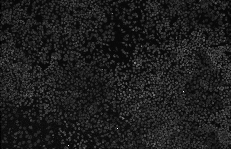
Application: ImmunocytochemistrySample Tested: R1 wild type Mouse embryonic Stem CellsSpecies: MouseVerified Customer | Posted 08/04/2016Tbx3 in R1 wild type mouse embryonic stem cells

There are no reviews that match your criteria.
Protocols
Find general support by application which include: protocols, troubleshooting, illustrated assays, videos and webinars.
- Appropriate Fixation of IHC/ICC Samples
- Cellular Response to Hypoxia Protocols
- ClariTSA™ Fluorophore Kits
- Detection & Visualization of Antibody Binding
- ICC Cell Smear Protocol for Suspension Cells
- ICC Immunocytochemistry Protocol Videos
- ICC for Adherent Cells
- Immunocytochemistry (ICC) Protocol
- Immunocytochemistry Troubleshooting
- Immunofluorescence of Organoids Embedded in Cultrex Basement Membrane Extract
- Immunohistochemistry (IHC) and Immunocytochemistry (ICC) Protocols
- Preparing Samples for IHC/ICC Experiments
- Preventing Non-Specific Staining (Non-Specific Binding)
- Primary Antibody Selection & Optimization
- Protocol for VisUCyte™ HRP Polymer Detection Reagent
- Protocol for the Fluorescent ICC Staining of Cell Smears - Graphic
- Protocol for the Fluorescent ICC Staining of Cultured Cells on Coverslips - Graphic
- Protocol for the Preparation and Fluorescent ICC Staining of Cells on Coverslips
- Protocol for the Preparation and Fluorescent ICC Staining of Non-adherent Cells
- Protocol for the Preparation and Fluorescent ICC Staining of Stem Cells on Coverslips
- Protocol for the Preparation of a Cell Smear for Non-adherent Cell ICC - Graphic
- R&D Systems Quality Control Western Blot Protocol
- TUNEL and Active Caspase-3 Detection by IHC/ICC Protocol
- The Importance of IHC/ICC Controls
- Troubleshooting Guide: Western Blot Figures
- Western Blot Conditions
- Western Blot Protocol
- Western Blot Protocol for Cell Lysates
- Western Blot Troubleshooting
- Western Blot Troubleshooting Guide
- View all Protocols, Troubleshooting, Illustrated assays and Webinars
Loading...