Thrombospondin-2 (TSP-2) is a 150 kDa calcium-binding protein that modulates cellular interactions with extracellular matrix. Thrombospondin-1 and -2 constitute subgroup A thrombospondin family members and form disulfide-linked homotrimers, whereas Thrombospondin-3, -4, and -5/COMP constitute subgroup B and form homopentamers (1-4). The human TSP-2 cDNA encodes a 1172 amino acid (aa) precursor that includes an 18 aa signal sequence followed by an N-terminal
heparin-binding domain, an oligomerization motif, one vWF-C domain, three TSP type-1 repeats, three EGF-like repeats, seven TSP type-3 repeats, and a lectin-like TSP C‑terminal domain (5). Human TSP-2 shares 88-90% aa sequence identity with bovine, mouse, and rat TSP-2. Within the TSP type-3 repeats and TSP C‑terminal domain, human TSP-2 shares 80% aa sequence identity with human TSP-1 and approximately 60% aa sequence identity with human TSP-3, -4, and -5/COMP. TSP-2 regulates collagen matrix formation by altering fibroblast behavior during development and in areas of tissue remodeling in the adult (6, 7). Trimerization of TSP-2 is required for the calcium-dependent cell attachment and spreading functions, while the heparin-binding domain is responsible for the destabilization of focal adhesion sites (8-10). The heparin-binding domain also mediates binding to Integrins alpha 3 beta 1 and alpha 6 beta 1 on microvascular endothelial cells (EC) and Integrin alpha 4 beta 1 on large blood vessel EC (11, 12). A fragment of TSP-2 (heparin-binding domain, oligomerization motif, and vWF-C domain) promotes EC survival, proliferation, and chemotaxis (11). Inclusion of the three TSP type-1 domains results in a molecule that inhibits VEGF-induced EC migration and vascular tube formation (13, 14). In vivo, full length TSP-2 blocks tumor angiogenesis and induces vascular EC apoptosis (13, 15). HPRG functions as an apparent decoy receptor by preventing interaction of TSP-2 with CD36 on macrophages and microvasculature EC (14). TSP-2 also binds MMP-2 and facilitates MMP-2 clearance by the scavenger receptor LRP (16).
Human Thrombospondin-2 Antibody (230934)
R&D Systems | Catalog # MAB1635

Key Product Details
Species Reactivity
Validated:
Human
Cited:
Human, Mouse
Applications
Validated:
Western Blot
Cited:
Immunohistochemistry, Western Blot
Label
Unconjugated
Antibody Source
Monoclonal Mouse IgG2A Clone # 230934
Loading...
Product Specifications
Immunogen
Mouse myeloma cell line NS0-derived recombinant human Thrombospondin-2
Gly19-Ile1172
Accession # P35442
Gly19-Ile1172
Accession # P35442
Specificity
Detects human Thrombospondin-2 in direct ELISAs and Western blots. In direct ELISAs, this antibody does not cross-react with recombinant human Thrombospondin‑1, -3, or -4.
Clonality
Monoclonal
Host
Mouse
Isotype
IgG2A
Scientific Data Images for Human Thrombospondin-2 Antibody (230934)
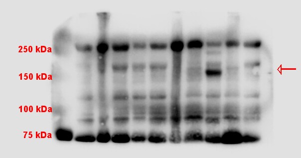
Detection of Mouse Thrombospondin-2 by Western Blot
Endothelial cells and TGF beta are not affected by cardiac Thbs1 overexpression.a Quantification of capillary number per mm2 of tissue from histological sections of tTA cont. and Thbs1 DTG hearts stained with isolectin B4 at 6 weeks of age. b Quantification of endothelial cell proliferation as measured by EdU incorporation co-labeled with CD31 in tTA cont. and Thbs1 DTG hearts at 6 weeks of age. c Quantification of endothelial cell apoptosis detected by TUNEL staining co-labeled with isolectin B4 in tTA cont. and Thbs1 DTG hearts at 6 weeks of age. d ELISA-based quantification of total TGF beta and e active TGF beta in protein extracts from tTA cont. or Thbs1 DTG hearts at 6 weeks of age. f Schematic diagram of WT Thbs1 domain structure and the Thbs1 delta t1 mutant lacking the Thbs1 type-1 repeat domain region. g Representative western blot analysis for Thbs1 from total protein extracts (Total) and extracellular matrix (ECM) extracts from hearts of tTA cont., Thbs1 DTG, and Thbs1 DTG delta t1 mice at 4 weeks of age. Vinculin is presented as cytosolic control. Coomassie stained (Coom.) gel is shown as loading control. h VW/BW ratio at 4 weeks of age in the indicated groups of mice. *P < 0.0001 versus tTA cont.; statistical analysis was performed using one-way ANOVA and Tukey multiple comparisons test. i Kaplan–Meier survival plot of tTA cont., Thbs1 DTG, and Thbs1 delta t1 DTG animals. *P < 0.0001 vs tTA cont. #P < 0.0001 vs Thbs1 DTG; both analyzed by two-tailed log-rank test. The same data from Fig. 2e are shown again here for tTA cont. and Thbs1 DTG mice (same strain and ages and sex ratio mix). j Representative western blots for Thbs2 and Gapdh as loading control, from heart protein extracts from tTA cont. and Thbs2 DTG mice at 8 weeks of age. k Heart weight (HW)/BW ratio, and l FS percentage at 8 weeks of age from tTA cont. and Thbs2 DTG mice. The number of biologically independent animals analyzed is indicated on each graph. Error bars are ±standard error of the mean. Source data are provApplications for Human Thrombospondin-2 Antibody (230934)
Application
Recommended Usage
Western Blot
1 µg/mL
Sample: Recombinant Human Thrombospondin‑2 (Catalog # 1635-T2)
Sample: Recombinant Human Thrombospondin‑2 (Catalog # 1635-T2)
Reviewed Applications
Read 1 review rated 4 using MAB1635 in the following applications:
Formulation, Preparation, and Storage
Purification
Protein A or G purified from hybridoma culture supernatant
Reconstitution
Reconstitute at 0.5 mg/mL in sterile PBS. For liquid material, refer to CoA for concentration.
Loading...
Formulation
Lyophilized from a 0.2 μm filtered solution in PBS with Trehalose. *Small pack size (SP) is supplied either lyophilized or as a 0.2 µm filtered solution in PBS.
Shipping
Lyophilized product is shipped at ambient temperature. Liquid small pack size (-SP) is shipped with polar packs. Upon receipt, store immediately at the temperature recommended below.
Stability & Storage
Use a manual defrost freezer and avoid repeated freeze-thaw cycles.
- 12 months from date of receipt, -20 to -70 °C as supplied.
- 1 month, 2 to 8 °C under sterile conditions after reconstitution.
- 6 months, -20 to -70 °C under sterile conditions after reconstitution.
Calculators
Background: Thrombospondin-2
References
- Elzie, C.A. and J.E. Murphy-Ullrich (2004) Int. J. Biochem. Cell Biol. 36:1090.
- Armstrong, L.C. and P. Bornstein (2003) Matrix Biol. 22:63.
- Murphy-Ullrich, J.E. (2001) J. Clin. Invest. 107:785.
- Bornstein, P. and E.H. Sage (2002) Curr. Opin. Cell Biol. 14:608.
- LaBell, T.L. and P.H. Byers (1993) Genomics 17:225.
- Kyriakides, T.R. et al. (1998) J. Histochem. Cytochem. 46:1007.
- Kyriakides, T.R. et al. (1998) J. Cell Biol. 140:419.
- Anilkumar, N. et al. (2002) J. Cell Sci. 115:2357.
- Misenheimer, T.M. et al. (2003) Biochemistry 42:5125.
- Murphy-Ullrich, J.E. et al. (1993) J. Biol. Chem. 268:26784.
- Calzada, M.J. et al. (2004) Circ. Res. 94:462.
- Calzada, M.J. et al. (2003) J. Biol. Chem. 278:40679.
- Noh, Y-H. et al. (2003) J. Invest. Dermatol. 121:1536.
- Simantov, R. et al. (2005) Matrix Biol. 24:27.
- Streit, M. et al. (1999) Proc. Natl. Acad. Sci. 96:14888.
- Yang, Z. et al. (2001) J. Biol. Chem. 276:8403.
Alternate Names
THBS2, Thrombospondin2, TSP-2
Entrez Gene IDs
7058 (Human)
Gene Symbol
THBS2
UniProt
Additional Thrombospondin-2 Products
Product Documents for Human Thrombospondin-2 Antibody (230934)
Certificate of Analysis
To download a Certificate of Analysis, please enter a lot or batch number in the search box below.
Note: Certificate of Analysis not available for kit components.
Product Specific Notices for Human Thrombospondin-2 Antibody (230934)
For research use only
Related Research Areas
Citations for Human Thrombospondin-2 Antibody (230934)
 Powered by Bioz
Customer Reviews for Human Thrombospondin-2 Antibody (230934) (1)
4 out of 5
1 Customer Rating
Have you used Human Thrombospondin-2 Antibody (230934)?
Submit a review and receive an Amazon gift card!
$25/€18/£15/$25CAN/¥2500 Yen for a review with an image
$10/€7/£6/$10CAN/¥1110 Yen for a review without an image
Submit a review
Customer Images
Showing
1
-
1 of
1 review
Showing All
Filter By:
-
Application: Western BlotSample Tested: Heparin PlasmaSpecies: HumanVerified Customer | Posted 06/30/2016Lanes in the WB are alternating between normal and diseased patients. Bands for thrombospondin-2 were resolved at the predicted MW; however, there was no difference between patients in this experiment. The results were clean, though, so I would consider using this for other applications in the future.

There are no reviews that match your criteria.
Protocols
Find general support by application which include: protocols, troubleshooting, illustrated assays, videos and webinars.
- Cellular Response to Hypoxia Protocols
- R&D Systems Quality Control Western Blot Protocol
- Troubleshooting Guide: Western Blot Figures
- Western Blot Conditions
- Western Blot Protocol
- Western Blot Protocol for Cell Lysates
- Western Blot Troubleshooting
- Western Blot Troubleshooting Guide
- View all Protocols, Troubleshooting, Illustrated assays and Webinars
Loading...
Associated Pathways
