Thrombospondin-4 (THSP4) is a 140 kDa calcium-binding protein that interacts with other extracellular matrix molecules and modulates the activity of various cell types. THSP1 and THSP2 constitute subgroup A and form disulfide-linked homotrimers, whereas THSP3, THSP4, and THSP5/COMP constitute subgroup B and form pentamers (1, 2). The human THSP4 cDNA encodes a 961 amino acid (aa) precursor that includes a 26 aa signal sequence followed by an N-terminal heparin-binding domain, a coiled-coil motif, four EGF-like repeats, seven THSP type-3 repeats (one with an RGD motif), and a THSP C‑terminal domain (3). Human THSP4 shares 93% aa sequence identity with mouse and rat THSP4. Within the THSP type-3 repeats and the THSP C‑terminal domain, human THSP4 shares 79% aa sequence identity with THSP3 and COMP, and 58% aa sequence identity with THSP1 and THSP2. The coiled-coil motif mediates pentamer formation with COMP, either homotypically or heterotypically (3-6). THSP4 binds a variety of matrix proteins including collagens I, II, III, V, laminin-1, fibronectin, and matrilin-2 (4). Interactions of THSP4 with non-collagenous proteins are independent of divalent cations, while interactions with collagenous proteins are enhanced in the presence of zinc (4). THSP4 is expressed in heart, skeletal muscle, vascular smooth muscle, and vascular endothelial cells (7-9). It accumulates at neuromuscular junctions and synapse-rich regions and is upregulated in muscle by experimental denervation (8). THSP4 mediates the adhesion of motor and sensory neurons and promotes neurite outgrowth (8). A polymorphism of THSP4 (A387P) is associated with early coronary artery disease (10-12). Unlike wild type THSP4, the A387P variant does not support HUVEC attachment and spreading (9). Integrin alpha M/ beta 2 enables activated neutrophil adhesion to both the variant A387P and wild type THSP4, although the A387P variant induces a greater release of pro-inflammatory molecules (13).

Key Product Details
Validated by
Biological Validation
Species Reactivity
Validated:
Human
Cited:
Human, Mouse, Rat, Primate - Macaca mulatta (Rhesus Macaque), Primate - Macaca nemestrina (Southern Pig-tailed Macaque), Primate - Pan troglodytes (Chimpanzee)
Applications
Validated:
Western Blot, Simple Western
Cited:
Immunohistochemistry, Immunohistochemistry-Paraffin, Immunohistochemistry-Frozen, Western Blot
Label
Unconjugated
Antibody Source
Polyclonal Goat IgG
Loading...
Product Specifications
Immunogen
Mouse myeloma cell line NS0-derived recombinant human Thrombospondin-4
Ala22-Asn961 (Pro276Ala, Ala420Val)
Accession # P35443
Ala22-Asn961 (Pro276Ala, Ala420Val)
Accession # P35443
Specificity
Detects human Thrombospondin-4 in direct ELISAs and Western blots. In direct ELISAs, approximately 50% cross-reactivity with recombinant mouse Thrombospondin-4 is observed and less than 5% cross-reactivity with recombinant human (rh) Thrombospondin-1, rhThrombospondin-2, and rhThrombospondin-3 is observed.
Clonality
Polyclonal
Host
Goat
Isotype
IgG
Scientific Data Images for Human Thrombospondin-4 Antibody
Detection of Human Thrombospondin‑4 by Western Blot.
Western blot shows lysates of human heart tissue. PVDF membrane was probed with 0.2 µg/mL of Goat Anti-Human Thrombospondin-4 Antigen Affinity-purified Polyclonal Antibody (Catalog # AF2390) followed by HRP-conjugated Anti-Goat IgG Secondary Antibody (Catalog # HAF109). A specific band was detected for Thrombospondin-4 at approximately 130kDa (as indicated). This experiment was conducted under reducing conditions and using Immunoblot Buffer Group 1.
Detection of Human Thrombospondin‑4 by Simple WesternTM.
Simple Western lane view shows lysates of human heart tissue, loaded at 0.2 mg/mL. A specific band was detected for Thrombospondin-4 at approximately 158 kDa (as indicated) using 2 µg/mL of Goat Anti-Human Thrombospondin-4 Antigen Affinity-purified Polyclonal Antibody (Catalog # AF2390) followed by 1:50 dilution of HRP-conjugated Anti-Goat IgG Secondary Antibody (Catalog # HAF109). This experiment was conducted under reducing conditions and using the 12-230 kDa separation system.
Detection of Mouse Thrombospondin-4 by Immunocytochemistry/Immunofluorescence
TSP-4 promotes accumulation of macrophages in peritoneal tissue of mice with LPS-induced peritonitis.a The number of macrophages in peritoneal cavity in mice with LPS-induced peritonitis. *p < 0.05, n = 5. b TSP-4 expression in macrophages from the peritoneal cavity lavage (left panel) and in the peritoneal tissue. QRT-PCR, fold increase (RQ) over the values in control mice injected with PBS; n = 3; *p < 0.05. c Macrophages and TSP-4 in peritoneal tissue of WT and P387-TSP-4-KI mice with LPS-induced peritonitis. Immunofluorescence; blue = nuclei (DAPI), green = macrophages (anti-CD68), red = TSP-4 (anti-TSP-4). Scale bar is 20 µm. Image collected and cropped by CiteAb from the following publication (https://pubmed.ncbi.nlm.nih.gov/31974349), licensed under a CC-BY license. Not internally tested by R&D Systems.
Detection of Mouse Thrombospondin-4 by Immunocytochemistry/Immunofluorescence
LPS induces TSP-4 in cultured macrophages.a Cultured macrophage-like RAW264.7 and cells and mouse bone-marrow-derived macrophages (BMDM) were treated with LPS (0.5 μg/ml for 1–24 h); western blotting with anti-TSP-4 Ab. b Cultured RAW264.7 (upper panels) and BMDM (lower panels) treated with LPS for 24 h were stained with anti-TSP-4 Ab; green = TSP-4, blue = nuclei, DAPI. Scale bar is 10 µm. c RAW 264.7 and BMDM cells were treated with LPS (0.5 μg/mL) for 24 h, and qRT-PCR was done (control = PBS treated); d RAW 264.7 cells were treated with CHX (25 μM) for 1–5 h and CHX (25 μM) + LPS (0.5 μg/mL) for 1–3 h followed by western blot detection of TSP-4 ( beta -actin as loading controls). CHX was added at time 0, followed by addition of LPS. Image collected and cropped by CiteAb from the following publication (https://pubmed.ncbi.nlm.nih.gov/31974349), licensed under a CC-BY license. Not internally tested by R&D Systems.Applications for Human Thrombospondin-4 Antibody
Application
Recommended Usage
Simple Western
2 µg/mL
Sample: Human heart tissue
Sample: Human heart tissue
Western Blot
0.2 µg/mL
Sample: Human heart tissue
Sample: Human heart tissue
Reviewed Applications
Read 6 reviews rated 4.3 using AF2390 in the following applications:
Formulation, Preparation, and Storage
Purification
Antigen Affinity-purified
Reconstitution
Reconstitute at 0.2 mg/mL in sterile PBS. For liquid material, refer to CoA for concentration.
Loading...
Formulation
Lyophilized from a 0.2 μm filtered solution in PBS with Trehalose. *Small pack size (SP) is supplied either lyophilized or as a 0.2 µm filtered solution in PBS.
Shipping
Lyophilized product is shipped at ambient temperature. Liquid small pack size (-SP) is shipped with polar packs. Upon receipt, store immediately at the temperature recommended below.
Stability & Storage
Use a manual defrost freezer and avoid repeated freeze-thaw cycles.
- 12 months from date of receipt, -20 to -70 °C as supplied.
- 1 month, 2 to 8 °C under sterile conditions after reconstitution.
- 6 months, -20 to -70 °C under sterile conditions after reconstitution.
Calculators
Background: Thrombospondin-4
References
- Adams, J.C. and J. Lawler (2004) Int. J. Biochem. Cell Biol. 36:961.
- Stenina, O.I. et al. (2004) Int. J. Biochem. Cell Biol. 36:1013.
- Lawler, J. et al. (1995) J. Biol. Chem. 270:2809.
- Narouz-Ott, L. et al. (2000) J. Biol. Chem. 275:37110.
- Hauser, N. et al. (1995) FEBS Lett. 368:307.
- Sodersten, F. et al. (2006) Connect. Tissue Res. 47:85.
- Lawler, J. et al. (1993) J. Cell Biochem. 120:1059.
- Arber, S. and P. Caroni (1995) J. Cell Biol. 131:1083.
- Stenina, O.I. et al. (2003) Circulation 108:1514.
- Topol, E.J. et al. (2001) Circulation 104:2641.
- Wessel, J. et al. (2004) Am. Heart J. 147:905.
- Stenina, O.I. et al. (2005) FASEB J. 19:1893.
- Pluskota, E. et al. (2005) Blood 106:3970.
Alternate Names
THBS4, Thrombospondin4, TSP-4
Gene Symbol
THBS4
UniProt
Additional Thrombospondin-4 Products
Product Documents for Human Thrombospondin-4 Antibody
Certificate of Analysis
To download a Certificate of Analysis, please enter a lot or batch number in the search box below.
Note: Certificate of Analysis not available for kit components.
Product Specific Notices for Human Thrombospondin-4 Antibody
For research use only
Related Research Areas
Citations for Human Thrombospondin-4 Antibody
 Powered by Bioz
Customer Reviews for Human Thrombospondin-4 Antibody (6)
4.3 out of 5
6 Customer Ratings
Have you used Human Thrombospondin-4 Antibody?
Submit a review and receive an Amazon gift card!
$25/€18/£15/$25CAN/¥2500 Yen for a review with an image
$10/€7/£6/$10CAN/¥1110 Yen for a review without an image
Submit a review
Customer Images
Showing
1
-
5 of
6 reviews
Showing All
Filter By:
-
Application: ELISASample Tested: Serum and PlasmaSpecies: HumanVerified Customer | Posted 07/01/2024

-
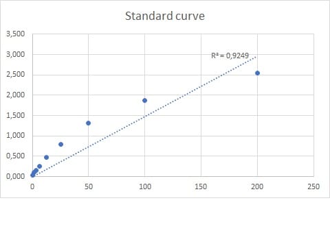
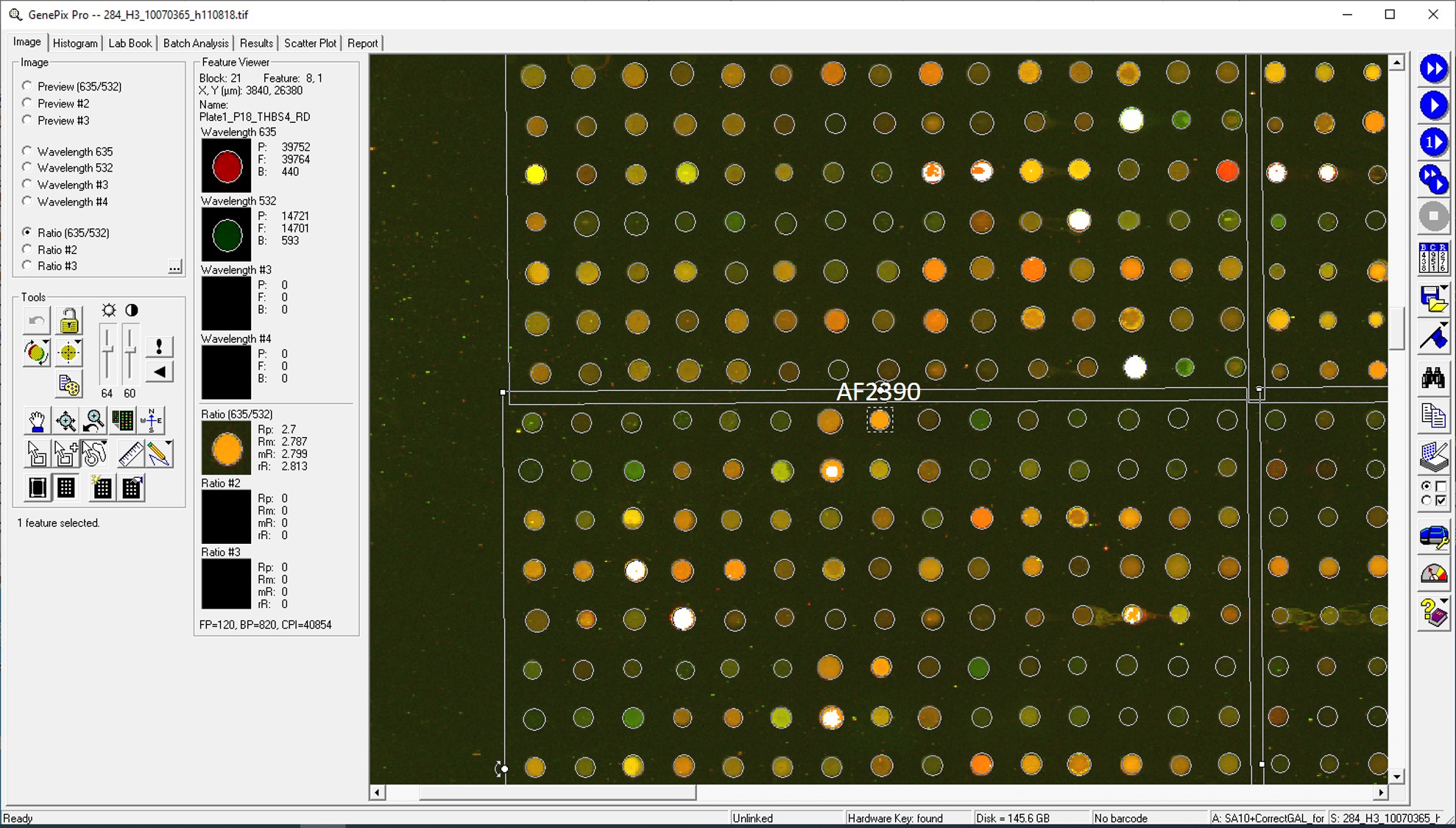
Application: MicroarraysSample Tested: EDTA PlasmaSpecies: HumanVerified Customer | Posted 01/14/2021
-
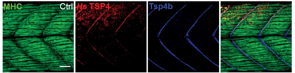
Application: ImmunohistochemistrySample Tested: EmbryoSpecies: ZebrafishVerified Customer | Posted 03/14/2020The antibody was used to detect human TSP4 protein (Red channel in the figure) that was injected into a zebrafish embryo. The antibody did not cross-react with zebrafish Tsp4 protein. The data has been published (eLife 2014;3:e02372).
-
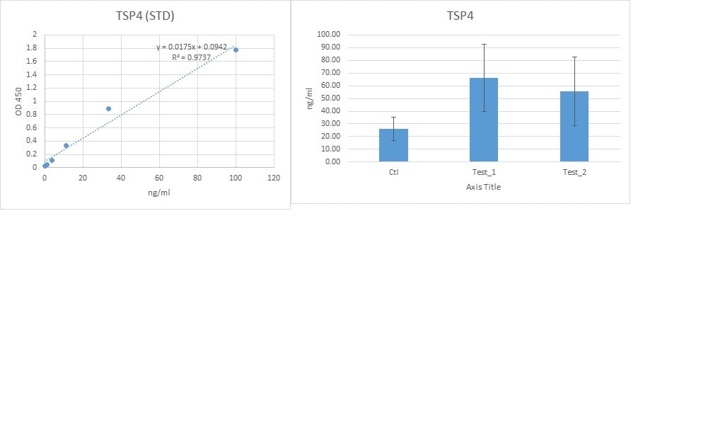
Application: ELISASample Tested: Serum and PlasmaSpecies: HumanVerified Customer | Posted 10/04/2019We used this antibody for a sandwich ELISA in combination with mAb (MAB2390)) and protein (2390-TH). This combination works very well for detecting the TSP4 in human serum and plasma
-
Application: LuminexSample Tested: SerumSpecies: HumanVerified Customer | Posted 11/08/2018
-
Application: MicroarraySample Tested: EDTA PlasmaSpecies: HumanVerified Customer | Posted 10/31/2018
There are no reviews that match your criteria.
Protocols
Find general support by application which include: protocols, troubleshooting, illustrated assays, videos and webinars.
- Cellular Response to Hypoxia Protocols
- R&D Systems Quality Control Western Blot Protocol
- Troubleshooting Guide: Western Blot Figures
- Western Blot Conditions
- Western Blot Protocol
- Western Blot Protocol for Cell Lysates
- Western Blot Troubleshooting
- Western Blot Troubleshooting Guide
- View all Protocols, Troubleshooting, Illustrated assays and Webinars
Loading...
Associated Pathways
