TSG-6 (TNF-stimulated Gene 6), also known as TNFIP6 is a secreted, 35‑39 kDa group A member of the LINK-Module superfamily of proteins (1‑4). Human TSG-6 is synthesized as a 277 amino acid (aa) precursor. It contains a 17 aa signal sequence and a 260 aa mature region (5, 6). The mature region shows an N-terminal LINK module (amino acids 36‑129) and a C-terminal CUB (C1s/C1r; urchin embryonic growth factor; BMP1) domain (amino acids 135‑247). Link modules are alpha -helical, beta ‑sheet structures that bind hyaluronan (HA) and participate in extracellular matrix (ECM) assembly (7). Mature human TSG-6 shares 94% aa identity with both mouse and canine TSG-6. Cells reported to express TSG-6 include activated fibroblasts, synoviocytes, chondrocytes, neutrophils, proximal tubular epithelium, bronchial epithelium, endothelium, and visceral, plus vascular smooth muscle (2, 8). TSG-6 has multiple functions, many of which involve the ECM. It is suggested to stabilize HA-rich ECM. It does so by serving as an intermediary, or link, between the individual subunits of extracellular decameric pentraxin 3 and the surrounding hyaluronan matrix (9). It also provides structure and organization to hyaluronan. This is accomplished by a TSG-6 mediated transfer of an 80‑85 kDa HC subunit from I alpha I (inter‑ alpha ‑inhibitor) to HA. I alpha I is a four-component, 225 kDa serine protease inhibitor. It contains a protease inhibitor subunit (bikunin), two independent, accompaning protein chains (HC1 and HC2), and a short chondroitin sulfate linking moiety. TSG-6 is a cation-dependent catalyst for the removal, transfer, and subsequent covalent linkage of HC 1/2 to surrounding HA. This provides substance and reinforcement to the ECM (1, 2, 10‑12). The disassembly of I alpha I also leads to free bikunin, which in the “free” state becomes a potent inhibitor of serine proteases (8).

Key Product Details
Species Reactivity
Validated:
Human
Cited:
Human, Mouse
Applications
Validated:
Western Blot, Immunocytochemistry
Cited:
Immunohistochemistry-Frozen, Western Blot
Label
Unconjugated
Antibody Source
Polyclonal Goat IgG
Loading...
Product Specifications
Immunogen
Mouse myeloma cell line NS0-derived recombinant human TSG-6
Trp18-Leu277
Accession # P98066
Trp18-Leu277
Accession # P98066
Specificity
Detects human TSG-6 in direct ELISAs and Western blots. In direct ELISAs and Western blots, approximately 70% cross-reactivity with recombinant mouse TSG-6 is observed and less than 2% cross-reactivity with recombinant human TSG-14 is observed.
Clonality
Polyclonal
Host
Goat
Isotype
IgG
Scientific Data Images for Human TSG-6 Antibody
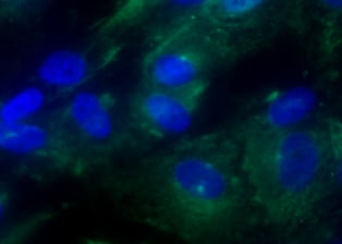
TSG‑6 in human PBMCs.
TSG-6 was detected in immersion fixed human peripheral blood mononuclear cells (PBMCs) using Goat Anti-Human TSG-6 Antigen Affinity-purified Polyclonal Antibody (Catalog # AF2104) at 15 µg/mL for 3 hours at room temperature. Cells were stained using the NorthernLights™ 557-conjugated Anti-Goat IgG Secondary Antibody (red; Catalog # NL001) and counterstained with DAPI (blue). Specific staining was localized to cytoplasm. View our protocol for Fluorescent ICC Staining of Cells on Coverslips.Applications for Human TSG-6 Antibody
Application
Recommended Usage
Immunocytochemistry
5-15 µg/mL
Sample: Immersion fixed human peripheral blood mononuclear cells (PBMCs)
Sample: Immersion fixed human peripheral blood mononuclear cells (PBMCs)
Western Blot
0.1 µg/mL
Sample: Recombinant Human TSG‑6 (Catalog # 2104-TS)
Sample: Recombinant Human TSG‑6 (Catalog # 2104-TS)
Reviewed Applications
Read 2 reviews rated 4.5 using AF2104 in the following applications:
Formulation, Preparation, and Storage
Purification
Antigen Affinity-purified
Reconstitution
Reconstitute at 0.2 mg/mL in sterile PBS. For liquid material, refer to CoA for concentration.
Loading...
Formulation
Lyophilized from a 0.2 μm filtered solution in PBS with Trehalose. *Small pack size (SP) is supplied either lyophilized or as a 0.2 µm filtered solution in PBS.
Shipping
Lyophilized product is shipped at ambient temperature. Liquid small pack size (-SP) is shipped with polar packs. Upon receipt, store immediately at the temperature recommended below.
Stability & Storage
Use a manual defrost freezer and avoid repeated freeze-thaw cycles.
- 12 months from date of receipt, -20 to -70 °C as supplied.
- 1 month, 2 to 8 °C under sterile conditions after reconstitution.
- 6 months, -20 to -70 °C under sterile conditions after reconstitution.
Calculators
Background: TSG-6
References
- Milner, C.M. et al. (2006) Biochem. Soc. Trans. 34:446.
- Milner, C.M. and A.J. Day (2003) J. Cell Sci. 116:1863.
- Wisnieewski, H-G. and J. Vilcek (2004) Cytokine Growth Factor Rev. 15:129.
- Blundell, C.D. et al. (2005) J. Biol. Chem. 280:18189.
- Lee, T.H. et al. (1990) Mol. Cell. Biol. 10:1982.
- Lee, T.H. et al. (1992) J. Cell Biol. 116:545.
- Kohda, D. et al. (1996) Cell 86:767.
- Forteza, R. et al. (2007) Am. J. Respir. Cell Mol. Biol. 36:20.
- Salustri, A. et al. (2003) Development 131:1577.
- Rugg, M.S. et al. (2005) J. Biol. Chem. 280:25674.
- Sanggaard, K.W. et al. (2006) Biochemistry 45:7661.
- Sanggaard, K.W. et al. (2005) J. Biol. Chem. 280:11936.
Long Name
Tumor Necrosis Factor-stimulated Gene Sequence 6
Alternate Names
TNFAIP6, TSG6
Gene Symbol
TNFAIP6
UniProt
Additional TSG-6 Products
Product Documents for Human TSG-6 Antibody
Certificate of Analysis
To download a Certificate of Analysis, please enter a lot or batch number in the search box below.
Note: Certificate of Analysis not available for kit components.
Product Specific Notices for Human TSG-6 Antibody
For research use only
Related Research Areas
Citations for Human TSG-6 Antibody
 Powered by Bioz
Customer Reviews for Human TSG-6 Antibody (2)
4.5 out of 5
2 Customer Ratings
Have you used Human TSG-6 Antibody?
Submit a review and receive an Amazon gift card!
$25/€18/£15/$25CAN/¥2500 Yen for a review with an image
$10/€7/£6/$10CAN/¥1110 Yen for a review without an image
Submit a review
Customer Images
Showing
1
-
2 of
2 reviews
Showing All
Filter By:
-
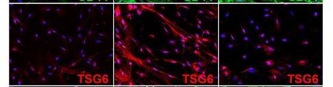
Application: Immunocytochemistry/ImmunofluorescenceSample Tested: mscSpecies: HumanVerified Customer | Posted 09/29/2022

-
Application: Immunocytochemistry/ImmunofluorescenceSample Tested: HUVEC human umbilical vein endothelial cellsSpecies: HumanVerified Customer | Posted 01/19/2019ICC on UMUC with 1 control and 2 conditions
There are no reviews that match your criteria.
Protocols
Find general support by application which include: protocols, troubleshooting, illustrated assays, videos and webinars.
- Appropriate Fixation of IHC/ICC Samples
- Cellular Response to Hypoxia Protocols
- ClariTSA™ Fluorophore Kits
- Detection & Visualization of Antibody Binding
- ICC Cell Smear Protocol for Suspension Cells
- ICC Immunocytochemistry Protocol Videos
- ICC for Adherent Cells
- Immunocytochemistry (ICC) Protocol
- Immunocytochemistry Troubleshooting
- Immunofluorescence of Organoids Embedded in Cultrex Basement Membrane Extract
- Immunohistochemistry (IHC) and Immunocytochemistry (ICC) Protocols
- Preparing Samples for IHC/ICC Experiments
- Preventing Non-Specific Staining (Non-Specific Binding)
- Primary Antibody Selection & Optimization
- Protocol for VisUCyte™ HRP Polymer Detection Reagent
- Protocol for the Fluorescent ICC Staining of Cell Smears - Graphic
- Protocol for the Fluorescent ICC Staining of Cultured Cells on Coverslips - Graphic
- Protocol for the Preparation and Fluorescent ICC Staining of Cells on Coverslips
- Protocol for the Preparation and Fluorescent ICC Staining of Non-adherent Cells
- Protocol for the Preparation and Fluorescent ICC Staining of Stem Cells on Coverslips
- Protocol for the Preparation of a Cell Smear for Non-adherent Cell ICC - Graphic
- R&D Systems Quality Control Western Blot Protocol
- TUNEL and Active Caspase-3 Detection by IHC/ICC Protocol
- The Importance of IHC/ICC Controls
- Troubleshooting Guide: Western Blot Figures
- Western Blot Conditions
- Western Blot Protocol
- Western Blot Protocol for Cell Lysates
- Western Blot Troubleshooting
- Western Blot Troubleshooting Guide
- View all Protocols, Troubleshooting, Illustrated assays and Webinars
Loading...