Thymic Stromal Lymphopoietin (TSLP) was originally identified as an activity from the conditioned medium of a mouse thymic stromal cell line that promoted the development of B cells (1-3). The activities of mouse TSLP overlap with, but are distinct from, those of mouse IL-7. Both mouse TSLP and IL-7 can co-stimulate growth of thymocytes and mature T cells, and support B lymphopoiesis in long-term cultures of fetal liver cells and bone-marrow cells. Whereas mouse IL-7 facilitates the development of B220+/IgM- pre-B cells, mouse TSLP promotes the development B220+/IgM+ B cells. Human TSLP was reported to preferentially stimulate myeloid cells; inducing the release of T cell-attracting chemokines from monocytes and enhancing the maturation of CD11c+ dendritic cells. Human TSLP cDNA encodes a 159 amino acid (aa) residue precursor protein with a 28 aa signal sequence (4, 5). Within the mature region, six of the seven cysteine residues present in the mouse TSLP involved in intramolecular disulfide bond formation are conserved in the human TSLP. Human TSLP shares approximately 43% aa sequence identity with mouse TSLP. By Northern blot analysis, human TSLP expression has been detected in many tissues with the highest expressions in heart, liver, testis and prostate. TSLP signals through a heterodimeric receptor complex that consists of IL-7 R alpha and the TSLP R, a member of the hemopoietin receptor family most closely related to R gamma c.

Key Product Details
Species Reactivity
Validated:
Human
Cited:
Human, Ovine
Applications
Validated:
Western Blot
Cited:
Immunohistochemistry-Frozen, Western Blot
Label
Unconjugated
Antibody Source
Monoclonal Mouse IgG2A Clone # 258136
Loading...
Product Specifications
Immunogen
E. coli-derived recombinant human TSLP
Tyr29-Gln159
Accession # Q969D9
Tyr29-Gln159
Accession # Q969D9
Specificity
Detects human TSLP in direct ELISAs and Western blots. Does not cross-react with recombinant mouse (rm) IL‑7 or rmTSLP.
Clonality
Monoclonal
Host
Mouse
Isotype
IgG2A
Scientific Data Images for Human TSLP Antibody (258136)
Detection of Human TSLP by Western Blot.
Western blot shows lysates of human lung and kidney tissue. PVDF membrane was probed with 2 µg/mL of Mouse Anti-Human TSLP Monoclonal Antibody (Catalog # MAB1398) followed by HRP-conjugated Anti-Mouse IgG Secondary Antibody (Catalog # HAF007). A specific band was detected for TSLP at approximately 18 kDa (as indicated). This experiment was conducted under non-reducing conditions only and using Immunoblot Buffer Group 1.
Detection of TSLP by Western Blot
Cytokine production induced by co-stimulation with TWEAK and TGF-beta 1 were steroid unresponsiveness. The mRNA levels of TSLP (A,C), CCL5 (D), and CCL17 (F) after 48 h of treatment and CCL2 (H) and IL-8 (J) after 2 h of treatment, analyzed by qRT–PCR. Data represent mean ± SD of two independent experiments. TSLP levels after 48 h of treatment assessed by immunoblotting (B, upper panel). The density of each band quantified by densitometry using ImageJ (version 6.1) (B, lower). The levels of CCL5 (E) and CCL17 (G) after 48 h of treatment, and CCL2 (I) and IL-8 (K) after 2 h of treatment in cell culture supernatants, analyzed by enzyme-linked immunosorbent assay (ELISA). Data represent the mean ± SD of three independent experiments. * p < 0.05, compared to untreated cultures as controls; † p < 0.05 compared with cultures in the absence of DEX. Image collected and cropped by CiteAb from the following open publication (https://www.mdpi.com/1422-0067/25/21/11625), licensed under a CC-BY license. Not internally tested by R&D Systems.
Detection of TSLP by Western Blot
MKP-1 is involved in steroid unresponsiveness induced by co-stimulation with TWEAK and TGF-beta 1 in BEAS-2B cells. The mRNA levels of MKP-1 (A), TSLP (C), and CCL5 (E), analyzed by qRT–PCR. Data represent mean ± SD of two independent experiments. MKP-1 and thymic stromal lymphopoietin (TSLP) expression after 48 h of treatment, assessed by immunoblotting (B,D, upper panel). Densitometric analysis of protein bands (B,D, lower). The level of CCL5 in cell culture supernatants after 48 h of treatment, analyzed using ELISA (F). Data represent mean ± SD of two independent experiments. * p < 0.05, compared to untreated cultures as controls; † p < 0.05 compared with cultures in the absence of DEX; ‡ p < 0.05 compared with control siRNA. Image collected and cropped by CiteAb from the following open publication (https://www.mdpi.com/1422-0067/25/21/11625), licensed under a CC-BY license. Not internally tested by R&D Systems.Applications for Human TSLP Antibody (258136)
Application
Recommended Usage
Western Blot
2 µg/mL
Sample: Human lung and kidney tissue under non-reducing conditions only
Sample: Human lung and kidney tissue under non-reducing conditions only
Reviewed Applications
Read 1 review rated 3 using MAB1398 in the following applications:
Formulation, Preparation, and Storage
Purification
Protein A or G purified from hybridoma culture supernatant
Reconstitution
Reconstitute at 0.5 mg/mL in sterile PBS. For liquid material, refer to CoA for concentration.
Loading...
Formulation
Lyophilized from a 0.2 μm filtered solution in PBS with Trehalose. *Small pack size (SP) is supplied either lyophilized or as a 0.2 µm filtered solution in PBS.
Shipping
Lyophilized product is shipped at ambient temperature. Liquid small pack size (-SP) is shipped with polar packs. Upon receipt, store immediately at the temperature recommended below.
Stability & Storage
Use a manual defrost freezer and avoid repeated freeze-thaw cycles.
- 12 months from date of receipt, -20 to -70 °C as supplied.
- 1 month, 2 to 8 °C under sterile conditions after reconstitution.
- 6 months, -20 to -70 °C under sterile conditions after reconstitution.
Calculators
Background: TSLP
References
- Sims, J.E. et al. (2000) J. Exp. Med. 192:671.
- Park, L.S. et al. (2000) J. Exp. Med. 192:659.
- Pandey, A. et al. (2000) Nature Immunol. 1:59.
- Reche, P.A. et al. (2001) J. Immunol. 167:336.
- Quentmeier, H. et al. (2001) Leukemia 15:1286.
Long Name
Thymic Stromal Lymphopoietin
Alternate Names
thymic stromal lymphopoietin
Gene Symbol
TSLP
UniProt
Additional TSLP Products
Product Documents for Human TSLP Antibody (258136)
Certificate of Analysis
To download a Certificate of Analysis, please enter a lot or batch number in the search box below.
Note: Certificate of Analysis not available for kit components.
Product Specific Notices for Human TSLP Antibody (258136)
For research use only
Citations for Human TSLP Antibody (258136)
 Powered by Bioz
Customer Reviews for Human TSLP Antibody (258136) (1)
3 out of 5
1 Customer Rating
Have you used Human TSLP Antibody (258136)?
Submit a review and receive an Amazon gift card!
$25/€18/£15/$25CAN/¥2500 Yen for a review with an image
$10/€7/£6/$10CAN/¥1110 Yen for a review without an image
Submit a review
Customer Images
Showing
1
-
1 of
1 review
Showing All
Filter By:
-
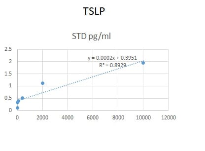
Application: ELISASample Tested: Serum and PlasmaSpecies: HumanVerified Customer | Posted 06/09/2020We used this antibody in an in-house ELISA along with pAb (AF1398) and protein (1398-TS-010) to quantify TSLP in human serum and plasma. This combination could not detect TSLP samples but generated a good standard curve.

There are no reviews that match your criteria.
Protocols
Find general support by application which include: protocols, troubleshooting, illustrated assays, videos and webinars.
- Cellular Response to Hypoxia Protocols
- R&D Systems Quality Control Western Blot Protocol
- Troubleshooting Guide: Western Blot Figures
- Western Blot Conditions
- Western Blot Protocol
- Western Blot Protocol for Cell Lysates
- Western Blot Troubleshooting
- Western Blot Troubleshooting Guide
- View all Protocols, Troubleshooting, Illustrated assays and Webinars
Loading...
Associated Pathways
