Vasoactive intestinal peptide (VIP) is a 28 amino acid (aa) peptide that exerts wide-ranging effects in embryonic and adult tissues through interactions with the 7‑transmembrane spanning receptors VIP R1 and VIP R2. The VIP proprotein is proteolytically processed to release bioactive VIP as well as the bioactive peptides PHV and PHM. VIP regulates appetite and feeding behavior and stimulates insulin and glucagon secretion in pancreatic islet cells. It also regulates astrocyte function, multiple aspects of cardiac function, and TLR function in innate immune responses. Mature VIP shares 100% aa sequence identity with mouse and rat VIP.

Key Product Details
Species Reactivity
Human
Applications
Immunohistochemistry
Label
Unconjugated
Antibody Source
Monoclonal Mouse IgG1 Clone # 576721
Loading...
Product Specifications
Immunogen
KLH-coupled human VIP synthetic peptides HSDAVFTDNYTR and KYLNSILN
Specificity
Detects recombinant human VIP in direct ELISAs. It does not detect the C-terminal region (aa 145-152).
Clonality
Monoclonal
Host
Mouse
Isotype
IgG1
Scientific Data Images for Human VIP Antibody (576721)
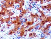
VIP in Human Stomach.
VIP was detected in immersion fixed paraffin-embedded sections of human stomach array using Mouse Anti-Human VIP Monoclonal Antibody (Catalog # MAB6079) at 15 µg/mL overnight at 4 °C. Tissue was stained using the Anti-Mouse HRP-DAB Cell & Tissue Staining Kit (brown; Catalog # CTS002) and counterstained with hematoxylin (blue). Lower panel shows a lack of labeling if primary antibodies are omitted and tissue is stained only with secondary antibody followed by incubation with detection reagents. View our protocol for Chromogenic IHC Staining of Paraffin-embedded Tissue Sections.Applications for Human VIP Antibody (576721)
Application
Recommended Usage
Immunohistochemistry
8-25 µg/mL
Sample: Immersion-fixed paraffin-embedded sections of human stomach and human stomach cancer tissue
Sample: Immersion-fixed paraffin-embedded sections of human stomach and human stomach cancer tissue
Reviewed Applications
Read 1 review rated 5 using MAB6079 in the following applications:
Formulation, Preparation, and Storage
Purification
Protein A or G purified from hybridoma culture supernatant
Reconstitution
Reconstitute at 0.5 mg/mL in sterile PBS. For liquid material, refer to CoA for concentration.
Loading...
Formulation
Lyophilized from a 0.2 μm filtered solution in PBS with Trehalose. *Small pack size (SP) is supplied either lyophilized or as a 0.2 µm filtered solution in PBS.
Shipping
Lyophilized product is shipped at ambient temperature. Liquid small pack size (-SP) is shipped with polar packs. Upon receipt, store immediately at the temperature recommended below.
Stability & Storage
Use a manual defrost freezer and avoid repeated freeze-thaw cycles.
- 12 months from date of receipt, -20 to -70 °C as supplied.
- 1 month, 2 to 8 °C under sterile conditions after reconstitution.
- 6 months, -20 to -70 °C under sterile conditions after reconstitution.
Calculators
Background: VIP
Long Name
Vasoactive Intestinal Peptide
Alternate Names
MGC13587
Entrez Gene IDs
7432 (Human)
Gene Symbol
VIP
Additional VIP Products
Product Documents for Human VIP Antibody (576721)
Certificate of Analysis
To download a Certificate of Analysis, please enter a lot or batch number in the search box below.
Note: Certificate of Analysis not available for kit components.
Product Specific Notices for Human VIP Antibody (576721)
For research use only
Related Research Areas
Citations for Human VIP Antibody (576721)
 Powered by Bioz
Customer Reviews for Human VIP Antibody (576721) (1)
5 out of 5
1 Customer Rating
Have you used Human VIP Antibody (576721)?
Submit a review and receive an Amazon gift card!
$25/€18/£15/$25CAN/¥2500 Yen for a review with an image
$10/€7/£6/$10CAN/¥1110 Yen for a review without an image
Submit a review
Customer Images
Showing
1
-
1 of
1 review
Showing All
Filter By:
-
Application: ImmunohistochemistrySample Tested: Stomach tissueSpecies: HumanVerified Customer | Posted 02/21/2022

There are no reviews that match your criteria.
Protocols
Find general support by application which include: protocols, troubleshooting, illustrated assays, videos and webinars.
- Antigen Retrieval Protocol (PIER)
- Antigen Retrieval for Frozen Sections Protocol
- Appropriate Fixation of IHC/ICC Samples
- Cellular Response to Hypoxia Protocols
- Chromogenic IHC Staining of Formalin-Fixed Paraffin-Embedded (FFPE) Tissue Protocol
- Chromogenic Immunohistochemistry Staining of Frozen Tissue
- ClariTSA™ Fluorophore Kits
- Detection & Visualization of Antibody Binding
- Fluorescent IHC Staining of Frozen Tissue Protocol
- Graphic Protocol for Heat-induced Epitope Retrieval
- Graphic Protocol for the Preparation and Fluorescent IHC Staining of Frozen Tissue Sections
- Graphic Protocol for the Preparation and Fluorescent IHC Staining of Paraffin-embedded Tissue Sections
- Graphic Protocol for the Preparation of Gelatin-coated Slides for Histological Tissue Sections
- IHC Sample Preparation (Frozen sections vs Paraffin)
- Immunofluorescent IHC Staining of Formalin-Fixed Paraffin-Embedded (FFPE) Tissue Protocol
- Immunohistochemistry (IHC) and Immunocytochemistry (ICC) Protocols
- Immunohistochemistry Frozen Troubleshooting
- Immunohistochemistry Paraffin Troubleshooting
- Preparing Samples for IHC/ICC Experiments
- Preventing Non-Specific Staining (Non-Specific Binding)
- Primary Antibody Selection & Optimization
- Protocol for Heat-Induced Epitope Retrieval (HIER)
- Protocol for Making a 4% Formaldehyde Solution in PBS
- Protocol for VisUCyte™ HRP Polymer Detection Reagent
- Protocol for the Preparation & Fixation of Cells on Coverslips
- Protocol for the Preparation and Chromogenic IHC Staining of Frozen Tissue Sections
- Protocol for the Preparation and Chromogenic IHC Staining of Frozen Tissue Sections - Graphic
- Protocol for the Preparation and Chromogenic IHC Staining of Paraffin-embedded Tissue Sections
- Protocol for the Preparation and Chromogenic IHC Staining of Paraffin-embedded Tissue Sections - Graphic
- Protocol for the Preparation and Fluorescent IHC Staining of Frozen Tissue Sections
- Protocol for the Preparation and Fluorescent IHC Staining of Paraffin-embedded Tissue Sections
- Protocol for the Preparation of Gelatin-coated Slides for Histological Tissue Sections
- TUNEL and Active Caspase-3 Detection by IHC/ICC Protocol
- The Importance of IHC/ICC Controls
- Troubleshooting Guide: Immunohistochemistry
- View all Protocols, Troubleshooting, Illustrated assays and Webinars
Loading...