Cathepsin E is an intracellular aspartic protease of the pepsin family (1, 2). Unlike Cathepsin D, another member of the same family and a lysosomal protease with relatively ubiquitous distribution, Cathepsin E is not a lysosomal enzyme and has a limited cell and tissue distribution. However, both Cathepsins D and E play an important role in the degradation of proteins, the generation of bioactive proteins, and antigen processing (3). Both enzymes are efficient in cleaving the Swedish mutant of amyloid precursor protein (APP) at the beta site but show almost no reactivity with the wild-type APP (4). Mouse Cathepsin E is synthesized as a precursor protein, consisting of a signal peptide (residues 1-18), a propeptide (residues 19-59), and a mature chain (residues 60-397) (1).

Key Product Details
Species Reactivity
Validated:
Mouse
Cited:
Mouse, Transgenic Mouse
Applications
Validated:
Immunohistochemistry, Western Blot, Immunoprecipitation
Cited:
Immunohistochemistry, Immunohistochemistry-Paraffin, Western Blot
Label
Unconjugated
Antibody Source
Polyclonal Goat IgG
Loading...
Product Specifications
Immunogen
Mouse myeloma cell line NS0-derived recombinant mouse Cathepsin E
Gln19-Pro397
Accession # P70269
Gln19-Pro397
Accession # P70269
Specificity
Detects both pro and mature mouse Cathepsin E in direct ELISAs and Western blots. In direct ELISAs and Western blots, approximately 40% cross-reactivity with recombinant human Cathepsin E and less than 1% cross-reactivity with recombinant mouse Cathepsin D is observed.
Clonality
Polyclonal
Host
Goat
Isotype
IgG
Scientific Data Images for Mouse Cathepsin E Antibody
Cathepsin E in Mouse Lung.
Cathepsin E was detected in perfusion fixed frozen sections of mouse lung using Goat Anti-Mouse Cathepsin E Antigen Affinity-purified Polyclonal Antibody (Catalog # AF1130) at 15 µg/mL overnight at 4 °C. Tissue was stained using the Anti-Goat HRP-DAB Cell & Tissue Staining Kit (brown; Catalog # CTS008) and counterstained with hematoxylin (blue). Specific labeling was localized to the plasma membrane of type II alveolar cells. View our protocol for Chromogenic IHC Staining of Frozen Tissue Sections.Applications for Mouse Cathepsin E Antibody
Application
Recommended Usage
Immunohistochemistry
5-15 µg/mL
Sample: Perfusion fixed frozen sections of mouse lung
Sample: Perfusion fixed frozen sections of mouse lung
Immunoprecipitation
25 µg/mL
Sample: Conditioned cell culture medium spiked with Recombinant Mouse Cathepsin E (Catalog # 1130-AS), see our available Western blot detection antibodies
Sample: Conditioned cell culture medium spiked with Recombinant Mouse Cathepsin E (Catalog # 1130-AS), see our available Western blot detection antibodies
Western Blot
0.1 µg/mL
Sample: Recombinant Mouse Cathepsin E (Catalog # 1130-AS)
Sample: Recombinant Mouse Cathepsin E (Catalog # 1130-AS)
Reviewed Applications
Read 1 review rated 5 using AF1130 in the following applications:
Formulation, Preparation, and Storage
Purification
Antigen Affinity-purified
Reconstitution
Reconstitute at 0.2 mg/mL in sterile PBS. For liquid material, refer to CoA for concentration.
Loading...
Formulation
Lyophilized from a 0.2 μm filtered solution in PBS with Trehalose. *Small pack size (SP) is supplied either lyophilized or as a 0.2 µm filtered solution in PBS.
Shipping
Lyophilized product is shipped at ambient temperature. Liquid small pack size (-SP) is shipped with polar packs. Upon receipt, store immediately at the temperature recommended below.
Stability & Storage
Use a manual defrost freezer and avoid repeated freeze-thaw cycles.
- 12 months from date of receipt, -20 to -70 °C as supplied.
- 1 month, 2 to 8 °C under sterile conditions after reconstitution.
- 6 months, -20 to -70 °C under sterile conditions after reconstitution.
Calculators
Background: Cathepsin E
References
- Tatnell, P.J. et al. (1997) FEBS Lett. 408:62.
- Kay, J. and P.J. Tatnell (2004) in Handbook of Proteolytic Enzymes (Barrett, A.J. et al., eds.), p. 33, Academic Press, San Diego.
- Tsukuba, T. et al. (2000) Mol. Cells 10:601.
- Gruninger-Leitch, F. et al. (2000) Nat. Biotechnol. 18:66.
Alternate Names
CTSE
Gene Symbol
CTSE
UniProt
Additional Cathepsin E Products
Product Documents for Mouse Cathepsin E Antibody
Certificate of Analysis
To download a Certificate of Analysis, please enter a lot or batch number in the search box below.
Note: Certificate of Analysis not available for kit components.
Product Specific Notices for Mouse Cathepsin E Antibody
For research use only
Related Research Areas
Citations for Mouse Cathepsin E Antibody
 Powered by Bioz
Customer Reviews for Mouse Cathepsin E Antibody (1)
5 out of 5
1 Customer Rating
Have you used Mouse Cathepsin E Antibody?
Submit a review and receive an Amazon gift card!
$25/€18/£15/$25CAN/¥2500 Yen for a review with an image
$10/€7/£6/$10CAN/¥1110 Yen for a review without an image
Submit a review
Customer Images
Showing
1
-
1 of
1 review
Showing All
Filter By:
-
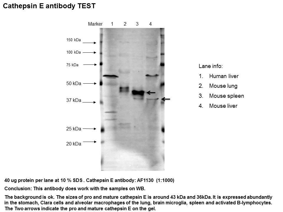
Application: Western BlotSample Tested: Liver tissue, Lung tissue and Spleen tissueSpecies: Human and MouseVerified Customer | Posted 11/01/2016

There are no reviews that match your criteria.
Protocols
Find general support by application which include: protocols, troubleshooting, illustrated assays, videos and webinars.
- Antigen Retrieval Protocol (PIER)
- Antigen Retrieval for Frozen Sections Protocol
- Appropriate Fixation of IHC/ICC Samples
- Cellular Response to Hypoxia Protocols
- Chromogenic IHC Staining of Formalin-Fixed Paraffin-Embedded (FFPE) Tissue Protocol
- Chromogenic Immunohistochemistry Staining of Frozen Tissue
- ClariTSA™ Fluorophore Kits
- Detection & Visualization of Antibody Binding
- Fluorescent IHC Staining of Frozen Tissue Protocol
- Graphic Protocol for Heat-induced Epitope Retrieval
- Graphic Protocol for the Preparation and Fluorescent IHC Staining of Frozen Tissue Sections
- Graphic Protocol for the Preparation and Fluorescent IHC Staining of Paraffin-embedded Tissue Sections
- Graphic Protocol for the Preparation of Gelatin-coated Slides for Histological Tissue Sections
- IHC Sample Preparation (Frozen sections vs Paraffin)
- Immunofluorescent IHC Staining of Formalin-Fixed Paraffin-Embedded (FFPE) Tissue Protocol
- Immunohistochemistry (IHC) and Immunocytochemistry (ICC) Protocols
- Immunohistochemistry Frozen Troubleshooting
- Immunohistochemistry Paraffin Troubleshooting
- Immunoprecipitation Protocol
- Preparing Samples for IHC/ICC Experiments
- Preventing Non-Specific Staining (Non-Specific Binding)
- Primary Antibody Selection & Optimization
- Protocol for Heat-Induced Epitope Retrieval (HIER)
- Protocol for Making a 4% Formaldehyde Solution in PBS
- Protocol for VisUCyte™ HRP Polymer Detection Reagent
- Protocol for the Preparation & Fixation of Cells on Coverslips
- Protocol for the Preparation and Chromogenic IHC Staining of Frozen Tissue Sections
- Protocol for the Preparation and Chromogenic IHC Staining of Frozen Tissue Sections - Graphic
- Protocol for the Preparation and Chromogenic IHC Staining of Paraffin-embedded Tissue Sections
- Protocol for the Preparation and Chromogenic IHC Staining of Paraffin-embedded Tissue Sections - Graphic
- Protocol for the Preparation and Fluorescent IHC Staining of Frozen Tissue Sections
- Protocol for the Preparation and Fluorescent IHC Staining of Paraffin-embedded Tissue Sections
- Protocol for the Preparation of Gelatin-coated Slides for Histological Tissue Sections
- R&D Systems Quality Control Western Blot Protocol
- TUNEL and Active Caspase-3 Detection by IHC/ICC Protocol
- The Importance of IHC/ICC Controls
- Troubleshooting Guide: Immunohistochemistry
- Troubleshooting Guide: Western Blot Figures
- Western Blot Conditions
- Western Blot Protocol
- Western Blot Protocol for Cell Lysates
- Western Blot Troubleshooting
- Western Blot Troubleshooting Guide
- View all Protocols, Troubleshooting, Illustrated assays and Webinars
Loading...