CD69, also known as Leu 23, AIM, EA-1 and MLR-3, is a type II transmembrane glycoprotein and is expressed on activated T cells, B cells, NK cells, neutrophils and eosinophils, Langerhans cells and platelets. The C-terminal extracellular domain of mouse CD69 shares approximately 57% amino acid sequence homology with the corresponding region of human CD69.

Key Product Details
Species Reactivity
Validated:
Mouse
Cited:
Human, Mouse
Applications
Validated:
Western Blot, Flow Cytometry, CyTOF-ready
Cited:
Immunohistochemistry, Immunohistochemistry-Frozen, Immunocytochemistry
Label
Unconjugated
Antibody Source
Polyclonal Goat IgG
Loading...
Product Specifications
Immunogen
Mouse myeloma cell line NS0-derived recombinant mouse CD69
Gly64-Arg199
Accession # P37217
Gly64-Arg199
Accession # P37217
Specificity
Detects mouse CD69 in direct ELISAs and Western blots. In direct ELISAs, less than 15% cross‑reactivity with recombinant human CD69 is observed.
Clonality
Polyclonal
Host
Goat
Isotype
IgG
Scientific Data Images for Mouse CD69 Antibody
Detection of CD69 in mouse splenocytes treated with 50ng/mL PMA and 200ng/mL Ionomycin overnight vs unstimulated mouse splenocytes by Flow Cytometry
Mouse splenocytes treated with 50ng/mL PMA and 200ng/mL Ionomycin overnight (A) vs unstimulated mouse splenocytes (B) were stained with Goat Anti-Mouse CD69 Antigen Affinity-purified Polyclonal Antibody (Catalog # AF2386) and Rat Anti-Mouse CD3 Fluorescein‑conjugated Monoclonal Antibody (Catalog # FAB4841F) followed by Allophycocyanin-conjugated Anti-Goat IgG Secondary Antibody (Catalog # F0108). View our protocol for Staining Membrane-associated Proteins.Applications for Mouse CD69 Antibody
Application
Recommended Usage
CyTOF-ready
Ready to be labeled using established conjugation methods. No BSA or other carrier proteins that could interfere with conjugation.
Flow Cytometry
0.25 µg/106 cells
Sample: - CTLL‑2 mouse cytotoxic T cell line- Mouse splenocytes treated with 50ng/mL PMA and 200ng/mL Ionomycin overnight vs unstimulated mouse splenocytes
Sample: - CTLL‑2 mouse cytotoxic T cell line- Mouse splenocytes treated with 50ng/mL PMA and 200ng/mL Ionomycin overnight vs unstimulated mouse splenocytes
Western Blot
0.1 µg/mL
Sample: Recombinant Mouse CD69
Sample: Recombinant Mouse CD69
Reviewed Applications
Read 1 review rated 4 using AF2386 in the following applications:
Flow Cytometry Panel Builder
Bio-Techne Knows Flow Cytometry
Save time and reduce costly mistakes by quickly finding compatible reagents using the Panel Builder Tool.
Advanced Features
- Spectra Viewer - Custom analysis of spectra from multiple fluorochromes
- Spillover Popups - Visualize the spectra of individual fluorochromes
- Antigen Density Selector - Match fluorochrome brightness with antigen density

Formulation, Preparation, and Storage
Purification
Antigen Affinity-purified
Reconstitution
Reconstitute at 0.2 mg/mL in sterile PBS. For liquid material, refer to CoA for concentration.
Loading...
Formulation
Lyophilized from a 0.2 μm filtered solution in PBS with Trehalose. See Certificate of Analysis for details.
*Small pack size (-SP) is supplied either lyophilized or as a 0.2 µm filtered solution in PBS.
*Small pack size (-SP) is supplied either lyophilized or as a 0.2 µm filtered solution in PBS.
Shipping
Lyophilized product is shipped at ambient temperature. Liquid small pack size (-SP) is shipped with polar packs. Upon receipt, store immediately at the temperature recommended below.
Stability & Storage
Use a manual defrost freezer and avoid repeated freeze-thaw cycles.
- 12 months from date of receipt, -20 to -70 °C as supplied.
- 1 month, 2 to 8 °C under sterile conditions after reconstitution.
- 6 months, -20 to -70 °C under sterile conditions after reconstitution.
Calculators
Background: CD69
Alternate Names
CD69, EA-1, Leu23, MLR-3, p60
Gene Symbol
CD69
UniProt
Additional CD69 Products
Product Documents for Mouse CD69 Antibody
Certificate of Analysis
To download a Certificate of Analysis, please enter a lot or batch number in the search box below.
Note: Certificate of Analysis not available for kit components.
Product Specific Notices for Mouse CD69 Antibody
For research use only
Citations for Mouse CD69 Antibody
 Powered by Bioz
Customer Reviews for Mouse CD69 Antibody (1)
4 out of 5
1 Customer Rating
Have you used Mouse CD69 Antibody?
Submit a review and receive an Amazon gift card!
$25/€18/£15/$25CAN/¥2500 Yen for a review with an image
$10/€7/£6/$10CAN/¥1110 Yen for a review without an image
Submit a review
Customer Images
Showing
1
-
1 of
1 review
Showing All
Filter By:
-
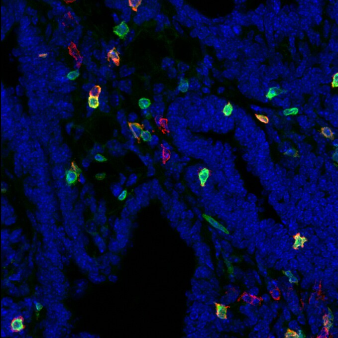
Application: Immunocytochemistry/ImmunofluorescenceSample Tested: Mammary gland tissueSpecies: MouseVerified Customer | Posted 07/09/2019Cryosections from PLP immersion fixed PyMT mammary gland tumors were stained for CD69 (1:100) for 4h @ RT. After washing, tissue was stained with Donkey anti-Goat alexa546 2ndary antibody for 1h at RT. CD3 in green, CD69 in red, nuclear staining (Hoechst) in blue.

There are no reviews that match your criteria.
Protocols
Find general support by application which include: protocols, troubleshooting, illustrated assays, videos and webinars.
- 7-Amino Actinomycin D (7-AAD) Cell Viability Flow Cytometry Protocol
- Cellular Response to Hypoxia Protocols
- Extracellular Membrane Flow Cytometry Protocol
- Flow Cytometry Protocol for Cell Surface Markers
- Flow Cytometry Protocol for Staining Membrane Associated Proteins
- Flow Cytometry Staining Protocols
- Flow Cytometry Troubleshooting Guide
- Intracellular Flow Cytometry Protocol Using Alcohol (Methanol)
- Intracellular Flow Cytometry Protocol Using Detergents
- Intracellular Nuclear Staining Flow Cytometry Protocol Using Detergents
- Intracellular Staining Flow Cytometry Protocol Using Alcohol Permeabilization
- Intracellular Staining Flow Cytometry Protocol Using Detergents to Permeabilize Cells
- Propidium Iodide Cell Viability Flow Cytometry Protocol
- Protocol for Liperfluo
- Protocol for the Characterization of Human Th22 Cells
- Protocol for the Characterization of Human Th9 Cells
- Protocol: Annexin V and PI Staining by Flow Cytometry
- Protocol: Annexin V and PI Staining for Apoptosis by Flow Cytometry
- R&D Systems Quality Control Western Blot Protocol
- Troubleshooting Guide: Fluorokine Flow Cytometry Kits
- Troubleshooting Guide: Western Blot Figures
- Western Blot Conditions
- Western Blot Protocol
- Western Blot Protocol for Cell Lysates
- Western Blot Troubleshooting
- Western Blot Troubleshooting Guide
- View all Protocols, Troubleshooting, Illustrated assays and Webinars
Loading...
Associated Pathways
