Crossveinless-2 (CV-2), also known as bone morphogenetic protein-binding endothelial cell precursor-derived regulator (BMPER), is a secreted chordin-like protein that modulates the BMP signaling pathway (1‑3). Mouse CV-2 is synthesized as a 685 amino acid (aa) residue precursor protein with a putative 39 aa signal peptide, five tandem chordin-like cysteine-rich (CR) domains, a partial von Willebrand factor type D domain (vWD), and a carboxyl trypsin inhibitor-like cysteine-rich domain (TIL) (1, 2, 4). Secreted CV-2 is reported to be proteolytically cleaved to generate two fragments that are disulfide-linked (1, 2). The GDPH sequence is conserved in CV-2 from other species. It is also found in multiple proteins that undergo a similar type of cleavage (5). Mouse CV-2 message is detected in many tissues, with the highest expression detected in the heart, lungs, and skin (2). It is also expressed in flk-1+ endothelial cell precursors and in primary chondrocytes (2). During embryonic development, CV-2 is expressed in the dorsal midline, regions of the telencephalon, migrating cells of the branchial neural crest and endothelial cells in the yolk sac (2). Mouse CV-2 shares 92% and 34% aa sequence identity with the human and Drosophila homologs, respectively (1, 4). Results from biochemical experiments using recombinant CV-2 show that CV-2 directly interacts with BMP-2, -4, and -6 to antagonize BMP signaling, which can regulate a wide range of differentiation processes (1, 2). In contrast, genetic data from Drosophila suggest that CV-2 potentiates BMP-signaling (6). It is possible that like TSG, CV-2 can positively and negatively modulate BMP signal transduction depending on the cell context (7).
Mouse Crossveinless-2/CV-2 Antibody
R&D Systems | Catalog # AF2299

Key Product Details
Species Reactivity
Validated:
Mouse
Cited:
Mouse, Fish - Danio rerio (Zebrafish), Transgenic Mouse
Applications
Validated:
Immunohistochemistry, Western Blot
Cited:
Immunohistochemistry, Immunohistochemistry-Frozen, Western Blot, Immunoprecipitation
Label
Unconjugated
Antibody Source
Polyclonal Goat IgG
Loading...
Product Specifications
Immunogen
Mouse myeloma cell line NS0-derived recombinant mouse Crossveinless-2/CV-2 (R&D Systems, Catalog # 2299-CV)
Ala39-Arg685
Accession # AAH66153
Ala39-Arg685
Accession # AAH66153
Specificity
Detects mouse Crossveinless-2/CV-2 in direct ELISAs and Western blots. In Western blots, approximately 50% cross-reactivity with recombinant human CV-2 is observed.
Clonality
Polyclonal
Host
Goat
Isotype
IgG
Scientific Data Images for Mouse Crossveinless-2/CV-2 Antibody
Crossveinless‑2/CV‑2 in Mouse Embryo.
Crossveinless-2/CV-2 was detected in immersion fixed frozen sections of mouse embryo (E13.5) using Mouse Crossveinless-2/CV-2 Antigen Affinity-purified Polyclonal Antibody (Catalog # AF2299) at 10 µg/mL overnight at 4 °C. Tissue was stained using the NorthernLights™ 557-conjugated Anti-Goat IgG Secondary Antibody (red; Catalog # NL001) and counterstained with DAPI (blue). Specific staining was localized to the epithelium surrounding the nasal cavity. View our protocol for Fluorescent IHC Staining of Frozen Tissue Sections.
Detection of Mouse Crossveinless-2/CV-2/BMPER by Immunocytochemistry/Immunofluorescence
Negative correlation between distributions of BMPER and pSMAD1/5/8 along with BMP4-induced expression suggests BMPER regulates BMP signaling through negative feedback. (A–C) Localization of pSMAD1/5/8 and BMPER protein in the AGM region from E9.5 (A), E10.5 (B), and E11.5 (C) embryos. Magenta, BMPER; cyan, pSMAD1/5/8; green, CD31; blue, DAPI. Bars, 100 µm. Boxes highlight regions of complementarity between BMPER and pSMAD1/5/8 shown at higher magnification in D and Fig. S5 A. UGR, one of two indicated. (D) Higher magnification of E10.5 ventro-lateral region highlighted in B. Magenta, BMPER; cyan, pSMAD1/5/8; green, CD31; blue, DAPI. Bar, 50 µm. Dotted line indicates the boundary around regions with high BMPER protein. (E) Higher magnification view of intra-aortic cluster highlighted in box “E” of B. Magenta, BMPER; cyan, pSMAD1/5/8; green, CD31; blue, DAPI. Arrowheads indicate intra-aortic cluster. Asterisks indicate subendothelial cells with strong nuclear pSMAD1/5/8 signal. Bar, 50 µm. (F) Quantification of pSMAD1/5/8 staining intensity (mean gray values) over an 80-µm band around the dorsal aorta on transverse embryo sections from E9.5, E10.5, and E11.5 (shown in Fig. S5 C). Quantification was on sections from at least two embryos (littermates). Significance measured by Student’s t test: **, P = 0.0015. (G) Expression of Bmper and Bmp4 in AGMs dissected from E9.5, E10.5, and E11.5 stage embryos normalized to Tbp. Each point represents one embryo (littermates). Significance measured by Student’s t test: ****, Bmper E9.5 versus E11.5 P = 8 × 10⁵. (H) Expression of Bmper in E11.5 AGM explants after 24-h culture with BMP4 at displayed dose, without cytokines or serum normalized to Tbp. Experiments were performed twice. Error bars represent SD from the mean. Significance measured by Student’s t test: *, P = 0.035. (I) Expression of Bmper in OP9-BMP4 after reaggregation and culture with doxycycline to induce Bmp4 overexpression. Expression was normalized to Tbp. For ea
Detection of Mouse Crossveinless-2/CV-2/BMPER by Immunocytochemistry/Immunofluorescence
Perivascular cells and subaortic mesenchyme are the main source of BMPER within the AGM region. (A) Distribution of BMPER protein in a transverse section of E10.5 AGM measured by immunostaining. Green, CD31; magenta, BMPER; cyan, RUNX1; blue, DAPI. gut, hindgut; nc, notochord. Bar, 50 µm. (B) Bmper mRNA in transverse section of the E10.5 AGM region by in situ hybridization. Bar, 50 µm. (C) Quantification of the mean immunostaining signal intensity (mean gray values) of DAPI (blue) and BMPER (magenta) along a box (not depicted) drawn over the dorsal-ventral axis of the AGM region from A. Distance is from the notochord (dorsal), position 0, to the intersection with the gut and the AGM region (ventral), position 500. (D) Higher magnification of the highlighted region from A showing the aortic endothelium and perivascular population. Green, CD31; magenta, BMPER; cyan, RUNX1; blue, DAPI. Bar, 50 µm. (E) Higher magnification of the region highlighted in B showing Bmper mRNA around the lining of the aorta. Bar, 50 µm. (F) Expression level of Bmper relative to Tbp in each sorted population: Lin−VC−CD45+, representing hematopoietic cells; Lin−VC+CD45−, endothelial cells; Lin−VC−CD45−CD146+, putative perivascular cells; and Lin−VC−CD45−CD146−, remaining stroma. Expression was normalized to the Lin−VC−CD45−CD146+ population. Each population as percentage of Lin− cells indicated below. Sorting was performed twice, first from one pool of embryos from four to five litters and second from two pools of embryos from four to five litters. Error bars represent SD from the mean (n = 3). Significance calculated by t test: **, P = 0.0016; ***, perivascular versus stroma, P = 0.0006; perivascular versus hematopoietic, P = 0.0002. (G) The distribution of nonendothelial, CD146-positive cells and endothelial CD146-positive cells in transverse section of the E10.5 AGM region. Green, CD31; red, CD146; blue, DAPI. Bar, 50 µm. (H) Higher magnification view of the region highlighted in G showing
Detection of Mouse Crossveinless-2/CV-2/BMPER by Immunocytochemistry/Immunofluorescence
Negative correlation between distributions of BMPER and pSMAD1/5/8 along with BMP4-induced expression suggests BMPER regulates BMP signaling through negative feedback. (A–C) Localization of pSMAD1/5/8 and BMPER protein in the AGM region from E9.5 (A), E10.5 (B), and E11.5 (C) embryos. Magenta, BMPER; cyan, pSMAD1/5/8; green, CD31; blue, DAPI. Bars, 100 µm. Boxes highlight regions of complementarity between BMPER and pSMAD1/5/8 shown at higher magnification in D and Fig. S5 A. UGR, one of two indicated. (D) Higher magnification of E10.5 ventro-lateral region highlighted in B. Magenta, BMPER; cyan, pSMAD1/5/8; green, CD31; blue, DAPI. Bar, 50 µm. Dotted line indicates the boundary around regions with high BMPER protein. (E) Higher magnification view of intra-aortic cluster highlighted in box “E” of B. Magenta, BMPER; cyan, pSMAD1/5/8; green, CD31; blue, DAPI. Arrowheads indicate intra-aortic cluster. Asterisks indicate subendothelial cells with strong nuclear pSMAD1/5/8 signal. Bar, 50 µm. (F) Quantification of pSMAD1/5/8 staining intensity (mean gray values) over an 80-µm band around the dorsal aorta on transverse embryo sections from E9.5, E10.5, and E11.5 (shown in Fig. S5 C). Quantification was on sections from at least two embryos (littermates). Significance measured by Student’s t test: **, P = 0.0015. (G) Expression of Bmper and Bmp4 in AGMs dissected from E9.5, E10.5, and E11.5 stage embryos normalized to Tbp. Each point represents one embryo (littermates). Significance measured by Student’s t test: ****, Bmper E9.5 versus E11.5 P = 8 × 10⁵. (H) Expression of Bmper in E11.5 AGM explants after 24-h culture with BMP4 at displayed dose, without cytokines or serum normalized to Tbp. Experiments were performed twice. Error bars represent SD from the mean. Significance measured by Student’s t test: *, P = 0.035. (I) Expression of Bmper in OP9-BMP4 after reaggregation and culture with doxycycline to induce Bmp4 overexpression. Expression was normalized to Tbp. For ea
Detection of Mouse Crossveinless-2/CV-2/BMPER by Immunocytochemistry/Immunofluorescence
Perivascular cells and subaortic mesenchyme are the main source of BMPER within the AGM region. (A) Distribution of BMPER protein in a transverse section of E10.5 AGM measured by immunostaining. Green, CD31; magenta, BMPER; cyan, RUNX1; blue, DAPI. gut, hindgut; nc, notochord. Bar, 50 µm. (B) Bmper mRNA in transverse section of the E10.5 AGM region by in situ hybridization. Bar, 50 µm. (C) Quantification of the mean immunostaining signal intensity (mean gray values) of DAPI (blue) and BMPER (magenta) along a box (not depicted) drawn over the dorsal-ventral axis of the AGM region from A. Distance is from the notochord (dorsal), position 0, to the intersection with the gut and the AGM region (ventral), position 500. (D) Higher magnification of the highlighted region from A showing the aortic endothelium and perivascular population. Green, CD31; magenta, BMPER; cyan, RUNX1; blue, DAPI. Bar, 50 µm. (E) Higher magnification of the region highlighted in B showing Bmper mRNA around the lining of the aorta. Bar, 50 µm. (F) Expression level of Bmper relative to Tbp in each sorted population: Lin−VC−CD45+, representing hematopoietic cells; Lin−VC+CD45−, endothelial cells; Lin−VC−CD45−CD146+, putative perivascular cells; and Lin−VC−CD45−CD146−, remaining stroma. Expression was normalized to the Lin−VC−CD45−CD146+ population. Each population as percentage of Lin− cells indicated below. Sorting was performed twice, first from one pool of embryos from four to five litters and second from two pools of embryos from four to five litters. Error bars represent SD from the mean (n = 3). Significance calculated by t test: **, P = 0.0016; ***, perivascular versus stroma, P = 0.0006; perivascular versus hematopoietic, P = 0.0002. (G) The distribution of nonendothelial, CD146-positive cells and endothelial CD146-positive cells in transverse section of the E10.5 AGM region. Green, CD31; red, CD146; blue, DAPI. Bar, 50 µm. (H) Higher magnification view of the region highlighted in G showingApplications for Mouse Crossveinless-2/CV-2 Antibody
Application
Recommended Usage
Immunohistochemistry
5-15 µg/mL
Sample: Immersion fixed frozen sections of mouse embryo (E13.5)
Sample: Immersion fixed frozen sections of mouse embryo (E13.5)
Western Blot
0.1 µg/mL
Sample: Recombinant Mouse Crossveinless‑2/CV‑2 (Catalog # 2299-CV)
Sample: Recombinant Mouse Crossveinless‑2/CV‑2 (Catalog # 2299-CV)
Reviewed Applications
Read 2 reviews rated 4.5 using AF2299 in the following applications:
Formulation, Preparation, and Storage
Purification
Antigen Affinity-purified
Reconstitution
Reconstitute at 0.2 mg/mL in sterile PBS. For liquid material, refer to CoA for concentration.
Loading...
Formulation
Lyophilized from a 0.2 μm filtered solution in PBS with Trehalose. *Small pack size (SP) is supplied either lyophilized or as a 0.2 µm filtered solution in PBS.
Shipping
Lyophilized product is shipped at ambient temperature. Liquid small pack size (-SP) is shipped with polar packs. Upon receipt, store immediately at the temperature recommended below.
Stability & Storage
Use a manual defrost freezer and avoid repeated freeze-thaw cycles.
- 12 months from date of receipt, -20 to -70 °C as supplied.
- 1 month, 2 to 8 °C under sterile conditions after reconstitution.
- 6 months, -20 to -70 °C under sterile conditions after reconstitution.
Calculators
Background: Crossveinless-2/CV-2
References
- Binnerts, M.E. et al. (2004) Biochem Biophys Res Commun. 315:272.
- Moser, M. et al. (2003) Mol Cell Biol. 23:5664.
- Garcia-Abreu, J. et al. (2002) Gene 287:39.
- Coffinier, C. et al. (2002) Mech Dev. 119:S179.
- Lidell, M.E. et al. (2003) J. Biol. Chem. 278:13944.
- Conley, C.A. et al. (2000) Development 127:3947.
- Kamimura, M. et al. (2004) Developmental Dynamics 230:434.
Long Name
BMP-binding Endothelial Regulator Protein
Alternate Names
BMPER
Gene Symbol
BMPER
UniProt
Additional Crossveinless-2/CV-2 Products
Product Documents for Mouse Crossveinless-2/CV-2 Antibody
Certificate of Analysis
To download a Certificate of Analysis, please enter a lot or batch number in the search box below.
Note: Certificate of Analysis not available for kit components.
Product Specific Notices for Mouse Crossveinless-2/CV-2 Antibody
For research use only
Related Research Areas
Citations for Mouse Crossveinless-2/CV-2 Antibody
 Powered by Bioz
Customer Reviews for Mouse Crossveinless-2/CV-2 Antibody (2)
4.5 out of 5
2 Customer Ratings
Have you used Mouse Crossveinless-2/CV-2 Antibody?
Submit a review and receive an Amazon gift card!
$25/€18/£15/$25CAN/¥2500 Yen for a review with an image
$10/€7/£6/$10CAN/¥1110 Yen for a review without an image
Submit a review
Customer Images
Showing
1
-
2 of
2 reviews
Showing All
Filter By:
-
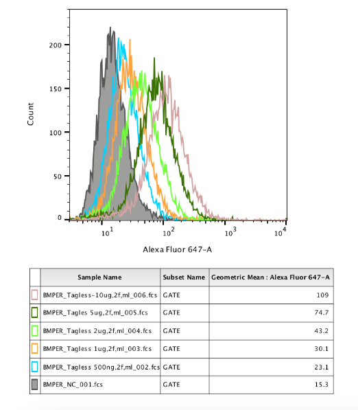
Application: Flow CytometrySample Tested: 3T3-L1 mouse embryonic fibroblast adipose-like cell lineSpecies: MouseVerified Customer | Posted 09/11/2023

-
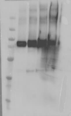
Application: Western BlotSample Tested: Recombinant proteinSpecies: MouseVerified Customer | Posted 08/14/2023
There are no reviews that match your criteria.
Protocols
Find general support by application which include: protocols, troubleshooting, illustrated assays, videos and webinars.
- Antigen Retrieval Protocol (PIER)
- Antigen Retrieval for Frozen Sections Protocol
- Appropriate Fixation of IHC/ICC Samples
- Cellular Response to Hypoxia Protocols
- Chromogenic IHC Staining of Formalin-Fixed Paraffin-Embedded (FFPE) Tissue Protocol
- Chromogenic Immunohistochemistry Staining of Frozen Tissue
- ClariTSA™ Fluorophore Kits
- Detection & Visualization of Antibody Binding
- Fluorescent IHC Staining of Frozen Tissue Protocol
- Graphic Protocol for Heat-induced Epitope Retrieval
- Graphic Protocol for the Preparation and Fluorescent IHC Staining of Frozen Tissue Sections
- Graphic Protocol for the Preparation and Fluorescent IHC Staining of Paraffin-embedded Tissue Sections
- Graphic Protocol for the Preparation of Gelatin-coated Slides for Histological Tissue Sections
- IHC Sample Preparation (Frozen sections vs Paraffin)
- Immunofluorescent IHC Staining of Formalin-Fixed Paraffin-Embedded (FFPE) Tissue Protocol
- Immunohistochemistry (IHC) and Immunocytochemistry (ICC) Protocols
- Immunohistochemistry Frozen Troubleshooting
- Immunohistochemistry Paraffin Troubleshooting
- Preparing Samples for IHC/ICC Experiments
- Preventing Non-Specific Staining (Non-Specific Binding)
- Primary Antibody Selection & Optimization
- Protocol for Heat-Induced Epitope Retrieval (HIER)
- Protocol for Making a 4% Formaldehyde Solution in PBS
- Protocol for VisUCyte™ HRP Polymer Detection Reagent
- Protocol for the Preparation & Fixation of Cells on Coverslips
- Protocol for the Preparation and Chromogenic IHC Staining of Frozen Tissue Sections
- Protocol for the Preparation and Chromogenic IHC Staining of Frozen Tissue Sections - Graphic
- Protocol for the Preparation and Chromogenic IHC Staining of Paraffin-embedded Tissue Sections
- Protocol for the Preparation and Chromogenic IHC Staining of Paraffin-embedded Tissue Sections - Graphic
- Protocol for the Preparation and Fluorescent IHC Staining of Frozen Tissue Sections
- Protocol for the Preparation and Fluorescent IHC Staining of Paraffin-embedded Tissue Sections
- Protocol for the Preparation of Gelatin-coated Slides for Histological Tissue Sections
- R&D Systems Quality Control Western Blot Protocol
- TUNEL and Active Caspase-3 Detection by IHC/ICC Protocol
- The Importance of IHC/ICC Controls
- Troubleshooting Guide: Immunohistochemistry
- Troubleshooting Guide: Western Blot Figures
- Western Blot Conditions
- Western Blot Protocol
- Western Blot Protocol for Cell Lysates
- Western Blot Troubleshooting
- Western Blot Troubleshooting Guide
- View all Protocols, Troubleshooting, Illustrated assays and Webinars
Loading...