

Scientific images may be resized, cropped, or adjusted for brightness or contrast for presentation purposes. These updates are limited to presentation and do not affect the underlying scientific interpretation of the data. Where available, additional source data may be provided upon request.
Key Product Details
Species Reactivity
Validated:
Cited:
Applications
Validated:
Cited:
Label
Antibody Source
Product Specifications
Immunogen
Ser30-His272
Accession # O54908
Specificity
Clonality
Host
Isotype
Scientific Data Images for Mouse Dkk-1 Antibody
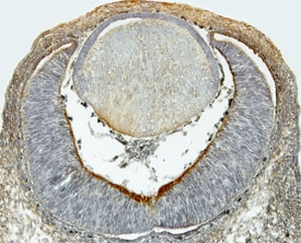
Dkk‑1 in Mouse Embryo.
Dkk‑1 was detected in immersion fixed frozen sections of mouse embryo (15 d.p.c., cross-section through the eye) using Goat Anti-Mouse Dkk‑1 Antigen Affinity-purified Polyclonal Antibody (Catalog # AF1765) at 15 µg/mL overnight at 4 °C. Tissue was stained using the Anti-Goat HRP-DAB Cell & Tissue Staining Kit (brown; Catalog # CTS008) and counterstained with hematoxylin (blue). View our protocol for Chromogenic IHC Staining of Frozen Tissue Sections.
Mouse Dkk-1 ELISA Standard Curve
Recombinant Mouse Dkk‑1 (Histidine-tagged) (Catalog # 1765-DK) was serially diluted and captured by Goat Anti-Mouse Dkk‑1 Antigen Affinity-purified Polyclonal Antibody (Catalog # AF1765) coated on a Clear Polystyrene Microplate (Catalog # DY990). Goat Anti-Mouse Dkk‑1 Antigen Affinity-purified Polyclonal Antibody (Catalog # AF1765) was biotinylated and incubated with the protein captured on the plate. Detection of the standard curve was achieved by incubating Streptavidin-HRP (Catalog # DY998)
Mouse Dkk-1 ELISA Standard Curve
Recombinant Mouse Dkk‑1 (Histidine-tagged) (Catalog # 1765-DK) was serially diluted and captured by Goat Anti-Mouse Dkk‑1 Antigen Affinity-purified Polyclonal Antibody (Catalog # AF1765) coated on a Clear Polystyrene Microplate (Catalog # DY990). Goat Anti-Mouse Dkk‑1 Antigen Affinity-purified Polyclonal Antibody (Catalog # AF1765) was biotinylated and incubated with the protein captured on the plate. Detection of the standard curve was achieved by incubating Streptavidin-HRP (Catalog # DY998)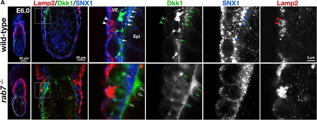
Detection of Dkk-1 by Immunohistochemistry
Rab7-Dependent Trafficking of Dkk1 Creates Wnt Signal Pattern in Peri-gastrulation Embryo(A) E6.0wild-type (n = 20) and Rab7-deficient embryos (n = 16) stained for Dkk1 (green) and sorting nexin 1 (SNX1, red). Arrows indicate colocalization of Dkk1 and SNX1 in the wild-type embryos. Outlined arrows indicate Dkk1 signals observed in Rab7 mutants, which colocalizes neither SNX1 nor Lamp2. Single-channel images are also shown.(B) E6.5 wild-type embryos (n = 21) stained for Dkk1 (green), Lamp2 (red), and mouse IgG (red). Arrows indicatethe Dkk1 signals overlapping with IgG or Lamp2.See also Videos S7 and S8. Image collected and cropped by CiteAb from the following open publication (https://pubmed.ncbi.nlm.nih.gov/32521258), licensed under a CC-BY license. Not internally tested by R&D Systems.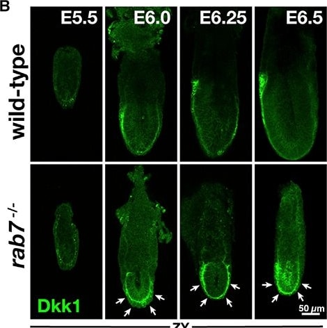
Detection of Dkk-1 by Immunohistochemistry
Accumulation of Wnt Antagonist, Dkk1 Proteins in Rab7-Deficient Embryos at Peri-gastrulation Stages(A) Schematic diagram of optical sections of a whole mounted embryo.(B) Localization of Dkk1 at E5.5 (wild type: n = 2 and rab7−/−: n = 5), E6.0 (wild type: n = 20 and rab7−/− n = 16), E6.25 (wild type: n = 10 and rab7−/−: n = 10), and E6.5 (wild type: n = 13 and rab7−/−: n = 6).(C) Elevated signal of Dkk1 in Rab7-deficient embryos between the visceral endoderm (VE) and Epi. E6.5 wild-type (n = 4) and Rab7-deficient embryos (n = 4) were stained for Dkk1 (green) and E-cadherin (red). The areas outlined by white dashed lines are magnified. Arrows indicate the Dkk1 signals outside the VE cells.(D) E6.5 wild-type (n = 10) and Rab7-deficient (n = 7) embryos stained for Dkk1 (green) and integrin beta 1 (red). The areas outlined by white dashed lines are magnified. Arrows indicate the Dkk1 signals outside the VE cells.See also Videos S1, S2, S3, S4, S5, and S6 for three-dimensional reconstitution of Dkk1 patterning. Image collected and cropped by CiteAb from the following open publication (https://pubmed.ncbi.nlm.nih.gov/32521258), licensed under a CC-BY license. Not internally tested by R&D Systems.Applications for Mouse Dkk-1 Antibody
Immunohistochemistry
Sample: Immersion fixed frozen sections of mouse embryo (E13.5-15.5)
Western Blot
Sample: Recombinant Mouse Dkk‑1 (Catalog # 1765-DK)
Mouse Dkk-1 Sandwich Immunoassay
Reviewed Applications
Read 2 reviews rated 4.5 using AF1765 in the following applications:
Formulation, Preparation, and Storage
Purification
Reconstitution
Reconstitute at 0.2 mg/mL in sterile PBS. For liquid material, refer to CoA for concentration.
Formulation
Shipping
Stability & Storage
- 12 months from date of receipt, -20 to -70 °C as supplied.
- 1 month, 2 to 8 °C under sterile conditions after reconstitution.
- 6 months, -20 to -70 °C under sterile conditions after reconstitution.
Calculators
Background: Dkk-1
References
- Glinka, A. et al. (1998) Nature 391:357.
- Niehrs, C. (2006) Oncogene 25:7469.
- Bullock, C.M. et al. (2004) Mol. Pharmacol. 65:582.
- Mao, B. et al. (2001) Nature 411:321.
- Mao, B. et al. (2002) Nature 417:664.
- Kemp, C. et al. (2005) Dev. Dyn. 233:1064.
- Adamska, M. et al. (2004) Dev. Biol. 272:134.
- Li, J. et al. (2006) Bone 36:754.
- Verani, R. et al. (2006) J. Neurochem. 101:242.
- Pinzone, J.J. et al. (2009) Blood 113:517.
- Morvan, F. et al. (2006) J. Bone Miner. Res. 21:934.
- Cappuccio, I. et al. (2005) J. Neurosci. 25:2647.
Long Name
Alternate Names
Gene Symbol
UniProt
Additional Dkk-1 Products
Product Documents for Mouse Dkk-1 Antibody
Certificate of Analysis
To download a Certificate of Analysis, please enter a lot or batch number in the search box below.
Note: Certificate of Analysis not available for kit components.
Product Specific Notices for Mouse Dkk-1 Antibody
For research use only
Related Research Areas
Citations for Mouse Dkk-1 Antibody
 Powered by Bioz
Customer Reviews for Mouse Dkk-1 Antibody (2)
Have you used Mouse Dkk-1 Antibody?
Submit a review and receive an Amazon gift card!
$25/€18/£15/$25CAN/¥2500 for a review with an image
$10/€7/£6/$10CAN/¥1110 for a review without an image
Submit a review
Customer Images
-
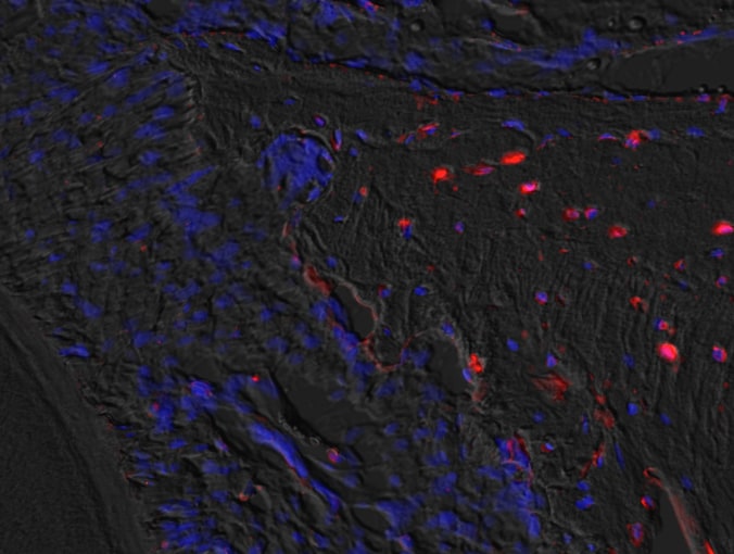
Application: ImmunohistochemistrySample Tested: mouse bone tissueSpecies: MouseVerified Customer | Posted 11/23/2020

-
Application: Immunohistochemistry-ParaffinSample Tested: See PMID 20050968Species: MouseVerified Customer | Posted 01/05/2015
There are no reviews that match your criteria.
Protocols
Find general support by application which include: protocols, troubleshooting, illustrated assays, videos and webinars.
- Antigen Retrieval Protocol (PIER)
- Antigen Retrieval for Frozen Sections Protocol
- Appropriate Fixation of IHC/ICC Samples
- Cellular Response to Hypoxia Protocols
- Chromogenic IHC Staining of Formalin-Fixed Paraffin-Embedded (FFPE) Tissue Protocol
- Chromogenic Immunohistochemistry Staining of Frozen Tissue
- ClariTSA™ Fluorophore Kits
- Detection & Visualization of Antibody Binding
- Fluorescent IHC Staining of Frozen Tissue Protocol
- Graphic Protocol for Heat-induced Epitope Retrieval
- Graphic Protocol for the Preparation and Fluorescent IHC Staining of Frozen Tissue Sections
- Graphic Protocol for the Preparation and Fluorescent IHC Staining of Paraffin-embedded Tissue Sections
- Graphic Protocol for the Preparation of Gelatin-coated Slides for Histological Tissue Sections
- IHC Sample Preparation (Frozen sections vs Paraffin)
- Immunofluorescent IHC Staining of Formalin-Fixed Paraffin-Embedded (FFPE) Tissue Protocol
- Immunohistochemistry (IHC) and Immunocytochemistry (ICC) Protocols
- Immunohistochemistry Frozen Troubleshooting
- Immunohistochemistry Paraffin Troubleshooting
- Preparing Samples for IHC/ICC Experiments
- Preventing Non-Specific Staining (Non-Specific Binding)
- Primary Antibody Selection & Optimization
- Protocol for Heat-Induced Epitope Retrieval (HIER)
- Protocol for Making a 4% Formaldehyde Solution in PBS
- Protocol for VisUCyte™ HRP Polymer Detection Reagent
- Protocol for the Preparation & Fixation of Cells on Coverslips
- Protocol for the Preparation and Chromogenic IHC Staining of Frozen Tissue Sections
- Protocol for the Preparation and Chromogenic IHC Staining of Frozen Tissue Sections - Graphic
- Protocol for the Preparation and Chromogenic IHC Staining of Paraffin-embedded Tissue Sections
- Protocol for the Preparation and Chromogenic IHC Staining of Paraffin-embedded Tissue Sections - Graphic
- Protocol for the Preparation and Fluorescent IHC Staining of Frozen Tissue Sections
- Protocol for the Preparation and Fluorescent IHC Staining of Paraffin-embedded Tissue Sections
- Protocol for the Preparation of Gelatin-coated Slides for Histological Tissue Sections
- R&D Systems Quality Control Western Blot Protocol
- TUNEL and Active Caspase-3 Detection by IHC/ICC Protocol
- The Importance of IHC/ICC Controls
- Troubleshooting Guide: Immunohistochemistry
- Troubleshooting Guide: Western Blot Figures
- Western Blot Conditions
- Western Blot Protocol
- Western Blot Protocol for Cell Lysates
- Western Blot Troubleshooting
- Western Blot Troubleshooting Guide
- View all Protocols, Troubleshooting, Illustrated assays and Webinars
Associated Pathways
