
Mouse ICOS (inducible co-stimulator), also called AILIM (activiation-inducible lymphocyte immunomediatory molecule) and CRP-1 (CD28-related protein-1), is a member of the CD28 family of immune costimulatory receptors. Other family members are CD28, CTLA-4 and PD-1. Mouse ICOS is a homodimeric type I transmembrane protein consisting of 200 amino acids (aa) with a putative 20 aa signal sequence, a 122 aa extracellular domain, a 23 aa transmembrane region, and a 35 aa cytoplasmic domain. ICOS shares approximately 39% aa similarity with CD 28 and CTLA-4. Mouse and human ICOS share approximately 72% aa identity. ICOS is expressed on most CD45RO+ cells. ICOS expression is upregulated within approximately 24‑48 hours of activation on Th primed cells. B7-H2, a member of the B7 family of costimulatory ligands, has been identified as the ICOS ligand. The B7-H2/ICOS interaction appears to play roles in T cell dependent B cell activation and Th differentiation.

Scientific images may be resized, cropped, or adjusted for brightness or contrast for presentation purposes. These updates are limited to presentation and do not affect the underlying scientific interpretation of the data. Where available, additional source data may be provided upon request.
Key Product Details
Species Reactivity
Applications
Label
Antibody Source
Product Specifications
Immunogen
Met1-Leu142
Accession # CAB71153
Specificity
Clonality
Host
Isotype
Endotoxin Level
Scientific Data Images for Mouse ICOS Antibody
Detection of ICOS by Flow Cytometry
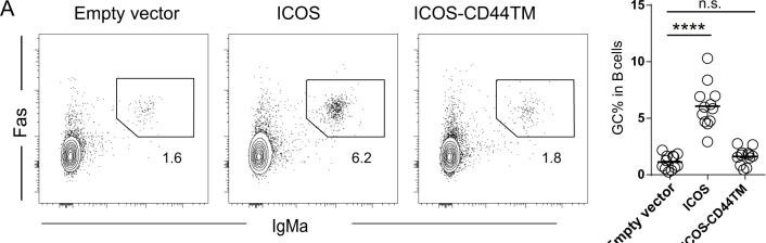
ICOS transmembrane domain-mediated helper functions. a, b Representative contour plots and frequencies of IgMa+Fashi GC B cells among the total CD19+ cells (a) and CXCR5hiPD-1hi TFH cells among the transferred OT-II cells (b). Each symbol in scatter plots represents one mouse, and the lines denote the means. The data presented were pooled from 3 independent experiments (**P < 0.01 and **** P < 0.0001, as determined by Mann–Whitney tests). c Surface CAR staining of Jurkat cells lentivirally transduced to express CD19-reactive BBz CAR, which has the original CD8 transmembrane domain, or ICOSTM-BBz CAR, which has the ICOS transmembrane domain. d Representative FACS profiles (left; the numbers indicate the event percentages in the gates) and summarized frequencies of CAR-expressing Jurkat cells in conjugation with TAMARA-labeled CD19+ Farage B cells (right). Each line connecting two dots represents one independent experiment (****P < 0.0001, as determined by paired t-test) Image collected and cropped by CiteAb from the following open publication (https://pubmed.ncbi.nlm.nih.gov/30523347), licensed under a CC-BY license. Not internally tested by R&D Systems.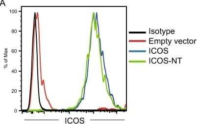
Detection of ICOS by Flow Cytometry
The ICOS transmembrane domain, but not the cytoplasmic domain, mediates the costimulation of calcium mobilization. a Surface ICOS staining of Jurkat cells lentivirally transduced with an empty vector or vectors expressing full-length ICOS or ICOS-NT. b Example of gating GFP+ stably transduced Jurkat cells and GFP− spike-in nontransduced control Jurkat cells for the calcium mobilization assay. c Calcium mobilization in Jurkat cells of the indicated types and the respective internal GFP- control cells after stimulation with biotinylated anti-CD3 (0.2 μg/ml) and anti-ICOS (2 μg/ml) antibodies coligated by streptavidin. The data represent the results of three independent experiments. d Surface ICOS staining of Jurkat cells lentivirally transduced with vectors expressing mutant ICOS molecules of the indicated types. e Surface ICOS staining after normalization of expression between ICOS-NT and ICOS-CD44TM-NT. f Calcium mobilization in Jurkat cells of the indicated types conducted as for c. The data represent the results of three independent experiments Image collected and cropped by CiteAb from the following open publication (https://pubmed.ncbi.nlm.nih.gov/30523347), licensed under a CC-BY license. Not internally tested by R&D Systems.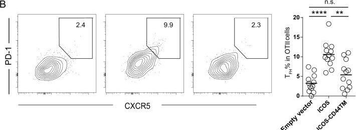
Detection of ICOS by Flow Cytometry
ICOS transmembrane domain-mediated helper functions. a, b Representative contour plots and frequencies of IgMa+Fashi GC B cells among the total CD19+ cells (a) and CXCR5hiPD-1hi TFH cells among the transferred OT-II cells (b). Each symbol in scatter plots represents one mouse, and the lines denote the means. The data presented were pooled from 3 independent experiments (**P < 0.01 and **** P < 0.0001, as determined by Mann–Whitney tests). c Surface CAR staining of Jurkat cells lentivirally transduced to express CD19-reactive BBz CAR, which has the original CD8 transmembrane domain, or ICOSTM-BBz CAR, which has the ICOS transmembrane domain. d Representative FACS profiles (left; the numbers indicate the event percentages in the gates) and summarized frequencies of CAR-expressing Jurkat cells in conjugation with TAMARA-labeled CD19+ Farage B cells (right). Each line connecting two dots represents one independent experiment (****P < 0.0001, as determined by paired t-test) Image collected and cropped by CiteAb from the following open publication (https://pubmed.ncbi.nlm.nih.gov/30523347), licensed under a CC-BY license. Not internally tested by R&D Systems.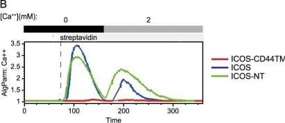
Detection of ICOS by Flow Cytometry
The ICOS transmembrane domain mediates calcium costimulation and is required for p85 recruitment. a Surface ICOS staining of Jurkat cells stably expressing ICOS, ICOS-CD44TM, or ICOS-NT molecules. b Calcium mobilization in Jurkat cells expressing ICOS molecules of the indicated types after stimulation with biotinylated anti-CD3 (0.2 μg/ml) and anti-ICOS (2 μg/ml) antibodies coligated by streptavidin. The data represent the results of three independent experiments. c Immunoblotting of phospho-ZAP70 and phospho-PLC gamma in Jurkat cells expressing the indicated ICOS molecules 30 s after stimulation with anti-CD3 (0.2 μg/ml) and anti-ICOS (2 μg/ml) antibodies coligated by streptavidin. The data represent the results of three independent experiments. d Input (3% total lysates) or anti-ICOS immunoprecipitates of the indicated Jurkat cells before or 1 minute after ICOS crosslinking stimulation were probed by Western blotting for p85 and ICOS. e Immunoblotting of the indicated molecules in ICOS or ICOS-CD44TM Jurkat cells following anti-ICOS crosslinking stimulation for the indicated periods of time. Left, representative pAkt patterns; right, normalized pAkt levels of three independent experiments Image collected and cropped by CiteAb from the following open publication (https://pubmed.ncbi.nlm.nih.gov/30523347), licensed under a CC-BY license. Not internally tested by R&D Systems.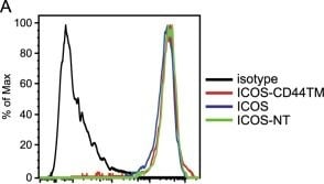
Detection of ICOS by Flow Cytometry
The ICOS transmembrane domain mediates calcium costimulation and is required for p85 recruitment. a Surface ICOS staining of Jurkat cells stably expressing ICOS, ICOS-CD44TM, or ICOS-NT molecules. b Calcium mobilization in Jurkat cells expressing ICOS molecules of the indicated types after stimulation with biotinylated anti-CD3 (0.2 μg/ml) and anti-ICOS (2 μg/ml) antibodies coligated by streptavidin. The data represent the results of three independent experiments. c Immunoblotting of phospho-ZAP70 and phospho-PLC gamma in Jurkat cells expressing the indicated ICOS molecules 30 s after stimulation with anti-CD3 (0.2 μg/ml) and anti-ICOS (2 μg/ml) antibodies coligated by streptavidin. The data represent the results of three independent experiments. d Input (3% total lysates) or anti-ICOS immunoprecipitates of the indicated Jurkat cells before or 1 minute after ICOS crosslinking stimulation were probed by Western blotting for p85 and ICOS. e Immunoblotting of the indicated molecules in ICOS or ICOS-CD44TM Jurkat cells following anti-ICOS crosslinking stimulation for the indicated periods of time. Left, representative pAkt patterns; right, normalized pAkt levels of three independent experiments Image collected and cropped by CiteAb from the following open publication (https://pubmed.ncbi.nlm.nih.gov/30523347), licensed under a CC-BY license. Not internally tested by R&D Systems.Applications for Mouse ICOS Antibody
Blockade of Receptor-ligand Interaction
Western Blot
Sample: Recombinant Mouse ICOS Fc Chimera (Catalog # 168-CS)
Formulation, Preparation, and Storage
Purification
Reconstitution
Reconstitute at 0.2 mg/mL in sterile PBS. For liquid material, refer to CoA for concentration.
Formulation
Shipping
Stability & Storage
- 12 months from date of receipt, -20 to -70 °C as supplied.
- 1 month, 2 to 8 °C under sterile conditions after reconstitution.
- 6 months, -20 to -70 °C under sterile conditions after reconstitution.
Calculators
Background: ICOS
References
- Coyle, A.J. and J.C. Gutierrez-Ramos (2001) Nat. Immunol. 2:203.
- Mages, H.W. et al. (2000) Eur. J. Immunol. 30:1040.
- Yoshinaga, S.K. et al. (1999) Nature 402:827.
- Hutloff, A. et al. (1999) Nature 397:263.
- Aicher, A. et al. (2000) J. Immunol. 164:4689.
- Coyle, A.J. et al. (2000) Immunity 13:95.
- Gonzalo, J.A. et al. (2001) J. Immunol. 166:1.
Long Name
Alternate Names
Gene Symbol
UniProt
Additional ICOS Products
Product Documents for Mouse ICOS Antibody
Certificate of Analysis
To download a Certificate of Analysis, please enter a lot or batch number in the search box below.
Note: Certificate of Analysis not available for kit components.
Product Specific Notices for Mouse ICOS Antibody
For research use only
Citations for Mouse ICOS Antibody
 Powered by Bioz
Customer Reviews for Mouse ICOS Antibody
There are currently no reviews for this product. Be the first to review Mouse ICOS Antibody and earn rewards!
Have you used Mouse ICOS Antibody?
Submit a review and receive an Amazon gift card!
$25/€18/£15/$25CAN/¥2500 for a review with an image
$10/€7/£6/$10CAN/¥1110 for a review without an image
Submit a review
Protocols
Find general support by application which include: protocols, troubleshooting, illustrated assays, videos and webinars.
- Cellular Response to Hypoxia Protocols
- R&D Systems Quality Control Western Blot Protocol
- Troubleshooting Guide: Western Blot Figures
- Western Blot Conditions
- Western Blot Protocol
- Western Blot Protocol for Cell Lysates
- Western Blot Troubleshooting
- Western Blot Troubleshooting Guide
- View all Protocols, Troubleshooting, Illustrated assays and Webinars
Associated Pathways
