Matrix metalloproteinases (MMPs) are a family of zinc and calcium dependent endopeptidases with the combined ability to degrade all the components of the extracellular matrix. MMP-7 (matrilysin) is expressed in epithelial cells of normal and diseased tissues, and is capable of digesting a large series of proteins of the extracellular matrix including collagen IV and X, gelatin, casein, laminin, aggrecan, entactin, elastin and versican (1). MMP-7 is implicated in the activation of other proteinases such as plasminogen, MMP-1, MMP-2, and MMP-9. In addition to its roles in connective tissue remodeling and cancer, MMP-7 also regulates intestinal alpha ‑defensin activation in innate host defense, releases tumor necrosis factor-alpha in a model of herniated disc resorption, and cleaves FasL to generate a soluble form in a model of prostate involution. Structurally, MMP-7 is the smallest of the MMPs and consists of two domains: a pro-domain that is cleaved upon activation and a catalytic domain containing the zinc-binding site.

Key Product Details
Species Reactivity
Validated:
Mouse
Cited:
Human, Mouse, Transgenic Mouse
Applications
Validated:
Immunohistochemistry
Cited:
Immunohistochemistry, Immunohistochemistry-Frozen, Western Blot, Neutralization
Label
Unconjugated
Antibody Source
Polyclonal Goat IgG
Loading...
Product Specifications
Immunogen
Mouse myeloma cell line NS0-derived recombinant mouse MMP‑7
Leu18-Leu264
Accession # AAA99983
Leu18-Leu264
Accession # AAA99983
Specificity
Detects mouse MMP-7 in direct ELISAs and Western blots. In direct ELISAs, approximately 10% cross-reactivity with recombinant human MMP‑7 is observed, and less than 2% cross-reactivity with recombinant mouse (rm) MMP-2, rmMMP-3, rmMMP-8, and rmMMP-9 is observed.
Clonality
Polyclonal
Host
Goat
Isotype
IgG
Scientific Data Images for Mouse MMP-7 Antibody
MMP‑7 in Mouse Intestine.
MMP‑7 was detected in perfusion fixed frozen sections of mouse small intestine using Goat Anti-Mouse MMP‑7 Antigen Affinity-purified Polyclonal Antibody (Catalog # AF2967) at 15 µg/mL overnight at 4 °C. Tissue was stained using the Anti-Goat HRP-DAB Cell & Tissue Staining Kit (brown; Catalog # CTS008) and counterstained with hematoxylin (blue). Specific labeling was localized to the cytoplasm of cells in intestinal glands. View our protocol for Chromogenic IHC Staining of Frozen Tissue Sections.Applications for Mouse MMP-7 Antibody
Application
Recommended Usage
Immunohistochemistry
5-15 µg/mL
Sample: Perfusion fixed frozen sections of mouse small intestine
Sample: Perfusion fixed frozen sections of mouse small intestine
Reviewed Applications
Read 1 review rated 5 using AF2967 in the following applications:
Formulation, Preparation, and Storage
Purification
Antigen Affinity-purified
Reconstitution
Reconstitute at 0.2 mg/mL in sterile PBS. For liquid material, refer to CoA for concentration.
Loading...
Formulation
Lyophilized from a 0.2 μm filtered solution in PBS with Trehalose. *Small pack size (SP) is supplied either lyophilized or as a 0.2 µm filtered solution in PBS.
Shipping
Lyophilized product is shipped at ambient temperature. Liquid small pack size (-SP) is shipped with polar packs. Upon receipt, store immediately at the temperature recommended below.
Stability & Storage
Use a manual defrost freezer and avoid repeated freeze-thaw cycles.
- 12 months from date of receipt, -20 to -70 °C as supplied.
- 1 month, 2 to 8 °C under sterile conditions after reconstitution.
- 6 months, -20 to -70 °C under sterile conditions after reconstitution.
Calculators
Background: MMP-7
References
- Woessner, J.F. (2004) in Handbook of Proteolytic Enzymes, Barrett, A.J. et al. eds. p. 532.
Long Name
Matrix Metalloproteinase 7
Alternate Names
Matrilysin, MMP7
Gene Symbol
MMP7
UniProt
Additional MMP-7 Products
Product Documents for Mouse MMP-7 Antibody
Certificate of Analysis
To download a Certificate of Analysis, please enter a lot or batch number in the search box below.
Note: Certificate of Analysis not available for kit components.
Product Specific Notices for Mouse MMP-7 Antibody
For research use only
Related Research Areas
Citations for Mouse MMP-7 Antibody
 Powered by Bioz
Customer Reviews for Mouse MMP-7 Antibody (1)
5 out of 5
1 Customer Rating
Have you used Mouse MMP-7 Antibody?
Submit a review and receive an Amazon gift card!
$25/€18/£15/$25CAN/¥2500 Yen for a review with an image
$10/€7/£6/$10CAN/¥1110 Yen for a review without an image
Submit a review
Customer Images
Showing
1
-
1 of
1 review
Showing All
Filter By:
-
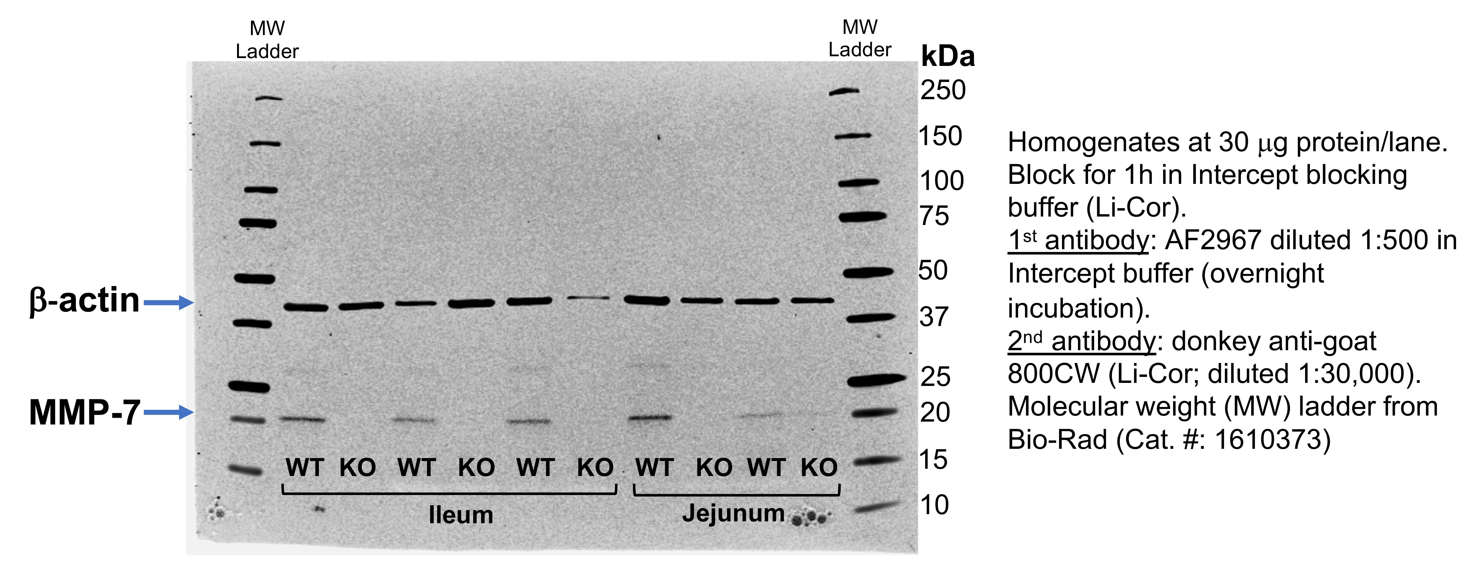
Application: Western BlotSample Tested: Ileum and jejunum homogenatesSpecies: MouseVerified Customer | Posted 09/03/2020

There are no reviews that match your criteria.
Protocols
Find general support by application which include: protocols, troubleshooting, illustrated assays, videos and webinars.
- Antigen Retrieval Protocol (PIER)
- Antigen Retrieval for Frozen Sections Protocol
- Appropriate Fixation of IHC/ICC Samples
- Cellular Response to Hypoxia Protocols
- Chromogenic IHC Staining of Formalin-Fixed Paraffin-Embedded (FFPE) Tissue Protocol
- Chromogenic Immunohistochemistry Staining of Frozen Tissue
- ClariTSA™ Fluorophore Kits
- Detection & Visualization of Antibody Binding
- Fluorescent IHC Staining of Frozen Tissue Protocol
- Graphic Protocol for Heat-induced Epitope Retrieval
- Graphic Protocol for the Preparation and Fluorescent IHC Staining of Frozen Tissue Sections
- Graphic Protocol for the Preparation and Fluorescent IHC Staining of Paraffin-embedded Tissue Sections
- Graphic Protocol for the Preparation of Gelatin-coated Slides for Histological Tissue Sections
- IHC Sample Preparation (Frozen sections vs Paraffin)
- Immunofluorescent IHC Staining of Formalin-Fixed Paraffin-Embedded (FFPE) Tissue Protocol
- Immunohistochemistry (IHC) and Immunocytochemistry (ICC) Protocols
- Immunohistochemistry Frozen Troubleshooting
- Immunohistochemistry Paraffin Troubleshooting
- Preparing Samples for IHC/ICC Experiments
- Preventing Non-Specific Staining (Non-Specific Binding)
- Primary Antibody Selection & Optimization
- Protocol for Heat-Induced Epitope Retrieval (HIER)
- Protocol for Making a 4% Formaldehyde Solution in PBS
- Protocol for VisUCyte™ HRP Polymer Detection Reagent
- Protocol for the Preparation & Fixation of Cells on Coverslips
- Protocol for the Preparation and Chromogenic IHC Staining of Frozen Tissue Sections
- Protocol for the Preparation and Chromogenic IHC Staining of Frozen Tissue Sections - Graphic
- Protocol for the Preparation and Chromogenic IHC Staining of Paraffin-embedded Tissue Sections
- Protocol for the Preparation and Chromogenic IHC Staining of Paraffin-embedded Tissue Sections - Graphic
- Protocol for the Preparation and Fluorescent IHC Staining of Frozen Tissue Sections
- Protocol for the Preparation and Fluorescent IHC Staining of Paraffin-embedded Tissue Sections
- Protocol for the Preparation of Gelatin-coated Slides for Histological Tissue Sections
- TUNEL and Active Caspase-3 Detection by IHC/ICC Protocol
- The Importance of IHC/ICC Controls
- Troubleshooting Guide: Immunohistochemistry
- View all Protocols, Troubleshooting, Illustrated assays and Webinars
Loading...