Tissue inhibitors of metalloproteinases or TIMPs are a family of homologous proteins that regulate the activity of matrix metalloproteinases (MMPs) (1, 2). There are four known members of the family, TIMP-1, TIMP-2, TIMP-3, and TIMP-4 that have been found to exhibit multiple functions, including inhibition of active MMPs, pro‑MMP activation, cell growth promotion, matrix binding, inhibition of angiogenesis and the induction of apoptosis. Structurally, TIMPs have two domains, an N‑terminal domain and a C-terminal domain. Each domain consists of three disulfide-bonded loops. TIMP-1 is a glycoprotein produced by a wide range of cell types. Through its N-terminal domain, TIMP-1 inhibits active MMPs by forming a non-covalent binary complex with the MMP active site. The C-terminal domain of TIMP‑1 interacts with the C-terminal domain of pro‑MMP‑9, which may play a role in regulating pro‑MMP‑9 activation.

Key Product Details
Species Reactivity
Validated:
Mouse
Cited:
Human, Mouse, Transgenic Mouse
Applications
Validated:
Immunohistochemistry, Western Blot, Neutralization, Immunoprecipitation
Cited:
Immunohistochemistry, Immunohistochemistry-Paraffin, Immunohistochemistry-Frozen, Western Blot, Neutralization, Immunocytochemistry, In vivo assay, Electron Microscopy
Label
Unconjugated
Antibody Source
Polyclonal Goat IgG
Loading...
Product Specifications
Immunogen
Mouse myeloma cell line NS0-derived recombinant mouse TIMP-1
Cys25-Arg205
Accession # P12032
Cys25-Arg205
Accession # P12032
Specificity
Detects mouse TIMP-1 in direct ELISAs and Western blots. In direct ELISAs, approximately 15% cross-reactivity with recombinant rat TIMP-1 is observed, less than 5% cross-reactivity with recombinant human (rh) TIMP-1 is observed, and less than 1% cross-reactivity with rhTIMP-2 is observed.
Clonality
Polyclonal
Host
Goat
Isotype
IgG
Scientific Data Images for Mouse TIMP-1 Antibody
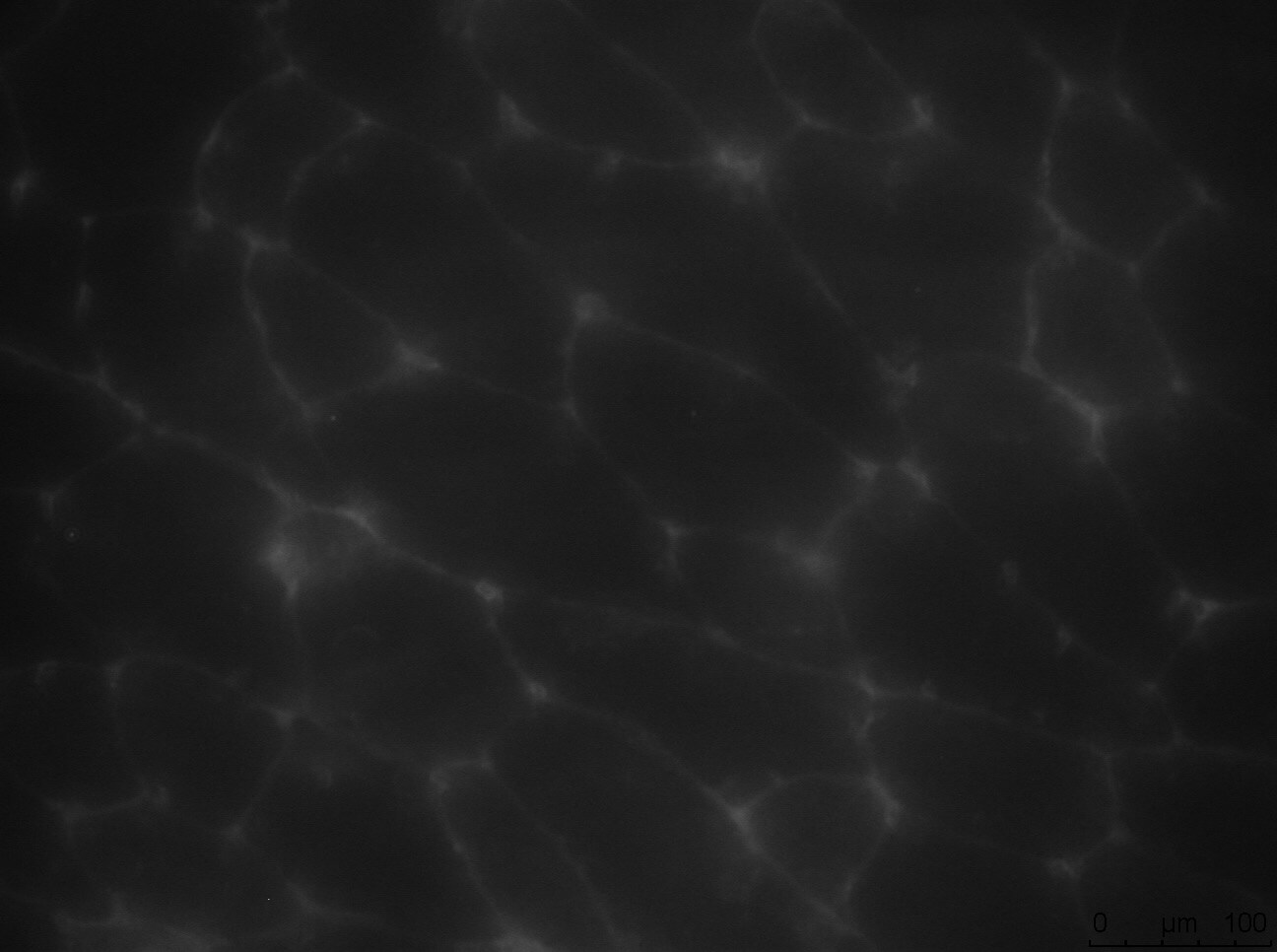
TIMP-1 in Mouse Ovary.
TIMP-1 was detected in perfusion fixed frozen sections of mouse ovary using Goat Anti-Mouse TIMP-1 Antigen Affinity-purified Polyclonal Antibody (red; Catalog # AF980) at 15 µg/mL overnight at 4 °C.
Detection of Mouse TIMP-1 by Western Blot
Conditioned media from G17-stimulated melanoma cells exhibit upregulation of MMP-2 and downregulation of TIMP-3 expression. Representative demonstration of the confirmation of proteomic data using Western blot analysis, with prosaposin serving as a benchmark for gastrin responsiveness (A). Mean and standard deviation (±SD) of the densitometric analysis results from three independent experiments (B). Statistical difference (* p < 0.05) between treated and untreated control groups (considered as 100%) are indicated as follows: prosaposin, TIMP-3 and TIMP-1 levels in G361 cells and MMP2 expressions in the SK-MEL-2 cell line. Image collected and cropped by CiteAb from the following open publication (https://www.mdpi.com/1422-0067/24/23/16851), licensed under a CC-BY license. Not internally tested by R&D Systems.
Mouse TIMP-1 ELISA Standard Curve
Recombinant Mouse TIMP‑1 (Catalog # 980-MT) was serially diluted and captured by Rat Anti-Mouse TIMP‑1 Monoclonal Antibody (Catalog # MAB980) coated on a Clear Polystyrene Microplate (Catalog # DY990). Goat Anti-Mouse TIMP‑1 Antigen Affinity-purified Polyclonal Antibody (Catalog # AF980) was biotinylated and incubated with the protein captured on the plate. Detection of the standard curve was achieved by incubating Streptavidin-HRP (Catalog # DY998)Applications for Mouse TIMP-1 Antibody
Application
Recommended Usage
Immunohistochemistry
5-15 µg/mL
Sample: Perfusion fixed frozen sections of mouse ovary
Sample: Perfusion fixed frozen sections of mouse ovary
Immunoprecipitation
25 µg/mL
Sample: Conditioned cell culture medium spiked with Recombinant Mouse TIMP‑1 (Catalog # 980‑MT), see our available Western blot detection antibodies
Sample: Conditioned cell culture medium spiked with Recombinant Mouse TIMP‑1 (Catalog # 980‑MT), see our available Western blot detection antibodies
Western Blot
0.1 µg/mL
Sample: Recombinant Mouse TIMP‑1 Western Blot Standard (Catalog # WBC022)
Sample: Recombinant Mouse TIMP‑1 Western Blot Standard (Catalog # WBC022)
Neutralization
Measured by its ability to neutralize Recombinant Mouse TIMP-1 (0.07 µg/mL, Catalog # 980-MT) inhibition of Recombinant Mouse/Rat MMP‑2 (0.2 µg/mL, Catalog # 924-MP) cleavage of the fluorogenic peptide substrate Mca-PLGL-Dpa-AR-NH2 (10 µM, Catalog # ES001). The Neutralization Dose (ND50) is typically 1.3 µg/mL.
Reviewed Applications
Read 3 reviews rated 4.3 using AF980 in the following applications:
Formulation, Preparation, and Storage
Purification
Antigen Affinity-purified
Reconstitution
Reconstitute at 0.2 mg/mL in sterile PBS. For liquid material, refer to CoA for concentration.
Loading...
Formulation
Lyophilized from a 0.2 μm filtered solution in PBS with Trehalose. *Small pack size (SP) is supplied either lyophilized or as a 0.2 µm filtered solution in PBS.
Shipping
Lyophilized product is shipped at ambient temperature. Liquid small pack size (-SP) is shipped with polar packs. Upon receipt, store immediately at the temperature recommended below.
Stability & Storage
Use a manual defrost freezer and avoid repeated freeze-thaw cycles.
- 12 months from date of receipt, -20 to -70 °C as supplied.
- 1 month, 2 to 8 °C under sterile conditions after reconstitution.
- 6 months, -20 to -70 °C under sterile conditions after reconstitution.
Calculators
Background: TIMP-1
References
- Murphy, G. and F. Willenbrock (1995) Methods Enzymol. 248:496.
- Brew, K. et al. (2000) Biochim. Biophys. Acta 1477:267.
Long Name
Tissue Inhibitors of Metalloproteinases 1
Alternate Names
TIMP1
Gene Symbol
TIMP1
UniProt
Additional TIMP-1 Products
Product Documents for Mouse TIMP-1 Antibody
Certificate of Analysis
To download a Certificate of Analysis, please enter a lot or batch number in the search box below.
Note: Certificate of Analysis not available for kit components.
Product Specific Notices for Mouse TIMP-1 Antibody
For research use only
Related Research Areas
Citations for Mouse TIMP-1 Antibody
 Powered by Bioz
Customer Reviews for Mouse TIMP-1 Antibody (3)
4.3 out of 5
3 Customer Ratings
Have you used Mouse TIMP-1 Antibody?
Submit a review and receive an Amazon gift card!
$25/€18/£15/$25CAN/¥2500 Yen for a review with an image
$10/€7/£6/$10CAN/¥1110 Yen for a review without an image
Submit a review
Customer Images
Showing
1
-
3 of
3 reviews
Showing All
Filter By:
-
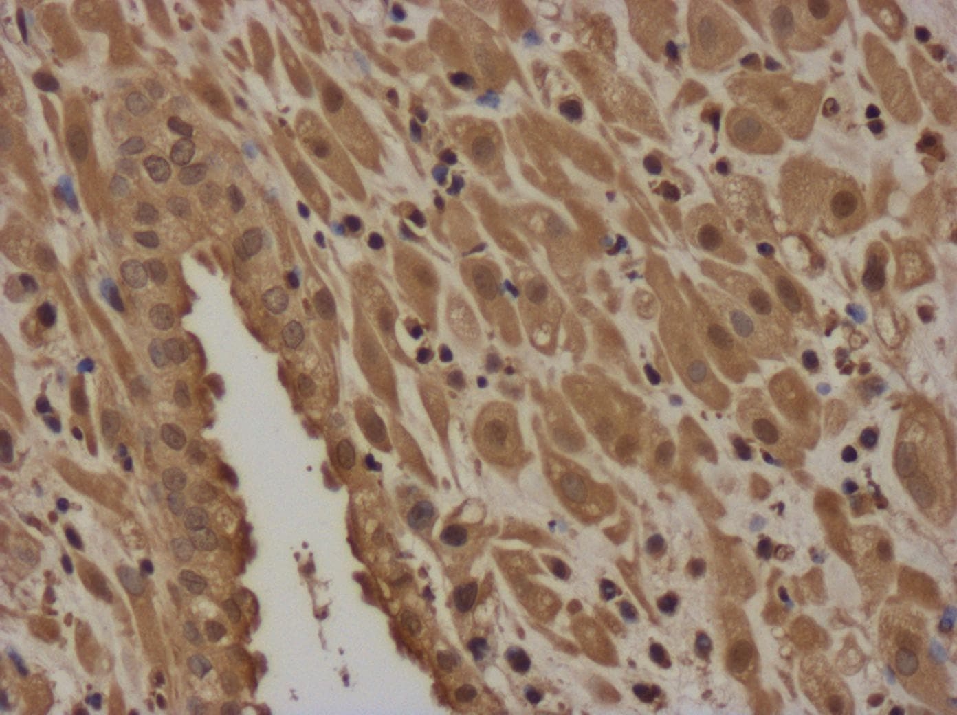
Application: ImmunohistochemistrySample Tested: Skeletal muscleSpecies: MouseVerified Customer | Posted 01/25/2024Fresh frozen skeletal muscle tissue from wt mouse (11 months old)

-
Application: Immunocytochemistry/ImmunofluorescenceSample Tested: activated mouse CD8 T cellSpecies: MouseVerified Customer | Posted 04/26/2017
-
Application: Immunohistochemistry-ParaffinSample Tested: First trimester human deciduaSpecies: GoatVerified Customer | Posted 01/20/201720X magnification. First trimester human decidua express TIMP-1.sodium citrate antigen retrieval
There are no reviews that match your criteria.
Protocols
Find general support by application which include: protocols, troubleshooting, illustrated assays, videos and webinars.
- Antigen Retrieval Protocol (PIER)
- Antigen Retrieval for Frozen Sections Protocol
- Appropriate Fixation of IHC/ICC Samples
- Cellular Response to Hypoxia Protocols
- Chromogenic IHC Staining of Formalin-Fixed Paraffin-Embedded (FFPE) Tissue Protocol
- Chromogenic Immunohistochemistry Staining of Frozen Tissue
- ClariTSA™ Fluorophore Kits
- Detection & Visualization of Antibody Binding
- Fluorescent IHC Staining of Frozen Tissue Protocol
- Graphic Protocol for Heat-induced Epitope Retrieval
- Graphic Protocol for the Preparation and Fluorescent IHC Staining of Frozen Tissue Sections
- Graphic Protocol for the Preparation and Fluorescent IHC Staining of Paraffin-embedded Tissue Sections
- Graphic Protocol for the Preparation of Gelatin-coated Slides for Histological Tissue Sections
- IHC Sample Preparation (Frozen sections vs Paraffin)
- Immunofluorescent IHC Staining of Formalin-Fixed Paraffin-Embedded (FFPE) Tissue Protocol
- Immunohistochemistry (IHC) and Immunocytochemistry (ICC) Protocols
- Immunohistochemistry Frozen Troubleshooting
- Immunohistochemistry Paraffin Troubleshooting
- Immunoprecipitation Protocol
- Preparing Samples for IHC/ICC Experiments
- Preventing Non-Specific Staining (Non-Specific Binding)
- Primary Antibody Selection & Optimization
- Protocol for Heat-Induced Epitope Retrieval (HIER)
- Protocol for Making a 4% Formaldehyde Solution in PBS
- Protocol for VisUCyte™ HRP Polymer Detection Reagent
- Protocol for the Preparation & Fixation of Cells on Coverslips
- Protocol for the Preparation and Chromogenic IHC Staining of Frozen Tissue Sections
- Protocol for the Preparation and Chromogenic IHC Staining of Frozen Tissue Sections - Graphic
- Protocol for the Preparation and Chromogenic IHC Staining of Paraffin-embedded Tissue Sections
- Protocol for the Preparation and Chromogenic IHC Staining of Paraffin-embedded Tissue Sections - Graphic
- Protocol for the Preparation and Fluorescent IHC Staining of Frozen Tissue Sections
- Protocol for the Preparation and Fluorescent IHC Staining of Paraffin-embedded Tissue Sections
- Protocol for the Preparation of Gelatin-coated Slides for Histological Tissue Sections
- R&D Systems Quality Control Western Blot Protocol
- TUNEL and Active Caspase-3 Detection by IHC/ICC Protocol
- The Importance of IHC/ICC Controls
- Troubleshooting Guide: Immunohistochemistry
- Troubleshooting Guide: Western Blot Figures
- Western Blot Conditions
- Western Blot Protocol
- Western Blot Protocol for Cell Lysates
- Western Blot Troubleshooting
- Western Blot Troubleshooting Guide
- View all Protocols, Troubleshooting, Illustrated assays and Webinars
Loading...