
CD4+ T Cells and the Adaptive Immune Response
CD4+ T Cells and the Adaptive Immune Response
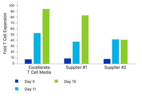
T Cell Culture
T Cell Culture
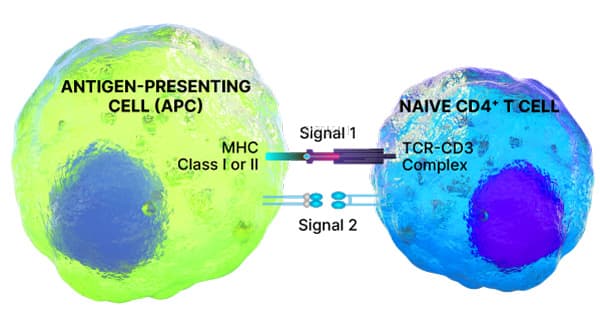
T Cell Activation
T Cell Activation

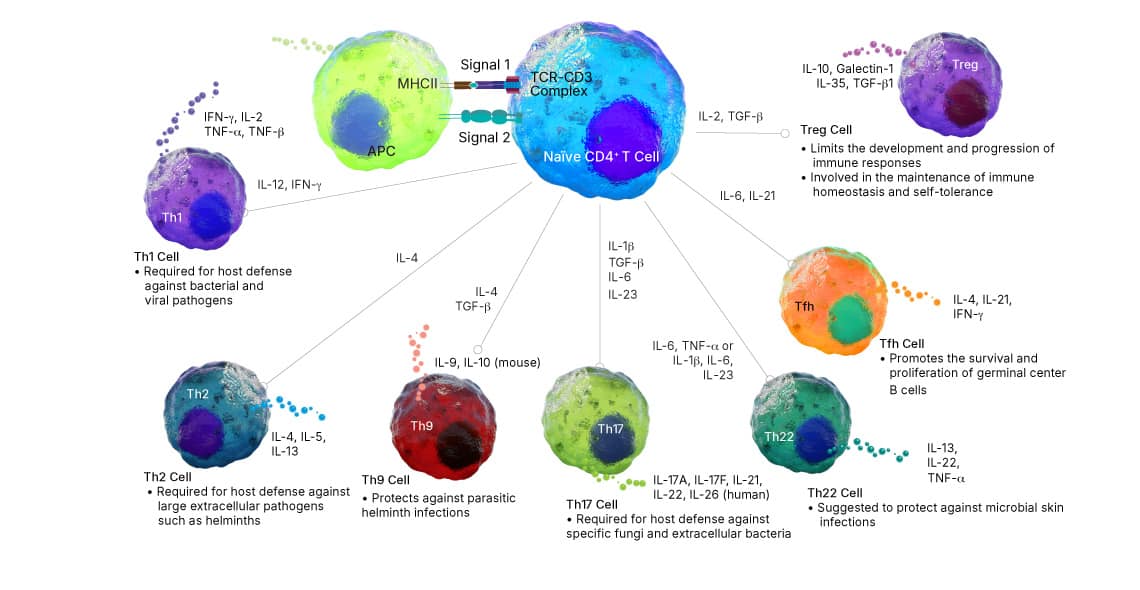
T Cell Differentiation
T Cell Differentiation
T Cell Markers
T Cell Markers


T Cell Cytokine Profiling
T Cell Cytokine Profiling
ELISpot Kits and ELISpot Development Modules
ELISpot Kits and ELISpot Development Modules
Multiplex Assays for Cytokine Secretion Analysis
Multiplex Assays for Cytokine Secretion Analysis


Additional Products for T Cell Culture and Analysis
Additional Products for T Cell Culture and Analysis
Featured Content
Featured Content
Featured Content

Are you interested in T cell subsets? Our scientifically informative and visually appealing poster will brighten up your lab.
Featured Content

Explore the benefits of IL-2 Heat Stable Agonist Protein, which can withstand high temperatures and extended culture durations.
Featured Content

Learn about different cytokine families. This poster will serve as a great reference tool and a colorful addition to your lab.
Featured Content

Explore a clear and easy to follow guide to flow cytometry suited for new researchers or experienced scientists.
Featured Content
Receive competitive discounts on bulk quantities of a single item or multiple items in one order.
Featured Content

Save space and costs with Simple Reader, a compact, cost-effective plate reader offering precise results.