Activin isoforms and other members of the TGF-beta superfamily exert their biological effects by binding to heteromeric complexes of a type I and a type II serine‑threonine kinase receptor, both of which are essential for signal transduction. Seven type I and five type II receptors, including the two type I and the two type II activin receptors, designated ActR-I(A), ActR-IB, ActR-II(A) and ActR-IIB, have been cloned from mammals. Through alternative mRNA splicing, multiple ActR-IIB isoforms can also be generated, adding to the complexity of the activin receptor system. Different activin isoforms bind with different high-affinities to the various type II isoforms. Type I activin receptors do not bind directly to activin, but will associate with the type II receptor-activin complex and initiate signal transduction. Besides the activin isoforms, ActR-II will also bind inhibin, BMP-2 and BMP-7 with lower affinities. ActR-I can also bind and form signaling complexes with the BMP‑2/7‑bound BMPR-II. Activin type II receptors are highly conserved. Human, mouse and rat type II activin receptors share greater than 98% amino acid sequence homology. Recombinant soluble activin type II receptors bind activin with high affinity, and are potent activin antagonists.

Key Product Details
Species Reactivity
Validated:
Human
Cited:
Human, Mouse, Rat, Feline, Ovine
Applications
Validated:
Immunohistochemistry, Western Blot, Blockade of Receptor-ligand Interaction
Cited:
Immunohistochemistry, Immunohistochemistry-Paraffin, Western Blot, Immunoprecipitation, Cell Culture
Label
Unconjugated
Antibody Source
Polyclonal Goat IgG
Loading...
Product Specifications
Immunogen
Mouse myeloma cell line NS0-derived recombinant human Activin RIIB
Ser19-Thr134
Accession # CAA54671
Ser19-Thr134
Accession # CAA54671
Specificity
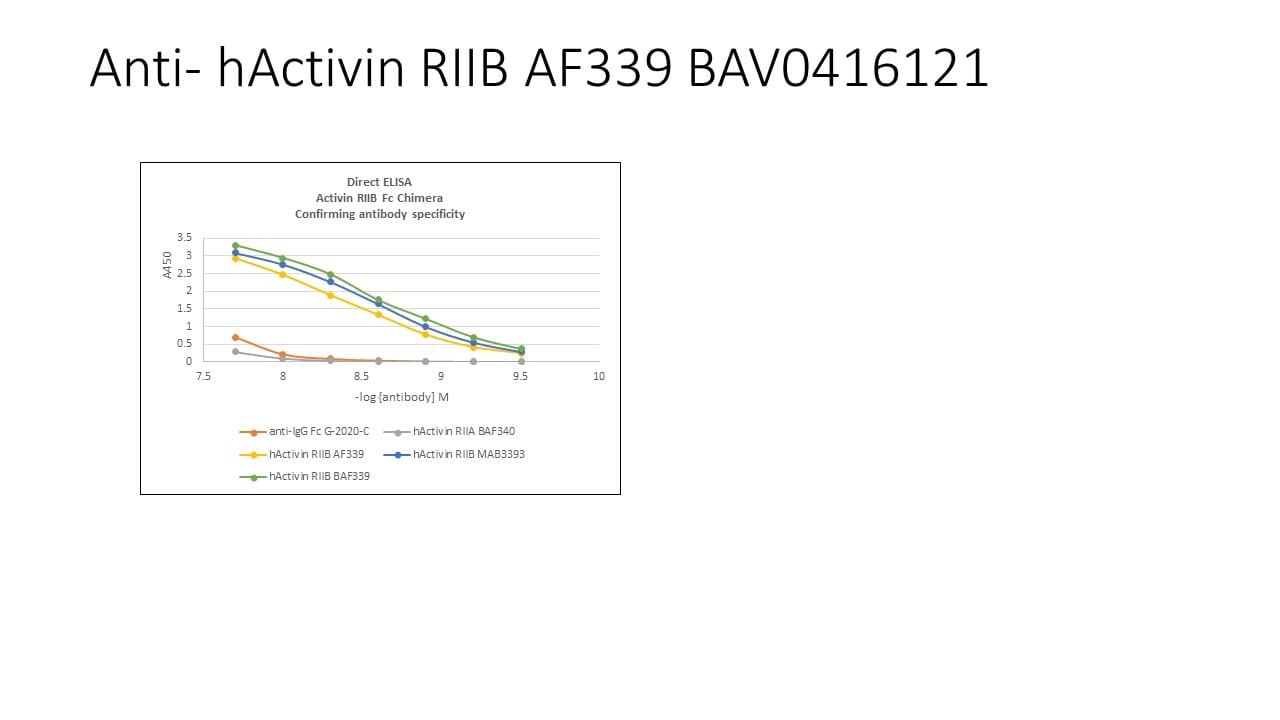
Detects human Activin RIIB in direct ELISAs and Western blots. In direct ELISAs, approximately 50% cross-reactivity with recombinant mouse Activin RIIB is observed, and less than 1% cross-reactivity with recombinant human (rh) Activin RIIA, rhActivin RIA, and rhActivin RIB is observed.
Clonality
Polyclonal
Host
Goat
Isotype
IgG
Endotoxin Level
<0.10 EU per 1 μg of the antibody by the LAL method.
Scientific Data Images for Human Activin RIIB Antibody
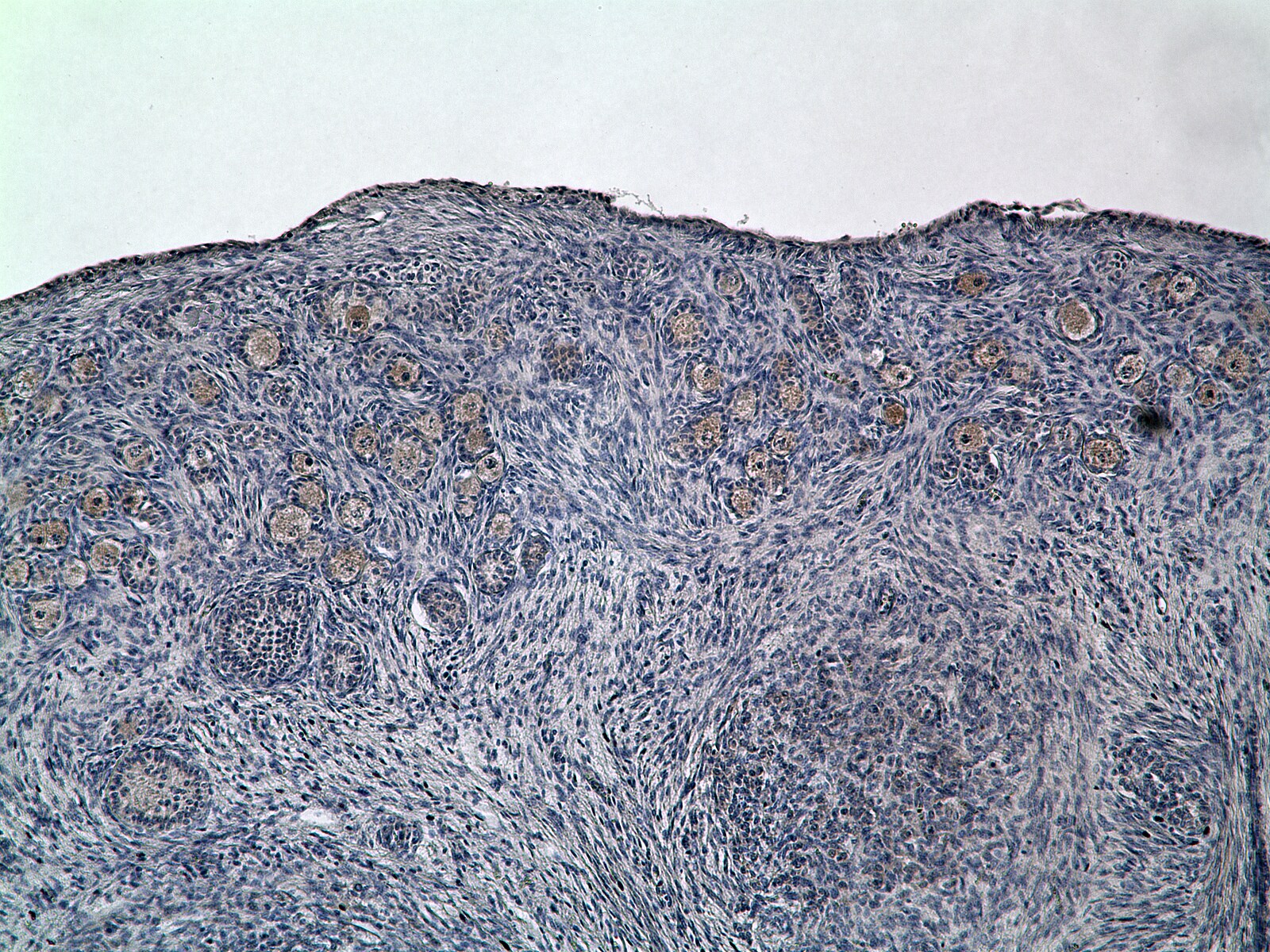
Activin RIIB in Human Placenta.
Activin RIIB was detected in immersion fixed paraffin-embedded sections of human placenta (cross-section of chorionic villi) using 5 µg/mL Goat Anti-Human Activin RIIB Antigen Affinity-purified Poly-clonal Antibody (Catalog # AF339) overnight at 4 °C. Tissue was stained with the Anti-Goat HRP-DAB Cell & Tissue Staining Kit (brown; Catalog # CTS008) and counterstained with hematoxylin (blue). View our protocol for Chromogenic IHC Staining of Paraffin-embedded Tissue Sections.
Detection of Porcine Activin RIIB by Western Blot
Myostatin signaling in longissimus dorsi muscle of low-birth-weight (LBWT) and normal-birth-weight (NBWT) neonatal pigs. (A): Representative western blots from four pairs of LBWT and NBWT pigs (N, NBWT; L, LBWT). (B–G): Protein abundance of myostain, ALK5 and ActRIIB, and protein abundance phosphorylation of smad2/3. Abundance was normalized to alpha -tubulin and phosphorylation normalized to the corresponding non-phospho-proteins. Results are means ± SE. n = 12. Values with different letters differ significantly (P ≤ 0.05). Image collected and cropped by CiteAb from the following open publication (https://journal.frontiersin.org/article/10.3389/fphys.2017.00482/full), licensed under a CC-BY license. Not internally tested by R&D Systems.Applications for Human Activin RIIB Antibody
Application
Recommended Usage
Blockade of Receptor-ligand Interaction
In a functional ELISA, 1-3 µg/mL of this antibody will block 50% of the binding of 30 ng/mL of Recombinant Biotinylated Human/Mouse/Rat Activin A to immobilized Recombinant Human Activin RIIB Fc Chimera (Catalog # 339-RB) coated at 2 µg/mL (100 µL/well). At 30 μg/mL, this antibody will block >90% of the binding.
Immunohistochemistry
5-15 µg/mL
Sample: Immersion fixed paraffin-embedded sections of human placenta (cross-section of chorionic villi)
Sample: Immersion fixed paraffin-embedded sections of human placenta (cross-section of chorionic villi)
Western Blot
0.1 µg/mL
Sample: Recombinant Human Activin RIIB Fc Chimera (Catalog # 339-RB)
Sample: Recombinant Human Activin RIIB Fc Chimera (Catalog # 339-RB)
Reviewed Applications
Read 2 reviews rated 4.5 using AF339 in the following applications:
Formulation, Preparation, and Storage
Purification
Antigen Affinity-purified
Reconstitution
Reconstitute at 0.2 mg/mL in sterile PBS. For liquid material, refer to CoA for concentration.
Loading...
Formulation
Lyophilized from a 0.2 μm filtered solution in PBS with Trehalose. *Small pack size (SP) is supplied either lyophilized or as a 0.2 µm filtered solution in PBS.
Shipping
Lyophilized product is shipped at ambient temperature. Liquid small pack size (-SP) is shipped with polar packs. Upon receipt, store immediately at the temperature recommended below.
Stability & Storage
Use a manual defrost freezer and avoid repeated freeze-thaw cycles.
- 12 months from date of receipt, -20 to -70 °C as supplied.
- 1 month, 2 to 8 °C under sterile conditions after reconstitution.
- 6 months, -20 to -70 °C under sterile conditions after reconstitution.
Calculators
Background: Activin RIIB
References
- Attisano, L. et al. (1996) Mol. and Cell Biol. 16:1066.
- Woodruff, T.K. (1998) Biochem. Pharmacology 55:953.
Long Name
Activin Receptor IIB
Alternate Names
ActivinRIIB, ACVR2B
Gene Symbol
ACVR2B
UniProt
Additional Activin RIIB Products
Product Documents for Human Activin RIIB Antibody
Certificate of Analysis
To download a Certificate of Analysis, please enter a lot or batch number in the search box below.
Note: Certificate of Analysis not available for kit components.
Product Specific Notices for Human Activin RIIB Antibody
For research use only
Related Research Areas
Citations for Human Activin RIIB Antibody
 Powered by Bioz
Customer Reviews for Human Activin RIIB Antibody (2)
4.5 out of 5
2 Customer Ratings
Have you used Human Activin RIIB Antibody?
Submit a review and receive an Amazon gift card!
$25/€18/£15/$25CAN/¥2500 Yen for a review with an image
$10/€7/£6/$10CAN/¥1110 Yen for a review without an image
Submit a review
Customer Images
Showing
1
-
2 of
2 reviews
Showing All
Filter By:
-
Application: ELISASample Tested: Human recombinant antibodySpecies: HumanVerified Customer | Posted 04/07/2020

-
Application: Immunohistochemistry-ParaffinSample Tested: bovine fetal ovarySpecies: OtherVerified Customer | Posted 04/02/2015Localization of Activin RIIB in bovine fetal ovaries
There are no reviews that match your criteria.
Protocols
Find general support by application which include: protocols, troubleshooting, illustrated assays, videos and webinars.
- Antigen Retrieval Protocol (PIER)
- Antigen Retrieval for Frozen Sections Protocol
- Appropriate Fixation of IHC/ICC Samples
- Cellular Response to Hypoxia Protocols
- Chromogenic IHC Staining of Formalin-Fixed Paraffin-Embedded (FFPE) Tissue Protocol
- Chromogenic Immunohistochemistry Staining of Frozen Tissue
- ClariTSA™ Fluorophore Kits
- Detection & Visualization of Antibody Binding
- Fluorescent IHC Staining of Frozen Tissue Protocol
- Graphic Protocol for Heat-induced Epitope Retrieval
- Graphic Protocol for the Preparation and Fluorescent IHC Staining of Frozen Tissue Sections
- Graphic Protocol for the Preparation and Fluorescent IHC Staining of Paraffin-embedded Tissue Sections
- Graphic Protocol for the Preparation of Gelatin-coated Slides for Histological Tissue Sections
- IHC Sample Preparation (Frozen sections vs Paraffin)
- Immunofluorescent IHC Staining of Formalin-Fixed Paraffin-Embedded (FFPE) Tissue Protocol
- Immunohistochemistry (IHC) and Immunocytochemistry (ICC) Protocols
- Immunohistochemistry Frozen Troubleshooting
- Immunohistochemistry Paraffin Troubleshooting
- Preparing Samples for IHC/ICC Experiments
- Preventing Non-Specific Staining (Non-Specific Binding)
- Primary Antibody Selection & Optimization
- Protocol for Heat-Induced Epitope Retrieval (HIER)
- Protocol for Making a 4% Formaldehyde Solution in PBS
- Protocol for VisUCyte™ HRP Polymer Detection Reagent
- Protocol for the Preparation & Fixation of Cells on Coverslips
- Protocol for the Preparation and Chromogenic IHC Staining of Frozen Tissue Sections
- Protocol for the Preparation and Chromogenic IHC Staining of Frozen Tissue Sections - Graphic
- Protocol for the Preparation and Chromogenic IHC Staining of Paraffin-embedded Tissue Sections
- Protocol for the Preparation and Chromogenic IHC Staining of Paraffin-embedded Tissue Sections - Graphic
- Protocol for the Preparation and Fluorescent IHC Staining of Frozen Tissue Sections
- Protocol for the Preparation and Fluorescent IHC Staining of Paraffin-embedded Tissue Sections
- Protocol for the Preparation of Gelatin-coated Slides for Histological Tissue Sections
- R&D Systems Quality Control Western Blot Protocol
- TUNEL and Active Caspase-3 Detection by IHC/ICC Protocol
- The Importance of IHC/ICC Controls
- Troubleshooting Guide: Immunohistochemistry
- Troubleshooting Guide: Western Blot Figures
- Western Blot Conditions
- Western Blot Protocol
- Western Blot Protocol for Cell Lysates
- Western Blot Troubleshooting
- Western Blot Troubleshooting Guide
- View all Protocols, Troubleshooting, Illustrated assays and Webinars
Loading...