Human ADAM15 Ectodomain Antibody (23G9)
R&D Systems | Catalog # MAB935

Key Product Details
Validated by
Biological Validation
Species Reactivity
Validated:
Human
Cited:
Human, Mouse
Applications
Validated:
Immunohistochemistry, Western Blot, Intracellular Staining by Flow Cytometry, Immunocytochemistry, Immunoprecipitation, CyTOF-ready
Cited:
Western Blot, Flow Cytometry, Immunocytochemistry
Label
Unconjugated
Antibody Source
Monoclonal Mouse IgG1 Clone # 23G9
Loading...
Product Specifications
Immunogen
COS-7 African green monkey SV40 transformed kidney fibroblast-like cell line-derived recombinant human ADAM15
Asp207-Thr696
Accession # Q13444
Asp207-Thr696
Accession # Q13444
Specificity
Detects the ectodomain of recombinant human (rh) ADAM15 in direct ELISAs and Western blots. In direct ELISAs, shows 100% cross‑reactivity with recombinant mouse (rm) ADAM15 but does not cross‑react with rhADAM8, 9, 17, 28, rmADAM9, or 10.
Clonality
Monoclonal
Host
Mouse
Isotype
IgG1
Scientific Data Images for Human ADAM15 Ectodomain Antibody (23G9)
ADAM15 in MCF‑7 Human Cell Line.
ADAM15 was detected in immersion fixed MCF-7 human breast cancer cell line using Human ADAM15 Ectodomain Monoclonal Antibody (Catalog # MAB935) at 10 µg/mL for 3 hours at room temperature. Cells were stained using the NorthernLights™ 557-conjugated Anti-Mouse IgG Secondary Antibody (red; NL007) and counterstained with DAPI (blue). View our protocol for Fluorescent ICC Staining of Cells on Coverslips.
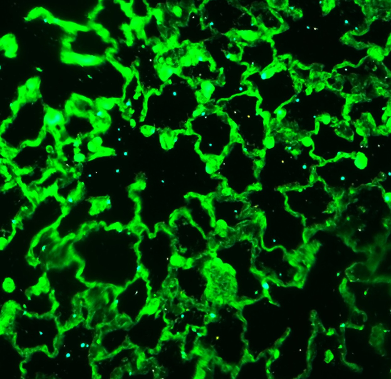
ADAM15 in Human Prostate Cancer Tissue.
ADAM15 was detected in immersion fixed paraffin-embedded sections of human prostate cancer tissue using 25 µg/mL Human ADAM15 Ectodomain Monoclonal Antibody (Catalog # MAB935) overnight at 4 °C. Tissue was stained with the Anti-Mouse HRP-DAB Cell & Tissue Staining Kit (brown; CTS002) and counterstained with hematoxylin (blue). View our protocol for Chromogenic IHC Staining of Paraffin-embedded Tissue Sections. & MOLT‑4 cells (Negative).")
Detection of ADAM15 in PC‑3 cells (Positive) & MOLT‑4 cells (Negative).
ADAM15 was detected in immersion fixed PC‑3 human prostate cancer cells (Positive) & absent in MOLT‑4 human acute lymphoblastic leukemia cells (Negative) using Mouse Anti-Human ADAM15 Ectodomain Monoclonal Antibody (Catalog # MAB935) at 8 µg/mL for 3 hours at room temperature. Cells were stained using the NorthernLights™ 557-conjugated Anti-Mouse IgG Secondary Antibody (red; Catalog # NL007) and counterstained with DAPI (blue). Specific staining was localized to Cytoplasmic. View our protocol for Fluorescent ICC Staining of Cells on Coverslips.
Detection of Human ADAM15 by Flow Cytometry
miR-147b down-regulates cell surface expression of ADAM15 in endothelial cells.At 48 h after transfection, live HUVECs were labeled with a monoclonal anti-ADAM15 ectodomain antibody and a secondary Cy3 conjugate. Cell surface expression was analyzed by flow cytometry (A–B). The bar graph shows quantitative cell surface ADAM15 expression in different treatment groups, each derived from 3 separate experiments (C). Data represent mean ± SEM. * P<0.05 vs. untreated control. Image collected and cropped by CiteAb from the following publication (https://pubmed.ncbi.nlm.nih.gov/25333931), licensed under a CC-BY license. Not internally tested by R&D Systems.
Detection of Human ADAM15 by Flow Cytometry
miR-147b down-regulates cell surface expression of ADAM15 in endothelial cells.At 48 h after transfection, live HUVECs were labeled with a monoclonal anti-ADAM15 ectodomain antibody and a secondary Cy3 conjugate. Cell surface expression was analyzed by flow cytometry (A–B). The bar graph shows quantitative cell surface ADAM15 expression in different treatment groups, each derived from 3 separate experiments (C). Data represent mean ± SEM. * P<0.05 vs. untreated control. Image collected and cropped by CiteAb from the following publication (https://pubmed.ncbi.nlm.nih.gov/25333931), licensed under a CC-BY license. Not internally tested by R&D Systems.Applications for Human ADAM15 Ectodomain Antibody (23G9)
Application
Recommended Usage
CyTOF-ready
Ready to be labeled using established conjugation methods. No BSA or other carrier proteins that could interfere with conjugation.
Immunocytochemistry
8-25 µg/mL
Sample: Immersion fixed MCF-7 human breast cancer cell line, PC‑3 human prostate cancer cells (Positive) & absent in MOLT‑4 human acute lymphoblastic leukemia cells (Negative)
Sample: Immersion fixed MCF-7 human breast cancer cell line, PC‑3 human prostate cancer cells (Positive) & absent in MOLT‑4 human acute lymphoblastic leukemia cells (Negative)
Immunohistochemistry
8-25 µg/mL
Sample: Immersion fixed paraffin-embedded sections of human prostate cancer tissue
Sample: Immersion fixed paraffin-embedded sections of human prostate cancer tissue
Immunoprecipitation
25 µg/mL
Sample: Conditioned cell culture medium spiked with Recombinant Human ADAM15, see our available Western blot detection antibodies
Sample: Conditioned cell culture medium spiked with Recombinant Human ADAM15, see our available Western blot detection antibodies
Intracellular Staining by Flow Cytometry
2.5 µg/106 cells
Sample: MCF‑7 human breast cancer cell line fixed with paraformaldehyde and permeabilized with saponin
Sample: MCF‑7 human breast cancer cell line fixed with paraformaldehyde and permeabilized with saponin
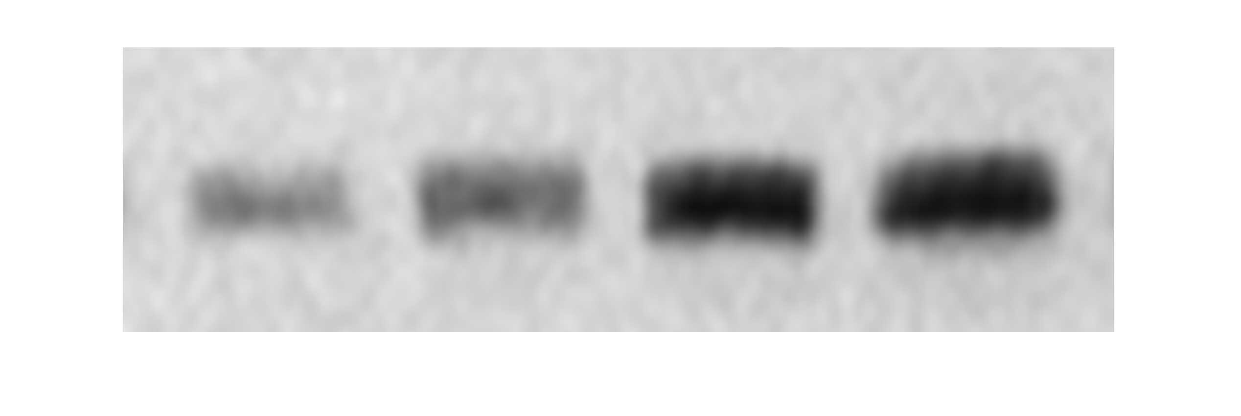
Western Blot
1 µg/mL
Sample: Recombinant Human ADAM15 Western Blot Standard (Catalog # WBC027)
under non-reducing conditions only
Sample: Recombinant Human ADAM15 Western Blot Standard (Catalog # WBC027)
under non-reducing conditions only
Reviewed Applications
Read 2 reviews rated 5 using MAB935 in the following applications:
Flow Cytometry Panel Builder
Bio-Techne Knows Flow Cytometry
Save time and reduce costly mistakes by quickly finding compatible reagents using the Panel Builder Tool.
Advanced Features
- Spectra Viewer - Custom analysis of spectra from multiple fluorochromes
- Spillover Popups - Visualize the spectra of individual fluorochromes
- Antigen Density Selector - Match fluorochrome brightness with antigen density

Formulation, Preparation, and Storage
Purification
Protein A or G purified from hybridoma culture supernatant
Reconstitution
Reconstitute at 0.5 mg/mL in sterile PBS. For liquid material, refer to CoA for concentration.
Loading...
Formulation
Lyophilized from a 0.2 μm filtered solution in PBS with Trehalose. *Small pack size (SP) is supplied either lyophilized or as a 0.2 µm filtered solution in PBS.
Shipping
Lyophilized product is shipped at ambient temperature. Liquid small pack size (-SP) is shipped with polar packs. Upon receipt, store immediately at the temperature recommended below.
Stability & Storage
Use a manual defrost freezer and avoid repeated freeze-thaw cycles.
- 12 months from date of receipt, -20 to -70 °C as supplied.
- 1 month, 2 to 8 °C under sterile conditions after reconstitution.
- 6 months, -20 to -70 °C under sterile conditions after reconstitution.
Calculators
Background: ADAM15
Long Name
A Disintegrin and Metalloprotease-like Domain 15
Alternate Names
MDC15, Metargidin
Gene Symbol
ADAM15
UniProt
Additional ADAM15 Products
Product Documents for Human ADAM15 Ectodomain Antibody (23G9)
Certificate of Analysis
To download a Certificate of Analysis, please enter a lot or batch number in the search box below.
Note: Certificate of Analysis not available for kit components.
Product Specific Notices for Human ADAM15 Ectodomain Antibody (23G9)
For research use only
Related Research Areas
Citations for Human ADAM15 Ectodomain Antibody (23G9)
 Powered by Bioz
Customer Reviews for Human ADAM15 Ectodomain Antibody (23G9) (2)
5 out of 5
2 Customer Ratings
Have you used Human ADAM15 Ectodomain Antibody (23G9)?
Submit a review and receive an Amazon gift card!
$25/€18/£15/$25CAN/¥2500 Yen for a review with an image
$10/€7/£6/$10CAN/¥1110 Yen for a review without an image
Submit a review
Customer Images
Showing
1
-
2 of
2 reviews
Showing All
Filter By:
-
Application: Western BlotSample Tested: THP-1 human acute monocytic leukemia cell line and Cell LysatesSpecies: HumanVerified Customer | Posted 09/14/2019

-
Application: Immunocytochemistry/ImmunofluorescenceSample Tested: Adult lungSpecies: HumanVerified Customer | Posted 09/30/2018
There are no reviews that match your criteria.
Protocols
Find general support by application which include: protocols, troubleshooting, illustrated assays, videos and webinars.
- 7-Amino Actinomycin D (7-AAD) Cell Viability Flow Cytometry Protocol
- Antigen Retrieval Protocol (PIER)
- Antigen Retrieval for Frozen Sections Protocol
- Appropriate Fixation of IHC/ICC Samples
- Cellular Response to Hypoxia Protocols
- Chromogenic IHC Staining of Formalin-Fixed Paraffin-Embedded (FFPE) Tissue Protocol
- Chromogenic Immunohistochemistry Staining of Frozen Tissue
- ClariTSA™ Fluorophore Kits
- Detection & Visualization of Antibody Binding
- Extracellular Membrane Flow Cytometry Protocol
- Flow Cytometry Protocol for Cell Surface Markers
- Flow Cytometry Protocol for Staining Membrane Associated Proteins
- Flow Cytometry Staining Protocols
- Flow Cytometry Troubleshooting Guide
- Fluorescent IHC Staining of Frozen Tissue Protocol
- Graphic Protocol for Heat-induced Epitope Retrieval
- Graphic Protocol for the Preparation and Fluorescent IHC Staining of Frozen Tissue Sections
- Graphic Protocol for the Preparation and Fluorescent IHC Staining of Paraffin-embedded Tissue Sections
- Graphic Protocol for the Preparation of Gelatin-coated Slides for Histological Tissue Sections
- ICC Cell Smear Protocol for Suspension Cells
- ICC Immunocytochemistry Protocol Videos
- ICC for Adherent Cells
- IHC Sample Preparation (Frozen sections vs Paraffin)
- Immunocytochemistry (ICC) Protocol
- Immunocytochemistry Troubleshooting
- Immunofluorescence of Organoids Embedded in Cultrex Basement Membrane Extract
- Immunofluorescent IHC Staining of Formalin-Fixed Paraffin-Embedded (FFPE) Tissue Protocol
- Immunohistochemistry (IHC) and Immunocytochemistry (ICC) Protocols
- Immunohistochemistry Frozen Troubleshooting
- Immunohistochemistry Paraffin Troubleshooting
- Immunoprecipitation Protocol
- Intracellular Flow Cytometry Protocol Using Alcohol (Methanol)
- Intracellular Flow Cytometry Protocol Using Detergents
- Intracellular Nuclear Staining Flow Cytometry Protocol Using Detergents
- Intracellular Staining Flow Cytometry Protocol Using Alcohol Permeabilization
- Intracellular Staining Flow Cytometry Protocol Using Detergents to Permeabilize Cells
- Preparing Samples for IHC/ICC Experiments
- Preventing Non-Specific Staining (Non-Specific Binding)
- Primary Antibody Selection & Optimization
- Propidium Iodide Cell Viability Flow Cytometry Protocol
- Protocol for Heat-Induced Epitope Retrieval (HIER)
- Protocol for Liperfluo
- Protocol for Making a 4% Formaldehyde Solution in PBS
- Protocol for VisUCyte™ HRP Polymer Detection Reagent
- Protocol for the Characterization of Human Th22 Cells
- Protocol for the Characterization of Human Th9 Cells
- Protocol for the Fluorescent ICC Staining of Cell Smears - Graphic
- Protocol for the Fluorescent ICC Staining of Cultured Cells on Coverslips - Graphic
- Protocol for the Preparation & Fixation of Cells on Coverslips
- Protocol for the Preparation and Chromogenic IHC Staining of Frozen Tissue Sections
- Protocol for the Preparation and Chromogenic IHC Staining of Frozen Tissue Sections - Graphic
- Protocol for the Preparation and Chromogenic IHC Staining of Paraffin-embedded Tissue Sections
- Protocol for the Preparation and Chromogenic IHC Staining of Paraffin-embedded Tissue Sections - Graphic
- Protocol for the Preparation and Fluorescent ICC Staining of Cells on Coverslips
- Protocol for the Preparation and Fluorescent ICC Staining of Non-adherent Cells
- Protocol for the Preparation and Fluorescent ICC Staining of Stem Cells on Coverslips
- Protocol for the Preparation and Fluorescent IHC Staining of Frozen Tissue Sections
- Protocol for the Preparation and Fluorescent IHC Staining of Paraffin-embedded Tissue Sections
- Protocol for the Preparation of Gelatin-coated Slides for Histological Tissue Sections
- Protocol for the Preparation of a Cell Smear for Non-adherent Cell ICC - Graphic
- Protocol: Annexin V and PI Staining by Flow Cytometry
- Protocol: Annexin V and PI Staining for Apoptosis by Flow Cytometry
- R&D Systems Quality Control Western Blot Protocol
- TUNEL and Active Caspase-3 Detection by IHC/ICC Protocol
- The Importance of IHC/ICC Controls
- Troubleshooting Guide: Fluorokine Flow Cytometry Kits
- Troubleshooting Guide: Immunohistochemistry
- Troubleshooting Guide: Western Blot Figures
- Western Blot Conditions
- Western Blot Protocol
- Western Blot Protocol for Cell Lysates
- Western Blot Troubleshooting
- Western Blot Troubleshooting Guide
- View all Protocols, Troubleshooting, Illustrated assays and Webinars
Loading...