ADAM8, also known as cell surface antigen MS2 or CD156a, is a member of the ADAM family that contains a disintegrin and metalloprotease-like domain (1, 2). ADAM8 can cleave a variety of substrates and has been shown as a sheddase for the low affinity IgE receptor CD23 and the neural recognition molecule CHL1 (3, 4). Expression and regulation studies suggest that ADAM8 is a novel osteoclast stimulating factor and may play a role in asthma (5, 6). The 824 amino acid precursor of human ADAM8 consists of a signal peptide (residues 1 to 16), a pro peptide (residues 17 to 199), a metaloprotease domain (residues 200 to 400), a disintegrin-like domain (residues 408 to 494), a cysteine-rich region (residues 497 to 613), an EGF-like domain (residues 614 to 640), a transmembrane region (residues 656 to 676) and a cytoplasmic domain (residues 677 to 824).

Key Product Details
Species Reactivity
Validated:
Human
Cited:
Human
Applications
Validated:
Western Blot, Immunocytochemistry, Immunoprecipitation
Cited:
Immunohistochemistry, Western Blot, Neutralization, Flow Cytometry, Immunocytochemistry, Immunoprecipitation, Native SDS-PAGE
Label
Unconjugated
Antibody Source
Polyclonal Goat IgG
Loading...
Product Specifications
Immunogen
S. frugiperda insect ovarian cell line Sf 21-derived recombinant human ADAM8 Ectodomain
Glu158-Ser653
Accession # P78325
Glu158-Ser653
Accession # P78325
Specificity
Detects human ADAM8 Ectodomain in direct ELISAs and Western blots. In direct ELISAs, approximately 50% cross-reactivity with recombinant mouse ADAM8 is observed and less than 1% cross-reactivity with recombinant human (rh) ADAM12, rhADAM19, and rhADAM33 is observed.
Clonality
Polyclonal
Host
Goat
Isotype
IgG
Scientific Data Images for Human ADAM8 Ectodomain Antibody
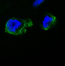
ADAM8 in Human PBMCs.
ADAM8 was detected in immersion fixed human peripheral blood mononuclear cells (PBMCs) using Goat Anti-Human ADAM8 Ectodomain Antigen Affinity-purified Polyclonal Antibody (Catalog # AF1031) at 15 µg/mL for 3 hours at room temperature. Cells were stained using the NorthernLights™ 557-conjugated Anti-Goat IgG Secondary Antibody (red; Catalog # NL001) and counterstained with DAPI (blue). Specific staining was localized to cytoplasmic. View our protocol for Fluorescent ICC Staining of Non-adherent Cells.Applications for Human ADAM8 Ectodomain Antibody
Application
Recommended Usage
Immunocytochemistry
5-15 µg/mL
Sample: Immersion fixed human peripheral blood mononuclear cells (PBMCs)
Sample: Immersion fixed human peripheral blood mononuclear cells (PBMCs)
Immunoprecipitation
25 µg/mL
Sample: Conditioned cell culture medium spiked with Recombinant Human ADAM8 aa 158-497 (Catalog # 1031-AD), see our available Western blot detection antibodies
Sample: Conditioned cell culture medium spiked with Recombinant Human ADAM8 aa 158-497 (Catalog # 1031-AD), see our available Western blot detection antibodies
Western Blot
0.1 µg/mL
Sample: Recombinant Human ADAM8 aa 158-497 (Catalog # 1031-AD)
Sample: Recombinant Human ADAM8 aa 158-497 (Catalog # 1031-AD)
Reviewed Applications
Read 3 reviews rated 4.3 using AF1031 in the following applications:
Formulation, Preparation, and Storage
Purification
Antigen Affinity-purified
Reconstitution
Reconstitute at 0.2 mg/mL in sterile PBS. For liquid material, refer to CoA for concentration.
Loading...
Formulation
Lyophilized from a 0.2 μm filtered solution in PBS with Trehalose. *Small pack size (SP) is supplied either lyophilized or as a 0.2 µm filtered solution in PBS.
Shipping
Lyophilized product is shipped at ambient temperature. Liquid small pack size (-SP) is shipped with polar packs. Upon receipt, store immediately at the temperature recommended below.
Stability & Storage
Use a manual defrost freezer and avoid repeated freeze-thaw cycles.
- 12 months from date of receipt, -20 to -70 °C as supplied.
- 1 month, 2 to 8 °C under sterile conditions after reconstitution.
- 6 months, -20 to -70 °C under sterile conditions after reconstitution.
Calculators
Background: ADAM8
References
- Yoshiyama, K. et al. (1997) Genomics 41:56.
- Moss, M.L. and J.W. Bartsch (2004) Biochemistry 43:7227.
- Fourie, A.M. et al. (2003) J. Biol. Chem. 278:30469.
- Naus, S. et al. (2004) J. Biol. Chem. 279:16083.
- Choi, S.J. et al. (2001) J. Bone Miner. Res. 16:814.
- King, N.E. et al. (2004) Am. J. Respir. Cell Mol. Biol. 31:257.
Long Name
A Disintegrin and Metalloprotease-like Domain 8
Alternate Names
CD156a, MS2
Entrez Gene IDs
101 (Human)
Gene Symbol
ADAM8
UniProt
Additional ADAM8 Products
Product Documents for Human ADAM8 Ectodomain Antibody
Certificate of Analysis
To download a Certificate of Analysis, please enter a lot or batch number in the search box below.
Note: Certificate of Analysis not available for kit components.
Product Specific Notices for Human ADAM8 Ectodomain Antibody
For research use only
Related Research Areas
Citations for Human ADAM8 Ectodomain Antibody
 Powered by Bioz
Customer Reviews for Human ADAM8 Ectodomain Antibody (3)
4.3 out of 5
3 Customer Ratings
Have you used Human ADAM8 Ectodomain Antibody?
Submit a review and receive an Amazon gift card!
$25/€18/£15/$25CAN/¥2500 Yen for a review with an image
$10/€7/£6/$10CAN/¥1110 Yen for a review without an image
Submit a review
Customer Images
Showing
1
-
3 of
3 reviews
Showing All
Filter By:
-
Application: Immunocytochemistry/ImmunofluorescenceSample Tested: THP-1 human acute monocytic leukemia cell lineSpecies: HumanVerified Customer | Posted 06/23/2017

-
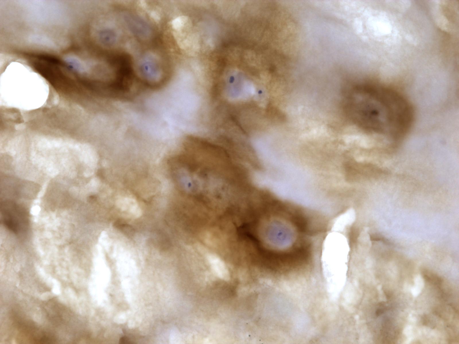
Application: Immunohistochemistry-FrozenSample Tested: Human intervetebral disc nucleus pulposusSpecies: HumanVerified Customer | Posted 01/27/2015Human nucleus pulposus ADAM8 IHC
-
Application: Immunohistochemistry-ParaffinSample Tested: See PMID 23101757Species: HumanVerified Customer | Posted 01/05/2015
There are no reviews that match your criteria.
Protocols
Find general support by application which include: protocols, troubleshooting, illustrated assays, videos and webinars.
- Appropriate Fixation of IHC/ICC Samples
- Cellular Response to Hypoxia Protocols
- ClariTSA™ Fluorophore Kits
- Detection & Visualization of Antibody Binding
- ICC Cell Smear Protocol for Suspension Cells
- ICC Immunocytochemistry Protocol Videos
- ICC for Adherent Cells
- Immunocytochemistry (ICC) Protocol
- Immunocytochemistry Troubleshooting
- Immunofluorescence of Organoids Embedded in Cultrex Basement Membrane Extract
- Immunohistochemistry (IHC) and Immunocytochemistry (ICC) Protocols
- Immunoprecipitation Protocol
- Preparing Samples for IHC/ICC Experiments
- Preventing Non-Specific Staining (Non-Specific Binding)
- Primary Antibody Selection & Optimization
- Protocol for VisUCyte™ HRP Polymer Detection Reagent
- Protocol for the Fluorescent ICC Staining of Cell Smears - Graphic
- Protocol for the Fluorescent ICC Staining of Cultured Cells on Coverslips - Graphic
- Protocol for the Preparation and Fluorescent ICC Staining of Cells on Coverslips
- Protocol for the Preparation and Fluorescent ICC Staining of Non-adherent Cells
- Protocol for the Preparation and Fluorescent ICC Staining of Stem Cells on Coverslips
- Protocol for the Preparation of a Cell Smear for Non-adherent Cell ICC - Graphic
- R&D Systems Quality Control Western Blot Protocol
- TUNEL and Active Caspase-3 Detection by IHC/ICC Protocol
- The Importance of IHC/ICC Controls
- Troubleshooting Guide: Western Blot Figures
- Western Blot Conditions
- Western Blot Protocol
- Western Blot Protocol for Cell Lysates
- Western Blot Troubleshooting
- Western Blot Troubleshooting Guide
- View all Protocols, Troubleshooting, Illustrated assays and Webinars
Loading...