
Key Product Details
Species Reactivity
Validated:
Human
Cited:
Human, Mouse, Rat, Transgenic Mouse, Xenograft
Applications
Validated:
Immunohistochemistry, Western Blot
Cited:
Immunohistochemistry, Immunohistochemistry-Paraffin, Western Blot, Neutralization, Immunocytochemistry
Label
Unconjugated
Antibody Source
Polyclonal Goat IgG
Loading...
Product Specifications
Immunogen
Mouse myeloma cell line NS0-derived recombinant human Angiopoietin-2
Asp68-Phe496
Accession # O15123
Asp68-Phe496
Accession # O15123
Specificity
Detects human Angiopoietin-2 in direct ELISAs and Western blots. In direct ELISAs, 100% cross-reactivity with recombinant mouse Angiopoietin-2 is observed and less than 5% cross-reactivity with recombinant human Angiopoietin-1 is observed.
Clonality
Polyclonal
Host
Goat
Isotype
IgG
Scientific Data Images for Human Angiopoietin-2 Antibody
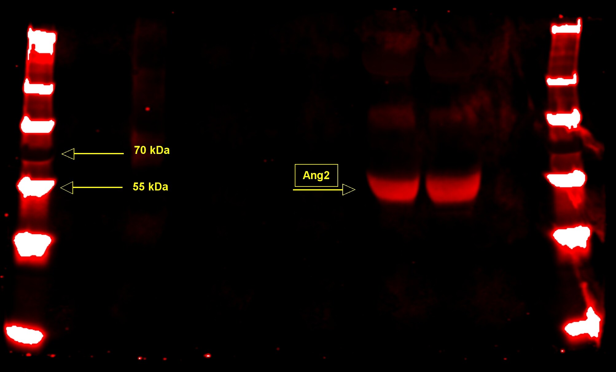
Detection of Human Angiopoietin‑2 by Western Blot.
Western blot shows lysates of HepG2 human hepatocellular carcinoma cell line. PVDF Membrane was probed with 1 µg/mL of Goat Anti-Human Angiopoietin-2 Antigen Affinity-purified Polyclonal Antibody (Catalog # AF623) followed by HRP-conjugated Anti-Goat IgG Secondary Antibody (Catalog # HAF019). A specific band was detected for Angiopoietin-2 at approximately 65 kDa (as indicated). This experiment was conducted under reducing conditions and using Immunoblot Buffer Group 1.
Angiopoietin‑2 in Human Gastrointestinal Cancer Tissue.
Angiopoietin-2 was detected in immersion fixed paraffin-embedded sections of human gastrointestinal cancer tissue using 15 µg/mL Goat Anti-Human Angiopoietin-2 Antigen Affinity-purified Polyclonal Antibody (Catalog # AF623) overnight at 4 °C. Tissue was stained with the Anti-Goat HRP-DAB Cell & Tissue Staining Kit (brown; Catalog # CTS008) and counterstained with hematoxylin (blue). Lower panel shows a lack of labeling if primary antibodies are omitted and tissue is stained only with secondary antibody followed by incubation with detection reagents. View our protocol for Chromogenic IHC Staining of Paraffin-embedded Tissue Sections.
Angiopoietin‑2 in Human Liver Cancer Tissue.
Angiopoietin-2 was detected in immersion fixed paraffin-embedded sections of human liver cancer tissue using Goat Anti-Human Angiopoietin-2 Antigen Affinity-purified Polyclonal Antibody (Catalog # AF623) at 3 µg/mL overnight at 4 °C. Tissue was stained using the Anti-Goat HRP-DAB Cell & Tissue Staining Kit (brown; Catalog # CTS008) and counterstained with hematoxylin (blue). Specific staining was localized to cytoplasm in cancer cells. View our protocol for Chromogenic IHC Staining of Paraffin-embedded Tissue Sections.
Detection of Human Angiopoietin-2 by Immunohistochemistry
sAng-2 concentration positively relates with Ang-2 expression on the epithelia and MVD in cervical tissues.Representative immunohistochemical staining of Ang-1 (A), Ang-2 (B) and CD34 (C) in 25 cervical cancer tissue specimens and 10 normal controls. Black arrows denote positively stained epithelial cells, whereas red arrows denote positively staining endothelial cells, all appearing brown. Scale bar, 20 µm. (D) sAng-2 is significantly higher in the patients with positive Ang-2 expression on cervix epithelia than those with negative Ang-2 expression. The scatter diagrams show the correlations of sAng-1 (E), sAng-2 (F) and sAng-1/ sAng-2 ratio (G) to MVD in the 35 cervical tissue specimens. Image collected and cropped by CiteAb from the following open publication (https://pubmed.ncbi.nlm.nih.gov/28584715), licensed under a CC-BY license. Not internally tested by R&D Systems.
Detection of Human Angiopoietin-2 by Immunohistochemistry
Expression of sst5TMD4 and co-localization with angiogenic marker in GEP-NET(A) Analysis of expression of angiogenic molecules and sst5TMD4 by specific serial immunohistochemistry in a pancreatic NET. Original magnification ×100 and ×400 (insets). N: normal tissue; T: tumor tissue. For specific immunostaining techniques see the “Materials and methods” section. (B) Expression of sst5TMD4 and angiogenic molecules by triple immunofluorescence in a gastrointestinal NET sample. Original magnification ×400. N: normal tissue; T: tumor tissue. For specific immunofluorescence techniques see the “Materials and methods” section. Scale bar for 100 μm is represented with a line for each Figure. Image collected and cropped by CiteAb from the following open publication (https://pubmed.ncbi.nlm.nih.gov/26673010), licensed under a CC-BY license. Not internally tested by R&D Systems.Applications for Human Angiopoietin-2 Antibody
Application
Recommended Usage
Immunohistochemistry
5-15 µg/mL
Sample: Immersion fixed paraffin-embedded sections of human gastrointestinal cancer tissue and human liver cancer tissue
Sample: Immersion fixed paraffin-embedded sections of human gastrointestinal cancer tissue and human liver cancer tissue
Western Blot
1 µg/mL
Sample: HepG2 human hepatocellular carcinoma cell line
Sample: HepG2 human hepatocellular carcinoma cell line
Reviewed Applications
Read 3 reviews rated 4.3 using AF623 in the following applications:
Formulation, Preparation, and Storage
Purification
Antigen Affinity-purified
Reconstitution
Reconstitute at 0.2 mg/mL in sterile PBS. For liquid material, refer to CoA for concentration.
Loading...
Formulation
Lyophilized from a 0.2 μm filtered solution in PBS with Trehalose. *Small pack size (SP) is supplied either lyophilized or as a 0.2 µm filtered solution in PBS.
Shipping
Lyophilized product is shipped at ambient temperature. Liquid small pack size (-SP) is shipped with polar packs. Upon receipt, store immediately at the temperature recommended below.
Stability & Storage
Use a manual defrost freezer and avoid repeated freeze-thaw cycles.
- 12 months from date of receipt, -20 to -70 °C as supplied.
- 1 month, 2 to 8 °C under sterile conditions after reconstitution.
- 6 months, -20 to -70 °C under sterile conditions after reconstitution.
Calculators
Background: Angiopoietin-2
Alternate Names
ANGPT2
Entrez Gene IDs
Gene Symbol
ANGPT2
UniProt
Additional Angiopoietin-2 Products
Product Documents for Human Angiopoietin-2 Antibody
Certificate of Analysis
To download a Certificate of Analysis, please enter a lot or batch number in the search box below.
Note: Certificate of Analysis not available for kit components.
Product Specific Notices for Human Angiopoietin-2 Antibody
For research use only
Citations for Human Angiopoietin-2 Antibody
 Powered by Bioz
Customer Reviews for Human Angiopoietin-2 Antibody (3)
4.3 out of 5
3 Customer Ratings
Have you used Human Angiopoietin-2 Antibody?
Submit a review and receive an Amazon gift card!
$25/€18/£15/$25CAN/¥2500 Yen for a review with an image
$10/€7/£6/$10CAN/¥1110 Yen for a review without an image
Submit a review
Customer Images
Showing
1
-
3 of
3 reviews
Showing All
Filter By:
-
Application: ELISASample Tested: HUVEC human umbilical vein endothelial cellsSpecies: HumanVerified Customer | Posted 04/10/2024

-
Application: Western BlotSample Tested: PlasmaSpecies: HumanVerified Customer | Posted 03/26/2021
-
Application: Western BlotSample Tested: See PMID 22203053Species: MouseVerified Customer | Posted 01/09/2015
There are no reviews that match your criteria.
Protocols
Find general support by application which include: protocols, troubleshooting, illustrated assays, videos and webinars.
- Antigen Retrieval Protocol (PIER)
- Antigen Retrieval for Frozen Sections Protocol
- Appropriate Fixation of IHC/ICC Samples
- Cellular Response to Hypoxia Protocols
- Chromogenic IHC Staining of Formalin-Fixed Paraffin-Embedded (FFPE) Tissue Protocol
- Chromogenic Immunohistochemistry Staining of Frozen Tissue
- ClariTSA™ Fluorophore Kits
- Detection & Visualization of Antibody Binding
- Fluorescent IHC Staining of Frozen Tissue Protocol
- Graphic Protocol for Heat-induced Epitope Retrieval
- Graphic Protocol for the Preparation and Fluorescent IHC Staining of Frozen Tissue Sections
- Graphic Protocol for the Preparation and Fluorescent IHC Staining of Paraffin-embedded Tissue Sections
- Graphic Protocol for the Preparation of Gelatin-coated Slides for Histological Tissue Sections
- IHC Sample Preparation (Frozen sections vs Paraffin)
- Immunofluorescent IHC Staining of Formalin-Fixed Paraffin-Embedded (FFPE) Tissue Protocol
- Immunohistochemistry (IHC) and Immunocytochemistry (ICC) Protocols
- Immunohistochemistry Frozen Troubleshooting
- Immunohistochemistry Paraffin Troubleshooting
- Preparing Samples for IHC/ICC Experiments
- Preventing Non-Specific Staining (Non-Specific Binding)
- Primary Antibody Selection & Optimization
- Protocol for Heat-Induced Epitope Retrieval (HIER)
- Protocol for Making a 4% Formaldehyde Solution in PBS
- Protocol for VisUCyte™ HRP Polymer Detection Reagent
- Protocol for the Preparation & Fixation of Cells on Coverslips
- Protocol for the Preparation and Chromogenic IHC Staining of Frozen Tissue Sections
- Protocol for the Preparation and Chromogenic IHC Staining of Frozen Tissue Sections - Graphic
- Protocol for the Preparation and Chromogenic IHC Staining of Paraffin-embedded Tissue Sections
- Protocol for the Preparation and Chromogenic IHC Staining of Paraffin-embedded Tissue Sections - Graphic
- Protocol for the Preparation and Fluorescent IHC Staining of Frozen Tissue Sections
- Protocol for the Preparation and Fluorescent IHC Staining of Paraffin-embedded Tissue Sections
- Protocol for the Preparation of Gelatin-coated Slides for Histological Tissue Sections
- R&D Systems Quality Control Western Blot Protocol
- TUNEL and Active Caspase-3 Detection by IHC/ICC Protocol
- The Importance of IHC/ICC Controls
- Troubleshooting Guide: Immunohistochemistry
- Troubleshooting Guide: Western Blot Figures
- Western Blot Conditions
- Western Blot Protocol
- Western Blot Protocol for Cell Lysates
- Western Blot Troubleshooting
- Western Blot Troubleshooting Guide
- View all Protocols, Troubleshooting, Illustrated assays and Webinars
Loading...