B-cell activating factor (BAFF), also known as BlyS, TALL-1, TNAK, and zTNF4, is a TNF ligand superfamily member and has been designated TNFSF13B. Produced by macrophages, dendritic cells, and T lymphocytes, BAFF promotes the survival of B cells and is essential for B cell maturation (1-4). BAFF binds to three TNF receptor superfamily members: B-cell maturation antigen (BCMA/TNFRSF17), transmembrane activator and calcium-modulator and cyclophilin ligand interactor (TACI/TNFRSF13B) and BAFF receptor (BAFF R/BR3/TNFRSF13C). These receptors are type III transmembrane proteins that lack a signal peptide. Whereas TACI and BCMA bind BAFF and another TNF superfamily ligand, APRIL (a proliferation-inducing ligand), BAFF R selectively binds BAFF. The BAFF R extracellular domain lacks the TNF receptor canonical cysteine-rich domain (CRD) and contains only a partial CRD with four cysteine residues. Human and mouse BAFF R share 56% aa sequence identity. BAFF R is highly expressed in spleen, lymph node and resting B cells. It is also expressed at lower levels in activated B cell, in resting CD4+ T cells, in thymus and peripheral blood leukocytes. BAFF knockout mice lack mature B cells. Similarly, A/WySnJ mice that are defective in BAFF-R intracellular signaling also lack mature B cells, suggesting that BAFF R is the critical receptor for BAFF during B lymphopoiesis. In contrast, BCMA- or TACI-deficient mice have no major defect in B-cell development. While the function of BCMA is not defined, TACI has been shown to control B-cell homeostasis and T-cell-independent immune responses.
Human BAFFR/TNFRSF13C Antibody (2403C)
R&D Systems | Catalog # MAB1162
Recombinant Monoclonal Antibody.

Key Product Details
Species Reactivity
Human
Applications
Immunohistochemistry, Flow Cytometry, CyTOF-ready
Label
Unconjugated
Antibody Source
Recombinant Monoclonal Rabbit IgG Clone # 2403C
Loading...
Product Specifications
Immunogen
Mouse myeloma cell line, NS0-derived human BAFF R/TNFRSF13C
Ser7-Ala71
Accession # Q96RJ3
Ser7-Ala71
Accession # Q96RJ3
Specificity
Detects human BAFF R/TNFRSF13C in direct ELISAs.
Clonality
Monoclonal
Host
Rabbit
Isotype
IgG
Scientific Data Images for Human BAFFR/TNFRSF13C Antibody (2403C)
Detection of BAFF R/TNFRSF13C in PBMCs by Flow Cytometry.
Peripheral blood mononuclear cells (PBMCs) were stained with either (A) Rabbit Anti-Human BAFF R/TNFRSF13C Monoclonal Antibody (Catalog # MAB1162) or (B) Rabbit IgG Control Antibody (Catalog # MAB1050) followed by APC-conjugated anti-rabbit IgG secondary antibody (Catalog # F0111) and Mouse Anti-Human CD19 PE-conjugated Monoclonal Antibody (Catalog # FAB4867P). View our protocol for Staining Membrane-associated Proteins.
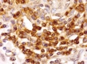
BAFF R/TNFRSF13C in Human Spleen.
BAFF R/TNFRSF13C was detected in immersion fixed paraffin-embedded sections of human spleen using Rabbit Anti-Human BAFF R/TNFRSF13C Monoclonal Antibody (Catalog # MAB1162) at 5 µg/mL for 1 hour at room temperature followed by incubation with the Anti-Rabbit IgG VisUCyte™ HRP Polymer Antibody (Catalog # VC003). Before incubation with the primary antibody, tissue was subjected to heat-induced epitope retrieval using Antigen Retrieval Reagent-Basic (Catalog # CTS013). Tissue was stained using DAB (brown) and counterstained with hematoxylin (blue). Specific staining was localized to cell surfaces in lymphocytes. View our protocol for IHC Staining with VisUCyte HRP Polymer Detection Reagents.Applications for Human BAFFR/TNFRSF13C Antibody (2403C)
Application
Recommended Usage
CyTOF-ready
Ready to be labeled using established conjugation methods. No BSA or other carrier proteins that could interfere with conjugation.
Flow Cytometry
0.25 µg/106 cells
Sample: Human PBMC
Sample: Human PBMC
Immunohistochemistry
5-25 µg/mL
Sample: Immersion fixed paraffin-embedded sections of human spleen
Sample: Immersion fixed paraffin-embedded sections of human spleen
Reviewed Applications
Read 3 reviews rated 4.3 using MAB1162 in the following applications:
Flow Cytometry Panel Builder
Bio-Techne Knows Flow Cytometry
Save time and reduce costly mistakes by quickly finding compatible reagents using the Panel Builder Tool.
Advanced Features
- Spectra Viewer - Custom analysis of spectra from multiple fluorochromes
- Spillover Popups - Visualize the spectra of individual fluorochromes
- Antigen Density Selector - Match fluorochrome brightness with antigen density

Formulation, Preparation, and Storage
Purification
Protein A or G purified from cell culture supernatant
Reconstitution
Reconstitute at 0.5 mg/mL in sterile PBS. For liquid material, refer to CoA for concentration.
Loading...
Formulation
Lyophilized from a 0.2 μm filtered solution in PBS with Trehalose. *Small pack size (SP) is supplied either lyophilized or as a 0.2 µm filtered solution in PBS.
Shipping
Lyophilized product is shipped at ambient temperature. Liquid small pack size (-SP) is shipped with polar packs. Upon receipt, store immediately at the temperature recommended below.
Stability & Storage
Use a manual defrost freezer and avoid repeated freeze-thaw cycles.
- 12 months from date of receipt, -20 to -70 °C as supplied.
- 1 month, 2 to 8 °C under sterile conditions after reconstitution.
- 6 months, -20 to -70 °C under sterile conditions after reconstitution.
Calculators
Background: BAFFR/TNFRSF13C
References
- Rolink, A.G. and F. Melcher (2002) Curr. Opin. Immunol. 14:266.
- Mackay F. and J.L. Browning (2002) Nature Reviews Immunology 2:464.
- Laabi, Y. et al. (2001) Current Biol. 11:R1013.
- Thompson, J.S. et al. (2001) Science 14:2108.
Long Name
B cell Activating Factor Receptor
Alternate Names
BAFF R, BR3, CD268, TNFRSF13C
Gene Symbol
TNFRSF13C
UniProt
Additional BAFFR/TNFRSF13C Products
Product Documents for Human BAFFR/TNFRSF13C Antibody (2403C)
Certificate of Analysis
To download a Certificate of Analysis, please enter a lot or batch number in the search box below.
Note: Certificate of Analysis not available for kit components.
Product Specific Notices for Human BAFFR/TNFRSF13C Antibody (2403C)
For research use only
Customer Reviews for Human BAFFR/TNFRSF13C Antibody (2403C) (3)
4.3 out of 5
3 Customer Ratings
Have you used Human BAFFR/TNFRSF13C Antibody (2403C)?
Submit a review and receive an Amazon gift card!
$25/€18/£15/$25CAN/¥2500 Yen for a review with an image
$10/€7/£6/$10CAN/¥1110 Yen for a review without an image
Submit a review
Customer Images
Showing
1
-
3 of
3 reviews
Showing All
Filter By:
-
Application: ImmunohistochemistrySample Tested: Pancreatic Ductal AdenocarcinomaSpecies: HumanVerified Customer | Posted 11/11/2021

-
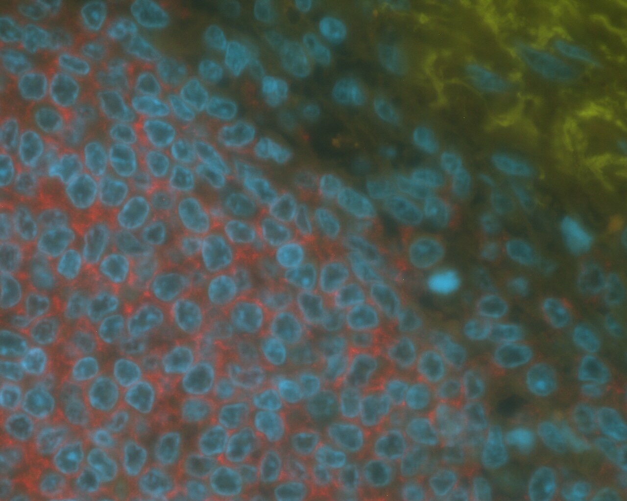
Application: Immunohistochemistry-ParaffinSample Tested: Human spleen, FFPESpecies: HumanVerified Customer | Posted 12/16/2019Spleen tissue stained with 1:50 MAB1162 shows strong membranous B-cell positivity (red fluorescence) in white pulp, but no positive staining in red pulp.Citrate buffer (pH 6) antigen unmasking. Antibody was used at a 1:50 concentration and detected with anti-rabbit AF546 (red fluor) secondary Ab.
-
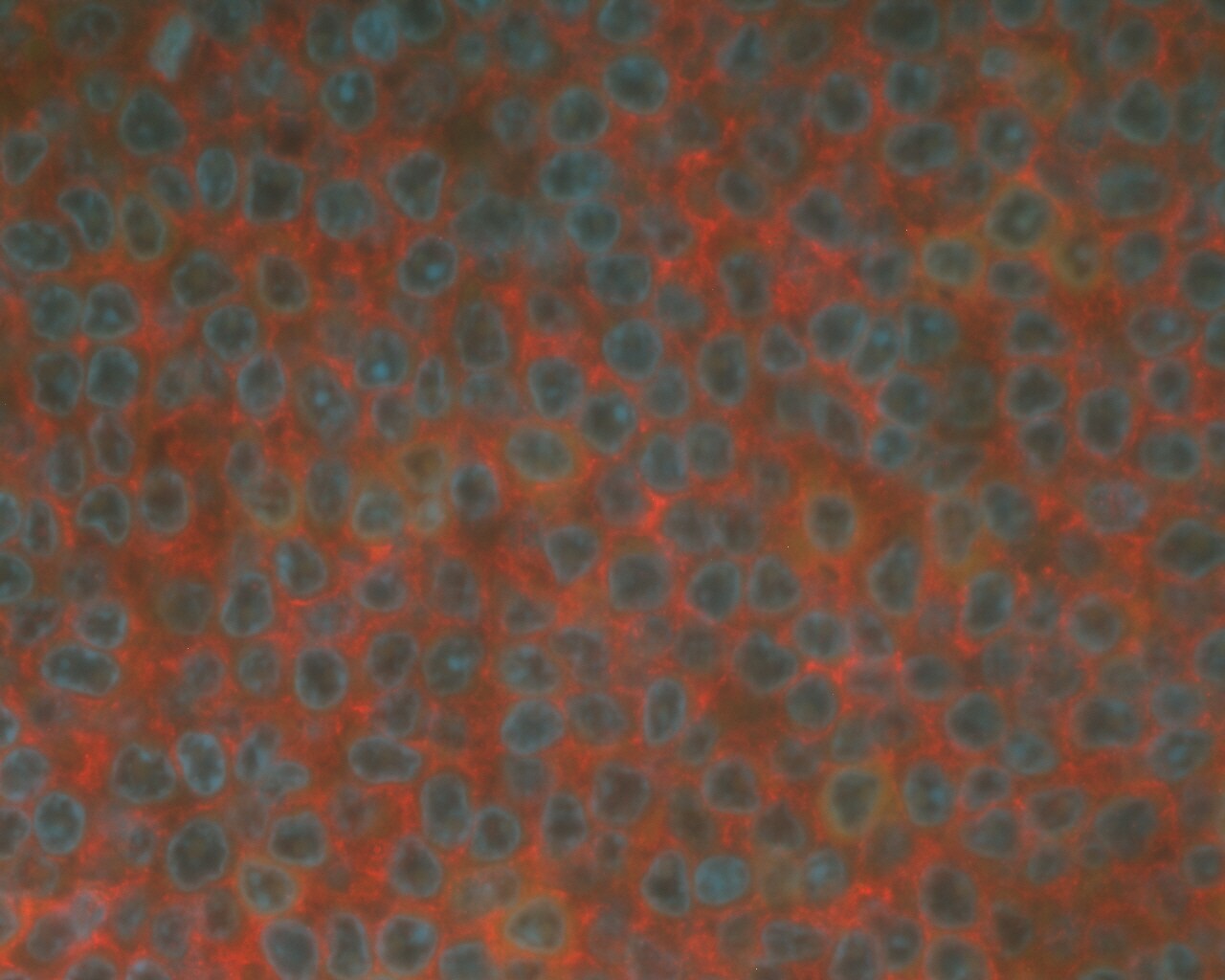
Application: ImmunohistochemistrySample Tested: Spleen tissueSpecies: HumanVerified Customer | Posted 11/04/2019Fluorescent immunohistochemistry with red-labelled secondary antibody on human spleen tissue, at 1:50 dilution. Next time would use higher dilution.
There are no reviews that match your criteria.
Protocols
Find general support by application which include: protocols, troubleshooting, illustrated assays, videos and webinars.
- 7-Amino Actinomycin D (7-AAD) Cell Viability Flow Cytometry Protocol
- Antigen Retrieval Protocol (PIER)
- Antigen Retrieval for Frozen Sections Protocol
- Appropriate Fixation of IHC/ICC Samples
- Cellular Response to Hypoxia Protocols
- Chromogenic IHC Staining of Formalin-Fixed Paraffin-Embedded (FFPE) Tissue Protocol
- Chromogenic Immunohistochemistry Staining of Frozen Tissue
- ClariTSA™ Fluorophore Kits
- Detection & Visualization of Antibody Binding
- Extracellular Membrane Flow Cytometry Protocol
- Flow Cytometry Protocol for Cell Surface Markers
- Flow Cytometry Protocol for Staining Membrane Associated Proteins
- Flow Cytometry Staining Protocols
- Flow Cytometry Troubleshooting Guide
- Fluorescent IHC Staining of Frozen Tissue Protocol
- Graphic Protocol for Heat-induced Epitope Retrieval
- Graphic Protocol for the Preparation and Fluorescent IHC Staining of Frozen Tissue Sections
- Graphic Protocol for the Preparation and Fluorescent IHC Staining of Paraffin-embedded Tissue Sections
- Graphic Protocol for the Preparation of Gelatin-coated Slides for Histological Tissue Sections
- IHC Sample Preparation (Frozen sections vs Paraffin)
- Immunofluorescent IHC Staining of Formalin-Fixed Paraffin-Embedded (FFPE) Tissue Protocol
- Immunohistochemistry (IHC) and Immunocytochemistry (ICC) Protocols
- Immunohistochemistry Frozen Troubleshooting
- Immunohistochemistry Paraffin Troubleshooting
- Intracellular Flow Cytometry Protocol Using Alcohol (Methanol)
- Intracellular Flow Cytometry Protocol Using Detergents
- Intracellular Nuclear Staining Flow Cytometry Protocol Using Detergents
- Intracellular Staining Flow Cytometry Protocol Using Alcohol Permeabilization
- Intracellular Staining Flow Cytometry Protocol Using Detergents to Permeabilize Cells
- Preparing Samples for IHC/ICC Experiments
- Preventing Non-Specific Staining (Non-Specific Binding)
- Primary Antibody Selection & Optimization
- Propidium Iodide Cell Viability Flow Cytometry Protocol
- Protocol for Heat-Induced Epitope Retrieval (HIER)
- Protocol for Liperfluo
- Protocol for Making a 4% Formaldehyde Solution in PBS
- Protocol for VisUCyte™ HRP Polymer Detection Reagent
- Protocol for the Characterization of Human Th22 Cells
- Protocol for the Characterization of Human Th9 Cells
- Protocol for the Preparation & Fixation of Cells on Coverslips
- Protocol for the Preparation and Chromogenic IHC Staining of Frozen Tissue Sections
- Protocol for the Preparation and Chromogenic IHC Staining of Frozen Tissue Sections - Graphic
- Protocol for the Preparation and Chromogenic IHC Staining of Paraffin-embedded Tissue Sections
- Protocol for the Preparation and Chromogenic IHC Staining of Paraffin-embedded Tissue Sections - Graphic
- Protocol for the Preparation and Fluorescent IHC Staining of Frozen Tissue Sections
- Protocol for the Preparation and Fluorescent IHC Staining of Paraffin-embedded Tissue Sections
- Protocol for the Preparation of Gelatin-coated Slides for Histological Tissue Sections
- Protocol: Annexin V and PI Staining by Flow Cytometry
- Protocol: Annexin V and PI Staining for Apoptosis by Flow Cytometry
- TUNEL and Active Caspase-3 Detection by IHC/ICC Protocol
- The Importance of IHC/ICC Controls
- Troubleshooting Guide: Fluorokine Flow Cytometry Kits
- Troubleshooting Guide: Immunohistochemistry
- View all Protocols, Troubleshooting, Illustrated assays and Webinars
Loading...
Associated Pathways
