Cathepsin A/lyososomal carboxypeptidase A is a member of the serine carboxypeptidase family (1). Cathepsin A is a multifunctional enzyme that expresses deaminidase and esterase activities at neutral pH and carboxypeptidase activity at acidic pH. Also known as protective protein, its association with beta -galactosidase ( beta -gal) and neuraminidase is essential for beta -gal stability and neuraminidase activation in the lysosomes. Inherited deficiency of Cathepsin A causes the lysosomal storage disorder galactosialidosis, characterized by a combined secondary deficiency of beta -gal and neuraminidase. Cathepsin A is capable of hydrolyzing a variety of bioactive peptide hormones including tachykinins, indicating that extralysosomal Cathepsin A plays a role in regulation of functions of these molecules (2). Cathepsin A is synthesized as a single-chain precursor and processed into heavy (32 kDa) and light (20 kDa) chains, which are linked by disulfide bonds.
Human Cathepsin A/Lysosomal Carboxypeptidase A Antibody
R&D Systems | Catalog # AF1049

Key Product Details
Species Reactivity
Validated:
Human
Cited:
Human, Mouse, Transgenic Mouse
Applications
Validated:
Immunohistochemistry, Western Blot
Cited:
Immunohistochemistry, Western Blot, Immunocytochemistry, Dot Blot
Label
Unconjugated
Antibody Source
Polyclonal Goat IgG
Loading...
Product Specifications
Immunogen
Mouse myeloma cell line NS0-derived recombinant human Cathepsin A/Lysosomal Carboxypeptidase A
Ala29-Tyr480
Accession # P10619
Ala29-Tyr480
Accession # P10619
Specificity
Detects both pro and active forms of human and mouse Cathepsin A/Lysosomal Carboxypeptidase A in direct ELISAs and Western blots. In Western blots, it recognizes all forms of recombinant human Cathepsin A: single chain (55 kDa), heavy chain (32 kDa), and light chain (20 kDa). Also in Western blots, less than 1% cross-reactivity with recombinant human (rh) Cathepsin B, rhCathepsin C, rhCathepsin D, rhCathepsin L, rhCathepsin L2/V, rhCathepsin O, rhCathepsin S and rhCathepsin X/Z/P is observed.
Clonality
Polyclonal
Host
Goat
Isotype
IgG
Scientific Data Images for Human Cathepsin A/Lysosomal Carboxypeptidase A Antibody
Cathepsin A/Lysosomal Carboxypeptidase A in Human Prostate.
Cathepsin A/Lysosomal Carboxypeptidase A was detected in immersion fixed paraffin-embedded sections of human prostate using Goat Anti-Human Cathepsin A/Lysosomal Carboxypeptidase A Antigen Affinity-purified Polyclonal Antibody (Catalog # AF1049) at 3 µg/mL overnight at 4 °C. Before incubation with the primary antibody, tissue was subjected to heat-induced epitope retrieval using Antigen Retrieval Reagent-Basic (Catalog # CTS013). Tissue was stained using the Anti-Goat HRP-DAB Cell & Tissue Staining Kit (brown; Catalog # CTS008) and counterstained with hematoxylin (blue). Specific staining was localized to cytoplasm in epithelial cells. View our protocol for Chromogenic IHC Staining of Paraffin-embedded Tissue Sections.Applications for Human Cathepsin A/Lysosomal Carboxypeptidase A Antibody
Application
Recommended Usage
Immunohistochemistry
5-15 µg/mL
Sample: Immersion fixed paraffin-embedded sections of human prostate subjected to Antigen Retrieval Reagent-Basic (Catalog # CTS013)
Sample: Immersion fixed paraffin-embedded sections of human prostate subjected to Antigen Retrieval Reagent-Basic (Catalog # CTS013)
Western Blot
0.1 µg/mL
Sample: Recombinant Human Cathepsin A/Lysosomal Carboxypeptidase A (Catalog # 1049-SE)
Sample: Recombinant Human Cathepsin A/Lysosomal Carboxypeptidase A (Catalog # 1049-SE)
Reviewed Applications
Read 1 review rated 4 using AF1049 in the following applications:
Formulation, Preparation, and Storage
Purification
Antigen Affinity-purified
Reconstitution
Reconstitute at 0.2 mg/mL in sterile PBS. For liquid material, refer to CoA for concentration.
Loading...
Formulation
Lyophilized from a 0.2 μm filtered solution in PBS with Trehalose. *Small pack size (SP) is supplied either lyophilized or as a 0.2 µm filtered solution in PBS.
Shipping
Lyophilized product is shipped at ambient temperature. Liquid small pack size (-SP) is shipped with polar packs. Upon receipt, store immediately at the temperature recommended below.
Stability & Storage
Use a manual defrost freezer and avoid repeated freeze-thaw cycles.
- 12 months from date of receipt, -20 to -70 °C as supplied.
- 1 month, 2 to 8 °C under sterile conditions after reconstitution.
- 6 months, -20 to -70 °C under sterile conditions after reconstitution.
Calculators
Background: Cathepsin A/Lysosomal Carboxypeptidase A
References
- Pshezhetsky, A.V. (2004) in Handbook of Proteolytic Enzymes (ed. Barrett, A.J. et al.) p. 1923, Academic Press, San Diego.
- Hiraiwa, M. (1999) Cell. Mol. Life. Sci. 56:894.
Alternate Names
CTSA, Lysosomal Carboxypeptidase A
Gene Symbol
CTSA
UniProt
Additional Cathepsin A/Lysosomal Carboxypeptidase A Products
- All Products for Cathepsin A/Lysosomal Carboxypeptidase A
- Cathepsin A/Lysosomal Carboxypeptidase A cDNA Clones
- Cathepsin A/Lysosomal Carboxypeptidase A ELISA Kits
- Cathepsin A/Lysosomal Carboxypeptidase A Lysates
- Cathepsin A/Lysosomal Carboxypeptidase A Primary Antibodies
- Cathepsin A/Lysosomal Carboxypeptidase A Proteins and Enzymes
Product Documents for Human Cathepsin A/Lysosomal Carboxypeptidase A Antibody
Certificate of Analysis
To download a Certificate of Analysis, please enter a lot or batch number in the search box below.
Note: Certificate of Analysis not available for kit components.
Product Specific Notices for Human Cathepsin A/Lysosomal Carboxypeptidase A Antibody
For research use only
Related Research Areas
Citations for Human Cathepsin A/Lysosomal Carboxypeptidase A Antibody
 Powered by Bioz
Customer Reviews for Human Cathepsin A/Lysosomal Carboxypeptidase A Antibody (1)
4 out of 5
1 Customer Rating
Have you used Human Cathepsin A/Lysosomal Carboxypeptidase A Antibody?
Submit a review and receive an Amazon gift card!
$25/€18/£15/$25CAN/¥2500 Yen for a review with an image
$10/€7/£6/$10CAN/¥1110 Yen for a review without an image
Submit a review
Customer Images
Showing
1
-
1 of
1 review
Showing All
Filter By:
-
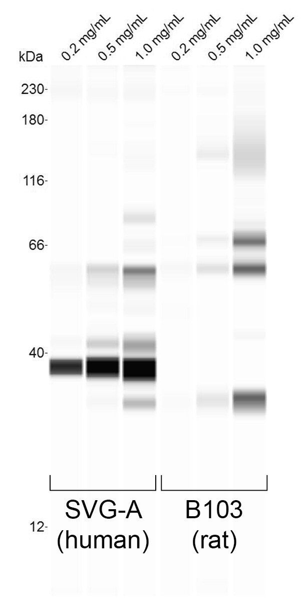
Application: Simple WesternSample Tested: B103 lysate and SVG-A lysateSpecies: Human and RatVerified Customer | Posted 08/30/2018CHAP lysates from the indicated cell lines, loaded at the indicated concentrations. Antibody was tested at 1:25 dil.

There are no reviews that match your criteria.
Protocols
Find general support by application which include: protocols, troubleshooting, illustrated assays, videos and webinars.
- Antigen Retrieval Protocol (PIER)
- Antigen Retrieval for Frozen Sections Protocol
- Appropriate Fixation of IHC/ICC Samples
- Cellular Response to Hypoxia Protocols
- Chromogenic IHC Staining of Formalin-Fixed Paraffin-Embedded (FFPE) Tissue Protocol
- Chromogenic Immunohistochemistry Staining of Frozen Tissue
- ClariTSA™ Fluorophore Kits
- Detection & Visualization of Antibody Binding
- Fluorescent IHC Staining of Frozen Tissue Protocol
- Graphic Protocol for Heat-induced Epitope Retrieval
- Graphic Protocol for the Preparation and Fluorescent IHC Staining of Frozen Tissue Sections
- Graphic Protocol for the Preparation and Fluorescent IHC Staining of Paraffin-embedded Tissue Sections
- Graphic Protocol for the Preparation of Gelatin-coated Slides for Histological Tissue Sections
- IHC Sample Preparation (Frozen sections vs Paraffin)
- Immunofluorescent IHC Staining of Formalin-Fixed Paraffin-Embedded (FFPE) Tissue Protocol
- Immunohistochemistry (IHC) and Immunocytochemistry (ICC) Protocols
- Immunohistochemistry Frozen Troubleshooting
- Immunohistochemistry Paraffin Troubleshooting
- Preparing Samples for IHC/ICC Experiments
- Preventing Non-Specific Staining (Non-Specific Binding)
- Primary Antibody Selection & Optimization
- Protocol for Heat-Induced Epitope Retrieval (HIER)
- Protocol for Making a 4% Formaldehyde Solution in PBS
- Protocol for VisUCyte™ HRP Polymer Detection Reagent
- Protocol for the Preparation & Fixation of Cells on Coverslips
- Protocol for the Preparation and Chromogenic IHC Staining of Frozen Tissue Sections
- Protocol for the Preparation and Chromogenic IHC Staining of Frozen Tissue Sections - Graphic
- Protocol for the Preparation and Chromogenic IHC Staining of Paraffin-embedded Tissue Sections
- Protocol for the Preparation and Chromogenic IHC Staining of Paraffin-embedded Tissue Sections - Graphic
- Protocol for the Preparation and Fluorescent IHC Staining of Frozen Tissue Sections
- Protocol for the Preparation and Fluorescent IHC Staining of Paraffin-embedded Tissue Sections
- Protocol for the Preparation of Gelatin-coated Slides for Histological Tissue Sections
- R&D Systems Quality Control Western Blot Protocol
- TUNEL and Active Caspase-3 Detection by IHC/ICC Protocol
- The Importance of IHC/ICC Controls
- Troubleshooting Guide: Immunohistochemistry
- Troubleshooting Guide: Western Blot Figures
- Western Blot Conditions
- Western Blot Protocol
- Western Blot Protocol for Cell Lysates
- Western Blot Troubleshooting
- Western Blot Troubleshooting Guide
- View all Protocols, Troubleshooting, Illustrated assays and Webinars
Loading...