Human CD27 Ligand/TNFSF7 Antibody (301731)
R&D Systems | Catalog # MAB2738

Key Product Details
Species Reactivity
Validated:
Human
Cited:
Human, Mouse
Applications
Validated:
Flow Cytometry
Cited:
Immunohistochemistry, Immunohistochemistry-Paraffin
Label
Unconjugated
Antibody Source
Monoclonal Mouse IgG2B Clone # 301731
Loading...
Product Specifications
Immunogen
Mouse myeloma cell line NS0-derived recombinant human CD27 Ligand/TNFSF7
Gln45-Pro193
Accession # AAA36175
Gln45-Pro193
Accession # AAA36175
Specificity
Detects human CD27 Ligand/TNFSF7 in direct ELISAs.
Clonality
Monoclonal
Host
Mouse
Isotype
IgG2B
Scientific Data Images for Human CD27 Ligand/TNFSF7 Antibody (301731)
Detection of CD27Ligand/TNFSF7 in HEK293 Human Cell Line Transfected with Human CD27 Ligand/TNFSF7 by Flow Cytometry.
HEK293 human embryonic kidney cell line transfected with human CD27 Ligand/TNFSF7 (filled histogram) or irrelevant protein (open histogram) was stained with Mouse Anti-Human CD27 Ligand/TNFSF7 Monoclonal Antibody (Catalog # MAB2738) followed by Goat anti-mouse PE-conjugated secondary antibody (F0102B). Staining was performed using our Staining Membrane-associated Proteins protocol.
Detection of CD27 Ligand/TNFSF7 by Western Blot
Experimental setup and the effect of PHD3 silencing on 786-O proteome. a siRNA-mediated silencing of PHD3 protein level in 786-O ccRCC cell line using two individual siRNA sequences in normoxic and in hypoxic (1% O2) condition. Quantification of three biological replicates, mean ± SEM, fold change to control (Scr). Asterisk indicates a statistically significant difference (*p < 0.05, **p < 0.01). b siRNA-mediated silencing of PHD3 mRNA expression, quantification of three individual experiments. Mean ± SEM, fold change to Scr (***p < 0.001). c Flow chart of the experimental procedure. 786-O cells were transfected with siPHD3#1 or with a non-targeting control siRNA (Scr) for 24 h followed by a hypoxic (1% O2) or normoxic (21% O2) exposure. Three independent experiments were performed; proteins were extracted, followed by in-gel digestion with trypsin. Purified peptides were ran through mass spectrometer. Protein identification was done with Mascot database search. Protein quantification was carried out with Progenesis QI, followed by testing the differential expression between sample groups using peptide-level expression change averaging (PECA). d Western blot validations of selected proteins in 786-O and RCC4 cell lines with two individual siRNA sequences targeting PHD3, representative analyses are shown Image collected and cropped by CiteAb from the following open publication (https://pubmed.ncbi.nlm.nih.gov/28680592), licensed under a CC-BY license. Not internally tested by R&D Systems.Applications for Human CD27 Ligand/TNFSF7 Antibody (301731)
Application
Recommended Usage
Flow Cytometry
0.25 µg/106 cells
Sample: HEK293 human embryonic kidney cell line transfected with human CD27 Ligand/TNFSF7
Sample: HEK293 human embryonic kidney cell line transfected with human CD27 Ligand/TNFSF7
Reviewed Applications
Read 2 reviews rated 4 using MAB2738 in the following applications:
Flow Cytometry Panel Builder
Bio-Techne Knows Flow Cytometry
Save time and reduce costly mistakes by quickly finding compatible reagents using the Panel Builder Tool.
Advanced Features
- Spectra Viewer - Custom analysis of spectra from multiple fluorochromes
- Spillover Popups - Visualize the spectra of individual fluorochromes
- Antigen Density Selector - Match fluorochrome brightness with antigen density

Formulation, Preparation, and Storage
Purification
Protein A or G purified from hybridoma culture supernatant
Reconstitution
Reconstitute at 0.5 mg/mL in sterile PBS. For liquid material, refer to CoA for concentration.
Loading...
Formulation
Lyophilized from a 0.2 μm filtered solution in PBS with Trehalose. *Small pack size (SP) is supplied either lyophilized or as a 0.2 µm filtered solution in PBS.
Shipping
Lyophilized product is shipped at ambient temperature. Liquid small pack size (-SP) is shipped with polar packs. Upon receipt, store immediately at the temperature recommended below.
Stability & Storage
Use a manual defrost freezer and avoid repeated freeze-thaw cycles.
- 12 months from date of receipt, -20 to -70 °C as supplied.
- 1 month, 2 to 8 °C under sterile conditions after reconstitution.
- 6 months, -20 to -70 °C under sterile conditions after reconstitution.
Calculators
Background: CD27 Ligand/TNFSF7
Alternate Names
CD70, TNFSF7
Gene Symbol
CD70
UniProt
Additional CD27 Ligand/TNFSF7 Products
Product Documents for Human CD27 Ligand/TNFSF7 Antibody (301731)
Certificate of Analysis
To download a Certificate of Analysis, please enter a lot or batch number in the search box below.
Note: Certificate of Analysis not available for kit components.
Product Specific Notices for Human CD27 Ligand/TNFSF7 Antibody (301731)
For research use only
Citations for Human CD27 Ligand/TNFSF7 Antibody (301731)
 Powered by Bioz
Customer Reviews for Human CD27 Ligand/TNFSF7 Antibody (301731) (2)
4 out of 5
2 Customer Ratings
Have you used Human CD27 Ligand/TNFSF7 Antibody (301731)?
Submit a review and receive an Amazon gift card!
$25/€18/£15/$25CAN/¥2500 Yen for a review with an image
$10/€7/£6/$10CAN/¥1110 Yen for a review without an image
Submit a review
Customer Images
Showing
1
-
2 of
2 reviews
Showing All
Filter By:
-
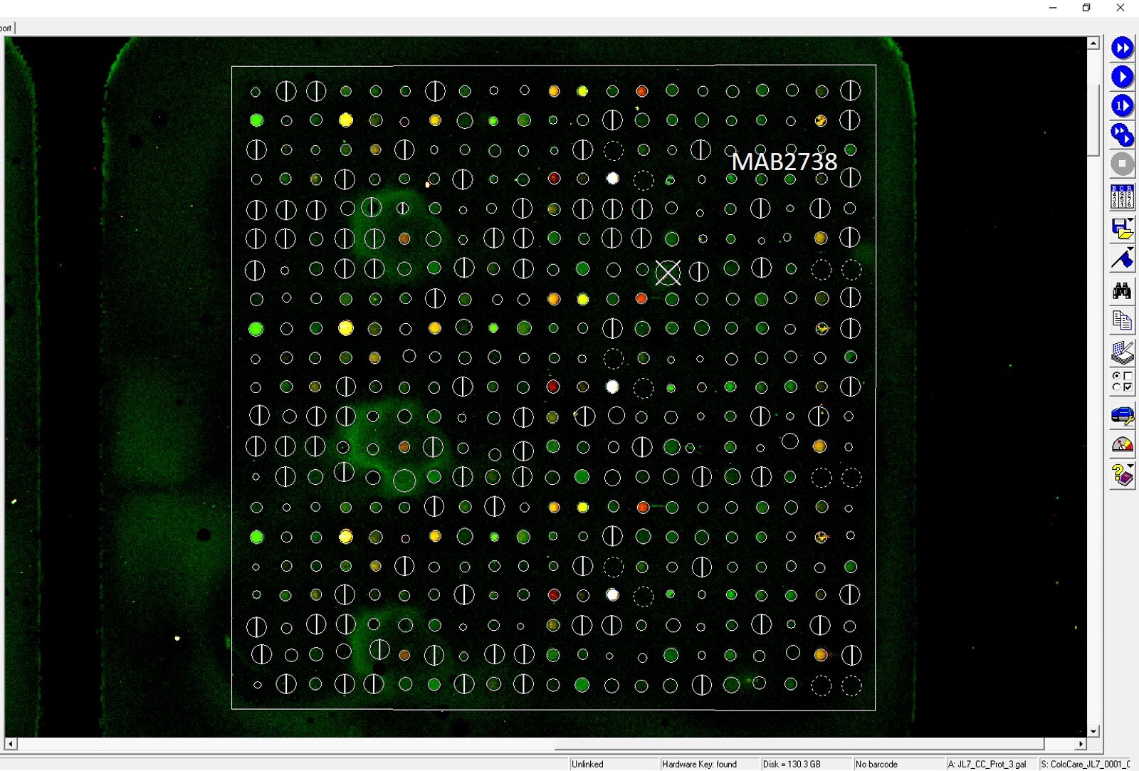
Application: MicroarraysSample Tested: EDTA PlasmaSpecies: HumanVerified Customer | Posted 03/19/2021

-
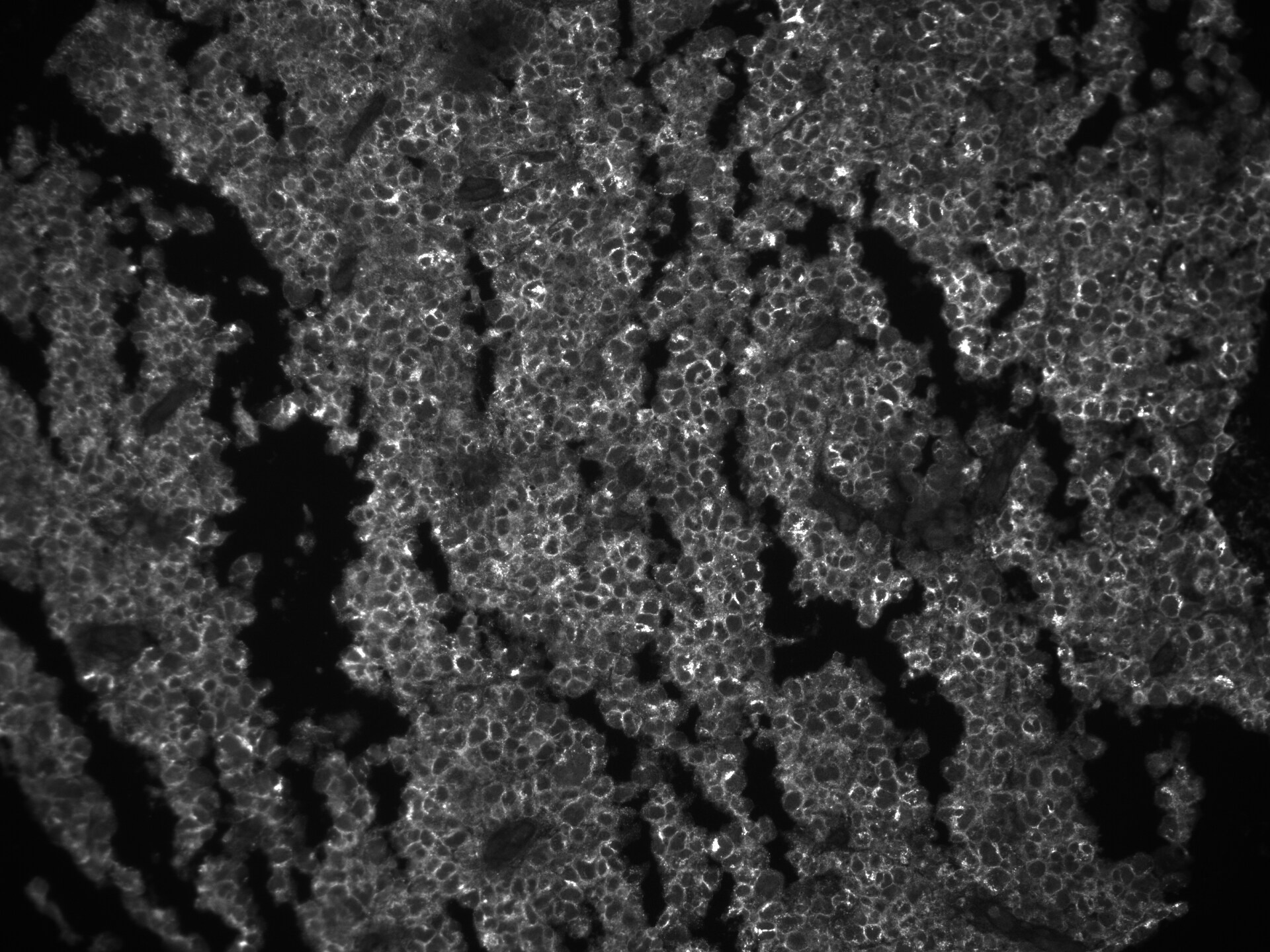
Application: Immunohistochemistry-ParaffinSample Tested: NK/T cell lymphomaSpecies: HumanVerified Customer | Posted 01/16/2020CD27L on human NK/T cell lymphoma, 1:200 dilutionpH 9 antigen retrieval
There are no reviews that match your criteria.
Protocols
Find general support by application which include: protocols, troubleshooting, illustrated assays, videos and webinars.
- 7-Amino Actinomycin D (7-AAD) Cell Viability Flow Cytometry Protocol
- Extracellular Membrane Flow Cytometry Protocol
- Flow Cytometry Protocol for Cell Surface Markers
- Flow Cytometry Protocol for Staining Membrane Associated Proteins
- Flow Cytometry Staining Protocols
- Flow Cytometry Troubleshooting Guide
- Intracellular Flow Cytometry Protocol Using Alcohol (Methanol)
- Intracellular Flow Cytometry Protocol Using Detergents
- Intracellular Nuclear Staining Flow Cytometry Protocol Using Detergents
- Intracellular Staining Flow Cytometry Protocol Using Alcohol Permeabilization
- Intracellular Staining Flow Cytometry Protocol Using Detergents to Permeabilize Cells
- Propidium Iodide Cell Viability Flow Cytometry Protocol
- Protocol for Liperfluo
- Protocol for the Characterization of Human Th22 Cells
- Protocol for the Characterization of Human Th9 Cells
- Protocol: Annexin V and PI Staining by Flow Cytometry
- Protocol: Annexin V and PI Staining for Apoptosis by Flow Cytometry
- Troubleshooting Guide: Fluorokine Flow Cytometry Kits
- View all Protocols, Troubleshooting, Illustrated assays and Webinars
Loading...
Associated Pathways
