Carcinoembryonic antigen-related cell adhesion molecule 6 (CEACAM-6), previously called nonspecific cross‑reacting antigen (NCA) or CD66c, is one of seven human CEACAM family members within the immunoglobulin superfamily (1-4). In humans, CEACAMs include type I transmembrane proteins (CEACAM-1, -3, and -4) and Glycosylphosphatidylinositol (GPI)-linked molecules (CEACAM-5 through -8) (1). There is no human CEACAM-2. Human CEACAM-6 is a 90 kDa, GPI-linked membrane protein that contains a 34 amino acid (aa) signal sequence, a 286 aa extracellular domain (ECD), and a 24 aa hydrophobic C-terminal propeptide. The GPI membrane anchor is attached at the C-terminus following cleavage of the propeptide. CEACAM-6 contains one N-terminal V-type Ig-like domain (N domain), followed by two C2-type Ig-like domains (2-4). It shows considerable glycosylation, including (sialyl) LewisX, which mediates binding to E-Selectin, Galectins and some bacterial fimbrae (1, 2). Mature human CEACAM-6 shows 84%, 85%, 80%, 87% and 51% aa identity to the equivalent extracellular regions of human CEACAMs -1, -5 (CEA) and -8, rhesus CEACAM-2, and bovine CEACAM-6, respectively. CEACAM-6 is expressed by granulocytes and their precursors. Activation enhances surface expression by mobilizing CEACAM-6 from storage in azurophilic granules (5, 6). It often shows aberrant expression in acute lymphocytic leukemias (10). CEACAM-6 is also expressed in epithelia of various organs and is upregulated in pancreatic and colon adenocarcinomas and hyperplastic polyps (7, 8). Over-expression confers resistance to adhesion-related apoptosis (anoikis) in tumor cells (8, 9). CEACAM-6 is an intercellular adhesion molecule, forming both homotypic, and heterotypic bonds with CEACAM-1, -5 and -8 through interaction of the V-type Ig-like domains (11, 12). Cross-linking of neutrophil CEACAM-6 augments Integrin alpha v beta 3 and beta 2-mediated adhesion, apparently by Src and Caveolin-mediated inside-out Integrin activation (8, 13, 14).
Human CEACAM-6/CD66c Antibody (439424)
R&D Systems | Catalog # MAB3934

Key Product Details
Species Reactivity
Validated:
Human
Cited:
Human
Applications
Validated:
Western Blot, Flow Cytometry, CyTOF-ready
Cited:
Flow Cytometry
Label
Unconjugated
Antibody Source
Monoclonal Mouse IgG2A Clone # 439424
Loading...
Product Specifications
Immunogen
Mouse myeloma cell line NS0-derived recombinant human CEACAM-6/CD66c
Lys35-Gly320
Accession # P40199
Lys35-Gly320
Accession # P40199
Specificity
Detects human CEACAM-6/CD66c in direct ELISAs and Western blots. In direct ELISAs and Western blots, no cross-reactivity with recombinant human CEACAM-1, -3, or -5 is observed.
Clonality
Monoclonal
Host
Mouse
Isotype
IgG2A
Scientific Data Images for Human CEACAM-6/CD66c Antibody (439424)
Detection of CEACAM‑6/CD66c in Human Granulocytes by Flow Cytometry.
Human whole blood granulocytes were stained with Mouse Anti-Human CEACAM-6/CD66c Monoclonal Antibody (Catalog # MAB3934, filled histogram) or isotype control antibody (Catalog # MAB003, open histogram), followed by Phycoerythrin-conjugated Anti-Mouse IgG F(ab')2Secondary Antibody (Catalog # F0102B).
Detection of CEACAM‑6/CD66c in HEK293 Human Cell Line Transfected with Human CEACAM-6/CD66c and eGFP by Flow Cytometry.
HEK293 human embryonic kidney cell line transfected with (A) human CEACAM-6/CD66c or (B) irrelevant transfectants and eGFP was stained with Mouse Anti-Human CEACAM-6/CD66c Monoclonal Antibody (Catalog # MAB3934) followed by Allophycocyanin-conjugated Anti-Mouse IgG Secondary Antibody (Catalog # F0101B). Quadrant markers were set based on control antibody staining (Catalog # MAB003).
Detection of CEACAM-6/CD66c by Flow Cytometry
SSc serum induced CEACAM6 expression on classical monocytes. CD14+16- monocytes were isolated from a healthy donor and were treated with serum from HCs (n=7) and patients with systemic sclerosis (n=14) for 15 h. The proportion of CEACAM-positive classical monocytes from patients with SSc who provided serum was also analyzed. (A) Dot plots show the correlation between the proportion of CEACAM+ monocytes from a healthy donor treated with SSc serum and that from patients with SSc obtained at the same time as the serum. Statistical analysis was performed using Spearman’s rank correlation test. (B) The frequency of serum-induced CEACAM+CD14+16- monocytes was compared among HCs (n=7), inactive SSc (n=7), and active SSc (n=7). Each analysis was performed in duplicates. The left figure shows a histogram of CEACAM. (C, D) Data show the frequency of CEACAM1, 3, 5, and 6+ monocytes treated with serum from HCs or patients with dcSSc. Gating of CEACAM6+ monocytes is shown in (C) Data are presented as mean ± standard deviation (SD). P values were calculated using an unpaired t-test. *P < 0.05, ***P < 0.001. Image collected and cropped by CiteAb from the following open publication (https://pubmed.ncbi.nlm.nih.gov/36341379), licensed under a CC-BY license. Not internally tested by R&D Systems.
Detection of CEACAM-6/CD66c by Flow Cytometry
SSc serum induced CEACAM6 expression on classical monocytes. CD14+16- monocytes were isolated from a healthy donor and were treated with serum from HCs (n=7) and patients with systemic sclerosis (n=14) for 15 h. The proportion of CEACAM-positive classical monocytes from patients with SSc who provided serum was also analyzed. (A) Dot plots show the correlation between the proportion of CEACAM+ monocytes from a healthy donor treated with SSc serum and that from patients with SSc obtained at the same time as the serum. Statistical analysis was performed using Spearman’s rank correlation test. (B) The frequency of serum-induced CEACAM+CD14+16- monocytes was compared among HCs (n=7), inactive SSc (n=7), and active SSc (n=7). Each analysis was performed in duplicates. The left figure shows a histogram of CEACAM. (C, D) Data show the frequency of CEACAM1, 3, 5, and 6+ monocytes treated with serum from HCs or patients with dcSSc. Gating of CEACAM6+ monocytes is shown in (C) Data are presented as mean ± standard deviation (SD). P values were calculated using an unpaired t-test. *P < 0.05, ***P < 0.001. Image collected and cropped by CiteAb from the following open publication (https://pubmed.ncbi.nlm.nih.gov/36341379), licensed under a CC-BY license. Not internally tested by R&D Systems.Applications for Human CEACAM-6/CD66c Antibody (439424)
Application
Recommended Usage
CyTOF-ready
Ready to be labeled using established conjugation methods. No BSA or other carrier proteins that could interfere with conjugation.
Flow Cytometry
2.5 µg/106 cells
Sample: Human whole blood granulocytes and HEK293 human embryonic kidney cell line transfected with human CEACAM-6/CD66c and eGFP
Sample: Human whole blood granulocytes and HEK293 human embryonic kidney cell line transfected with human CEACAM-6/CD66c and eGFP
Western Blot
1 µg/mL
Sample: Recombinant Human CEACAM‑6/CD66c (Catalog # 3934-CM)
Sample: Recombinant Human CEACAM‑6/CD66c (Catalog # 3934-CM)
Reviewed Applications
Read 1 review rated 3 using MAB3934 in the following applications:
Flow Cytometry Panel Builder
Bio-Techne Knows Flow Cytometry
Save time and reduce costly mistakes by quickly finding compatible reagents using the Panel Builder Tool.
Advanced Features
- Spectra Viewer - Custom analysis of spectra from multiple fluorochromes
- Spillover Popups - Visualize the spectra of individual fluorochromes
- Antigen Density Selector - Match fluorochrome brightness with antigen density

Formulation, Preparation, and Storage
Purification
Protein A or G purified from hybridoma culture supernatant
Reconstitution
Reconstitute at 0.5 mg/mL in sterile PBS. For liquid material, refer to CoA for concentration.
Loading...
Formulation
Lyophilized from a 0.2 μm filtered solution in PBS with Trehalose. *Small pack size (SP) is supplied either lyophilized or as a 0.2 µm filtered solution in PBS.
Shipping
Lyophilized product is shipped at ambient temperature. Liquid small pack size (-SP) is shipped with polar packs. Upon receipt, store immediately at the temperature recommended below.
Stability & Storage
Use a manual defrost freezer and avoid repeated freeze-thaw cycles.
- 12 months from date of receipt, -20 to -70 °C as supplied.
- 1 month, 2 to 8 °C under sterile conditions after reconstitution.
- 6 months, -20 to -70 °C under sterile conditions after reconstitution.
Calculators
Background: CEACAM-6/CD66c
References
- Beauchemin, N. et al. (1999) Exp. Cell Res. 252:243.
- Skubitz, K.M. et al. (1999) J. Biol. Regul. Homeost. Agents 13:244.
- Barnett, T. et al. (1988) Genomics 3:59.
- Tawaragi, Y. et al. (1988) Biochem. Biophys. Res. Comm. 150:89.
- Kuroki, M. et al. (1995) Immunol. Invest. 24:829.
- Ducker, T.P. and K.M. Skubitz (1992) J. Leukoc. Biol. 52:11.
- Scholzel, S. et al. (2000) Am. J. Pathol. 156:595.
- Duxbury, M.S. et al. (2004) J. Biol. Chem. 279:23176.
- Ilantzis, C. et al. (2002) Neoplasia 4:151.
- Kalina, T. et al. (2005) BMC Cancer 5:38.
- Oikawa, S. et al. (1992) Biochem. Biophys. Res. Commun. 186:881.
- Kuroki, M. et al. (2001) J. Leukoc. Biol. 70:543.
- Duxbury, M.S. et al. (2004) Biochem. Biophys. Res. Comm. 317:133.
- Skubitz, K.M. et al. (1999) J. Leukoc. Biol. 60:106.
Long Name
Carcinoembryonic Antigen-related Cell Adhesion Molecule 6
Alternate Names
CD66c, CEACAM6, CEAL, NCA
Gene Symbol
CEACAM6
UniProt
Additional CEACAM-6/CD66c Products
Product Documents for Human CEACAM-6/CD66c Antibody (439424)
Certificate of Analysis
To download a Certificate of Analysis, please enter a lot or batch number in the search box below.
Note: Certificate of Analysis not available for kit components.
Product Specific Notices for Human CEACAM-6/CD66c Antibody (439424)
For research use only
Citations for Human CEACAM-6/CD66c Antibody (439424)
 Powered by Bioz
Customer Reviews for Human CEACAM-6/CD66c Antibody (439424) (1)
3 out of 5
1 Customer Rating
Have you used Human CEACAM-6/CD66c Antibody (439424)?
Submit a review and receive an Amazon gift card!
$25/€18/£15/$25CAN/¥2500 Yen for a review with an image
$10/€7/£6/$10CAN/¥1110 Yen for a review without an image
Submit a review
Customer Images
Showing
1
-
1 of
1 review
Showing All
Filter By:
-
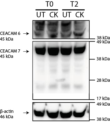
Application: Western BlotSample Tested: HT-29 human colon adenocarcinoma cell lineSpecies: HumanVerified Customer | Posted 08/09/2021

There are no reviews that match your criteria.
Protocols
Find general support by application which include: protocols, troubleshooting, illustrated assays, videos and webinars.
- 7-Amino Actinomycin D (7-AAD) Cell Viability Flow Cytometry Protocol
- Cellular Response to Hypoxia Protocols
- Extracellular Membrane Flow Cytometry Protocol
- Flow Cytometry Protocol for Cell Surface Markers
- Flow Cytometry Protocol for Staining Membrane Associated Proteins
- Flow Cytometry Staining Protocols
- Flow Cytometry Troubleshooting Guide
- Intracellular Flow Cytometry Protocol Using Alcohol (Methanol)
- Intracellular Flow Cytometry Protocol Using Detergents
- Intracellular Nuclear Staining Flow Cytometry Protocol Using Detergents
- Intracellular Staining Flow Cytometry Protocol Using Alcohol Permeabilization
- Intracellular Staining Flow Cytometry Protocol Using Detergents to Permeabilize Cells
- Propidium Iodide Cell Viability Flow Cytometry Protocol
- Protocol for Liperfluo
- Protocol for the Characterization of Human Th22 Cells
- Protocol for the Characterization of Human Th9 Cells
- Protocol: Annexin V and PI Staining by Flow Cytometry
- Protocol: Annexin V and PI Staining for Apoptosis by Flow Cytometry
- R&D Systems Quality Control Western Blot Protocol
- Troubleshooting Guide: Fluorokine Flow Cytometry Kits
- Troubleshooting Guide: Western Blot Figures
- Western Blot Conditions
- Western Blot Protocol
- Western Blot Protocol for Cell Lysates
- Western Blot Troubleshooting
- Western Blot Troubleshooting Guide
- View all Protocols, Troubleshooting, Illustrated assays and Webinars
Loading...
Associated Pathways
