CSL (CBF1/RBP-Jk; Su(H)/Suppressor of Hairless; Lag-1) is a 56 kDa member of the Su(H) family of transcription factors. It is constitutively bound to DNA where it participates in gene repression. Upon ligand binding, the intracellular domain of Notch translocates to the nucleus and interacts with CSL, allowing transcription of Notch target genes. Human CSL is 500 amino acids (aa) in length. It contains three DNA binding sites (aa 58‑65; 192‑201; and 265‑297) and one IPT domain (aa 355‑445). Multiple splice variants exist that involve the first 89 N-terminal amino acids. One variant (isoform 2) is 487 aa in length and shows a 6 aa substitution for amino acids 1‑19. Over amino acids 195‑487, human CSL isoform 2 shares more than 98% aa sequence identity with mouse, rat, and canine CSL.

Key Product Details
Species Reactivity
Human
Applications
Western Blot, Immunocytochemistry
Label
Unconjugated
Antibody Source
Polyclonal Goat IgG
Loading...
Product Specifications
Immunogen
E. coli-derived recombinant human CSL
Gly208-Ser500
Accession # Q06330
Gly208-Ser500
Accession # Q06330
Specificity
Detects human CSL in direct ELISAs and Western blots.
Clonality
Polyclonal
Host
Goat
Isotype
IgG
Scientific Data Images for Human CSL Antibody
CSL in U937 Human Cell Line.
CSL was detected in immersion fixed U937 human histiocytic lymphoma cell line using Goat Anti-Human CSL Antigen Affinity-purified Polyclonal Antibody (Catalog # AF4079) at 25 µg/mL for 3 hours at room temperature. Cells were stained using the NorthernLights™ 557-conjugated Anti-Goat IgG Secondary Antibody (red; NL001) and counterstained with DAPI (blue). Specific staining was localized to cytoplasm. Staining was performed using our protocol for Fluorescent ICC Staining of Non-adherent Cells.Applications for Human CSL Antibody
Application
Recommended Usage
Immunocytochemistry
5-25 µg/mL
Sample: Immersion fixed U937 human histiocytic lymphoma cell line
Sample: Immersion fixed U937 human histiocytic lymphoma cell line
Western Blot
0.1 µg/mL
Sample: Recombinant Human CSL
Sample: Recombinant Human CSL
Reviewed Applications
Read 1 review rated 5 using AF4079 in the following applications:
Formulation, Preparation, and Storage
Purification
Antigen Affinity-purified
Reconstitution
Reconstitute at 0.2 mg/mL in sterile PBS. For liquid material, refer to CoA for concentration.
Loading...
Formulation
Lyophilized from a 0.2 μm filtered solution in PBS with Trehalose. *Small pack size (SP) is supplied either lyophilized or as a 0.2 µm filtered solution in PBS.
Shipping
Lyophilized product is shipped at ambient temperature. Liquid small pack size (-SP) is shipped with polar packs. Upon receipt, store immediately at the temperature recommended below.
Stability & Storage
Use a manual defrost freezer and avoid repeated freeze-thaw cycles.
- 12 months from date of receipt, -20 to -70 °C as supplied.
- 1 month, 2 to 8 °C under sterile conditions after reconstitution.
- 6 months, -20 to -70 °C under sterile conditions after reconstitution.
Calculators
Background: CSL
Long Name
Recombining Binding Protein Supressor of Hairless [CBF1, CSL]
Alternate Names
CBF1, IGKJRB1, KBF2, RBP-JK, RBPJ, RBPSUH
Gene Symbol
RBPJ
UniProt
Additional CSL Products
Product Documents for Human CSL Antibody
Certificate of Analysis
To download a Certificate of Analysis, please enter a lot or batch number in the search box below.
Note: Certificate of Analysis not available for kit components.
Product Specific Notices for Human CSL Antibody
For research use only
Related Research Areas
Customer Reviews for Human CSL Antibody (1)
5 out of 5
1 Customer Rating
Have you used Human CSL Antibody?
Submit a review and receive an Amazon gift card!
$25/€18/£15/$25CAN/¥2500 Yen for a review with an image
$10/€7/£6/$10CAN/¥1110 Yen for a review without an image
Submit a review
Customer Images
Showing
1
-
1 of
1 review
Showing All
Filter By:
-
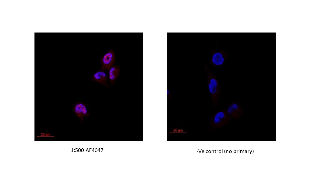
Application: Immunocytochemistry/ImmunofluorescenceSample Tested: Bladder tissueSpecies: HumanVerified Customer | Posted 02/08/2018Human T24 bladder cancer cell lines, overnight incubation at 4C. Concentration used 1:500. Secondary antigoat alexa 594, 1h incubation at room temperature.

There are no reviews that match your criteria.
Protocols
Find general support by application which include: protocols, troubleshooting, illustrated assays, videos and webinars.
- Appropriate Fixation of IHC/ICC Samples
- Cellular Response to Hypoxia Protocols
- ClariTSA™ Fluorophore Kits
- Detection & Visualization of Antibody Binding
- ICC Cell Smear Protocol for Suspension Cells
- ICC Immunocytochemistry Protocol Videos
- ICC for Adherent Cells
- Immunocytochemistry (ICC) Protocol
- Immunocytochemistry Troubleshooting
- Immunofluorescence of Organoids Embedded in Cultrex Basement Membrane Extract
- Immunohistochemistry (IHC) and Immunocytochemistry (ICC) Protocols
- Preparing Samples for IHC/ICC Experiments
- Preventing Non-Specific Staining (Non-Specific Binding)
- Primary Antibody Selection & Optimization
- Protocol for VisUCyte™ HRP Polymer Detection Reagent
- Protocol for the Fluorescent ICC Staining of Cell Smears - Graphic
- Protocol for the Fluorescent ICC Staining of Cultured Cells on Coverslips - Graphic
- Protocol for the Preparation and Fluorescent ICC Staining of Cells on Coverslips
- Protocol for the Preparation and Fluorescent ICC Staining of Non-adherent Cells
- Protocol for the Preparation and Fluorescent ICC Staining of Stem Cells on Coverslips
- Protocol for the Preparation of a Cell Smear for Non-adherent Cell ICC - Graphic
- R&D Systems Quality Control Western Blot Protocol
- TUNEL and Active Caspase-3 Detection by IHC/ICC Protocol
- The Importance of IHC/ICC Controls
- Troubleshooting Guide: Western Blot Figures
- Western Blot Conditions
- Western Blot Protocol
- Western Blot Protocol for Cell Lysates
- Western Blot Troubleshooting
- Western Blot Troubleshooting Guide
- View all Protocols, Troubleshooting, Illustrated assays and Webinars
Loading...
Associated Pathways

